Review
. 2012 Dec 7;11(6):751-64.
doi: 10.1016/j.stem.2012.11.001.

The harmonies played by TGF-β in stem cell biology

Affiliations
Review

The harmonies played by TGF-β in stem cell biology

Naoki Oshimori et al. Cell Stem Cell. .

Abstract

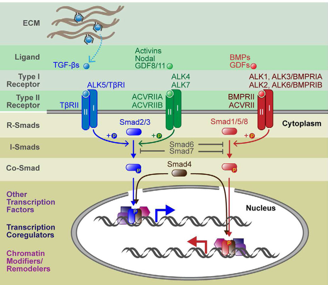
To rejuvenate tissues and/or repair wounds, stem cells must receive extrinsic signals from their surrounding environment and integrate them with their intrinsic abilities to self-renew and differentiate to make tissues. Increasing evidence suggests that the superfamily of transforming growth factor-βs (TGF-βs) constitute integral components in the intercellular crosstalk between stem cells and their microenvironment. In this review, we summarize recent advances in our understanding of TGF-β superfamily functions in embryonic and adult stem cells. We discuss how these pathways help to define the physiological environment where stem cells reside, and how perturbations in the signaling circuitry contribute to cancers.


Figures

Figure 1
Figure 1. Canonical Signaling Pathways of TGF-β Superfamily
When TGF-β propeptides are processed, the ligand associates noncovalently with the processed peptide. For some ligands, including the classical TGF-βs, activity is blocked by the peptide, and hence has been given the term latency associated peptide (LAP). LAPs can bind to latent TGF-β binding proteins (LTBPs) and other proteins within the extracellular matrix (ECM), and various activation mechanisms (dashed arrow) are then needed to extricate them from the ligand. Once liberated, ligands bind to specific combinations of type I and type II receptor serine/threonine kinases. In the ligand-receptor complexes, type II receptors phosphorylates and activates type I receptors, which in turn phosphorylate R-Smads and initiate intracellular signaling. R-Smad activation bifurcates the superfamily into two major branches: TGF-βs, activins and Nodal primarily use Smad2 and 3; BMPs and GDFs use Smad1, 5 and 8. Inhibitory Smads (Smad6 and 7) function by blocking type I receptor-mediated phosphorylation of Smad1/5/8 (Smad6) or both Smad2/3 and Smad1/5/8 (Smad7). Once phosphorylated, R-Smads couple with Co-Smad (Smad4) and translocate to the nucleus to act as a bipartite transcription factor. Generally, R-Smad-Smad4 transcription complexes have weak affinities to DNA and hence bind to other transcription factors for stable and specific binding to gene enhancers/promoters. R-Smad-Smad4 also associates with transcription coregulators, chromatin modifiers, and other chromatin remodelers.
Figure 2
Figure 2. Signaling Crosstalk between TGF-β and Other Pathways in Mouse and Human ESCs
(A) Self-renewal mechanisms in mouse ESCs. LIF and BMPs cooperate to maintain self-renewal. Tyrosine kinase receptor signaling, e.g. FGFs, typically activate downstream effectors ERK-MAPK, and induce differentiation. BMPs induce canonical Smad1/5/8 signaling pathways and one of their key targets for ESCs was recently shown to be the gene encoding ERK-specific phosphatase Dusp9. Dusp9 negatively affects ERK activity and hence support ESC self-renewal. Through the simultaneous induction of another BMP target gene, Id, and through activation of the STAT3 pathway by LIF, these two factors further promote ESC self-renewal by inhibiting mesendoderm and neuroectoderm differentiation, respectively. (B) Self-renewal mechanisms in human ESCs. Nodal/Activin and hyperactive PI3K/Akt signaling cooperate to maintain self-renewal. Nodal/Activin induce canonical Smad2/3 signaling pathways and a key target for ESCs is Nanog. Hyperactive PI3K/Akt signaling can be achieved by some growth factors, such as FGF and IGFs, and this leads to suppression of ERK activity. When PI3K/Akt signaling diminishes, ERK inhibition is relieved, which in turn, suppresses the GSK3β kinase. This results in stabilization of β-catenin, which apparently associates with pSmad2/3-Smad4 to influence the target genes that become transcribed, thereby switching the ESC from a self-renewing to differentiating status.
Figure 3
Figure 3. Molecular Mechanisms of ESC Differentiation and Lineage-specific Roles of TGF-β Signaling
(A) In ESCs, activated pSmad2/3-Smad4 complexes are recruited by the master regulator Oct4 to genes that maintain pluripotency. In myotubes and Pro-B lymphocyte progenitors, activated Smad complexes are recruited by different lineage-specific master regulators, MyoD and PU.1, respectively, which are bound to lineage-specific genes governing muscle and B-cell differentiation. (B) During mesendodermal fate specification of ESCs, signaling through TGF-βs, Activins and Nodal results in the generation of two distinct transcriptional complexes: pSmad2/3-Smad4 and pSmad2/3-TRIM33. Expression of homeostatic genes, such as Smad7 and Skil, are mediated only by pSmad2/3-Smad4. In contrast, master regulator genes of differentiation, such as Gsc and Mixl, exist in a transcriptionally poised state, as represented by the presence not only of RNA polymerase II at the transcription start site, but also the repressive histone modification, H3K9me3 and its binding associate HP1. In order to activate these poised genes, cooperative actions must occur between pSmad2/3-TRIM33 and pSmad2/3-Smad4 and an additional transcription cofactor FoxH1. In some way, this elicits an active chromatin conformation as revealed by histone acetyltransferase p300 and the added acetylation mark at lysine 18 on H3.
Figure 4
Figure 4. Diverse functions of TGF-β signaling in the control of adult stem cell behaviors
(A) The bone marrow niche consists of a variety of cell types, which include hematopoietic stem cells (HSCs), mesenchymal stem cells, osteoblasts, endothelial cells, CXCL12-abundant reticular (CAR) cells, sympathetic neurons and nonmyelinating Schwann cells. HSCs located on the endosteal side of the niche tend to be quiescent, a feature necessary for their self-renewal and maintenance of stemness, while HSCs residing on the perivascular side are more active, priming them for differentiation. HSC quiescence is dependent upon active TGF-β, which appears to be released from its associated LAP through a mechanism involving β8-integrin, expressed on the surface of the nonmyelinated Schwann cells. TGF-β signaling also exacerbates the functional differences between the myeloid and lymphoid HSC subtypes. TGF-β enhances My-HSC self-renewal at low levels and drives myeloid differentiation at higher levels. By contrast, even at low levels, TGF-β induces Ly-HSC differentiation and inhibits their self-renewal. (B) In the hair follicle, epithelial hair follicle stem cells (HFSCs) and melanocyte stem cells (McSCs) are located in a niche referred to as the bulge and hair germ (HG). Demarcating the dermis and epithelium is a basement membrane, and although the dermis contributes to niche signaling, HFSCs are restricted to the follicle side of the basement membrane. Sandwiched between the HFSC layer and the club hair is a layer of inner bulge cells (red circles) that emit high levels of BMP6 and FGF18 which maintain HFSC quiescence. Another key niche component is a transient one, the dermal papilla (DP), which only sits at the base of the bulge during the resting phase of the hair cycle, when HFSCs are in their most quiescent state. During this phase, crosstalk between HFSCs and DP change their transcriptional states and ultimately lead to TGF-β2 production by the DP, and pSmad2/3-Smad4 signaling by the adjacent HFSCs. A key downstream TGF-β2 signaling target is the gene encoding Tmeff1, which inhibits BMP signaling and lowers the threshold necessary to initiate HFSC self-renewal and fate specification necessary to launch a new round of hair growth.

Similar articles

Cited by

References

    1. Agricola E, Randall RA, Gaarenstroom T, Dupont S, Hill CS. Recruitment of TIF1γ to chromatin via its PHD finger-bromodomain activates its ubiquitin ligase and transcriptional repressor activities. Mol Cell. 2011;43:85–96. - PubMed
    1. Akhurst RJ. Large- and small-molecule inhibitors of transforming growth factor-beta signaling. Curr Opin Investig Drugs. 2006;7:513–521. - PubMed
    1. Annes JP, Munger JS, Rifkin DB. Making sense of latent TGFbeta activation. J Cell Sci. 2003;116:217–224. - PubMed
    1. Arnold SJ, Robertson EJ. Making a commitment: cell lineage allocation and axis patterning in the early mouse embryo. Nat Rev Mol Cell Biol. 2009;10:91–103. - PubMed
    1. Berge ten D, Kurek D, Blauwkamp T, Koole W, Maas A, Eroglu E, Siu RK, Nusse R. Embryonic stem cells require Wnt proteins to prevent differentiation to epiblast stem cells. Nat Cell Biol. 2011;13:1070–1075. - PMC - PubMed

Publication types

Substances