

Review
doi: 10.1016/j.virol.2012.08.022.
Cryo-electron tomography of bacterial viruses
Affiliations
- PMID: 23217626
- PMCID: PMC5119483
- DOI: 10.1016/j.virol.2012.08.022
Item in Clipboard
Review
Cryo-electron tomography of bacterial viruses
Virology.
.
Abstract
Bacteriophage particles contain both simple and complex macromolecular assemblages and machines that enable them to regulate the infection process under diverse environmental conditions with a broad range of bacterial hosts. Recent developments in cryo-electron tomography (cryo-ET) make it possible to observe the interactions of bacteriophages with their host cells under native-state conditions at unprecedented resolution and in three-dimensions. This review describes the application of cryo-ET to studies of bacteriophage attachment, genome ejection, assembly and egress. Current topics of investigation and future directions in the field are also discussed.
Copyright © 2012 Elsevier Inc. All rights reserved.
Figures
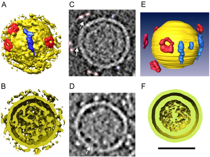
3D rendering of Φ12 bacteriophage structure. (A) Surface rendering of an individual virion depicting two types of protruding densities, labeled with red (donutshaped) and blue (elongated). (B) 7 nm thick section through a tomographic reconstruction of bacteriophage Φ12. The membrane and the two classes of protruding densities are clear. Red and blue lines represent the segmentation of donut-shaped and elongated protrusions, respectively. (C) 3D rendering resulting from the segmentation represented in panel ‘B’ with yellow label representing the membrane surface and red and blue the surface spikes. (D) Cross-section of the bacteriophage particle from panel ‘A’ illustrating two discrete shells. (E) Section through a Φ12 bacteriophage displaying the connection of the inner face of the envelope with the nucleocapsid (arrow). (F) Aligned average of 180 Φ12 bacteriophage particles clearly depicting two distinct shells. Reprinted from Virology, 372, Hu et al., Electron cryotomographic structure of cystovirus phi 12, 1–9, Copyright (2008), with permission from Elsevier. (For interpretation of the references to color in this figure legend, the reader is referred to the web version of this article.)
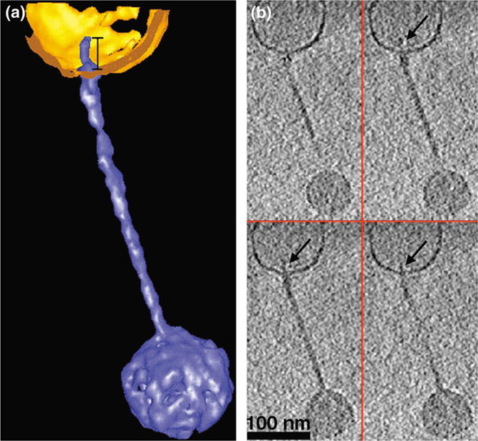
Binding of a T5 bacteriophage particle to a proteoliposome before DNA release. (a) Segmented 3D rendering of a T5 bacteriophage (depicted in blue) bound to a vesicle (displayed in gold) before DNA release. (b) Four XY slices (1.4 nm thick) through the same reconstruction depicted in (a). Arrows point to the tip of the bacteriophage tail inside the vesicle (Reprinted from Current Biology, 11/15, Böhm et al., FhuA-mediated phage genome transfer into liposomes: A cryo-electron tomography study, 1168–1175, Copyright (2001), with permission from Elsevier.) (For interpretation of the references to color in this figure legend, the reader is referred to the web version of this article.)
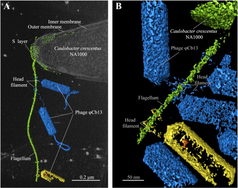
Segmented 3D volume from a cryo-electron tomogram of a ΦCb13-infected, C. crescentus cell illustrating head filament interacting with the flagellum (Reprinted from PNAS, 108/24, Guerrero-Ferreira et al., Alternative mechanism for bacteriophage adsorption to the motile bacterium Caulobacter crescentus, 9963–9968, Copyright (2011).
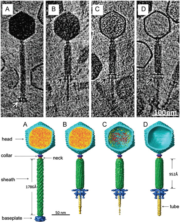
The top panel depicts cryo-ET of representative bacteriophage 8a particles at each of the four states of DNA ejection. Differences in genome content and tail sheath length are evident. Notice the difference in baseplate morphology indicated by the arrows. The bottom panel depicts the corresponding segmented volumes of the four states, indicating viral components and size differences between the extended and contracted tail. Modified from Fu et al. (2011). Reprinted from Virology, 410/2, Fu et al. The mechanism of DNA ejection in the Bacillus anthracis spore-binding phage 8a revealed by cryo-electron tomography, 141–148, Copyright (2011), with permission from Elsevier.
Similar articles
-
Bacteriophage-host interactions leading to genome internalization.Curr Opin Microbiol. 2011 Aug;14(4):492-6. doi: 10.1016/j.mib.2011.07.010. Epub 2011 Jul 21. Curr Opin Microbiol. 2011. PMID: 21783404 Review.
-
Bacteriophage electron microscopy.Adv Virus Res. 2012;82:1-32. doi: 10.1016/B978-0-12-394621-8.00017-0. Adv Virus Res. 2012. PMID: 22420849 Review.
-
Structure of epsilon15 bacteriophage reveals genome organization and DNA packaging/injection apparatus.Nature. 2006 Feb 2;439(7076):612-6. doi: 10.1038/nature04487. Nature. 2006. PMID: 16452981 Free PMC article.
-
The bacteriophage genome undergoes a succession of intracapsid phase transitions upon DNA ejection.J Mol Biol. 2010 Feb 19;396(2):384-95. doi: 10.1016/j.jmb.2009.11.047. Epub 2009 Nov 26. J Mol Biol. 2010. PMID: 19944702
-
Structural variability and complexity of the giant Pithovirus sibericum particle revealed by high-voltage electron cryo-tomography and energy-filtered electron cryo-microscopy.Sci Rep. 2017 Oct 16;7(1):13291. doi: 10.1038/s41598-017-13390-4. Sci Rep. 2017. PMID: 29038566 Free PMC article.
Cited by
-
Imaging Techniques for Detecting Prokaryotic Viruses in Environmental Samples.Viruses. 2021 Oct 21;13(11):2126. doi: 10.3390/v13112126. Viruses. 2021. PMID: 34834933 Free PMC article. Review.
-
Zernike phase-contrast electron cryotomography applied to marine cyanobacteria infected with cyanophages.Nat Protoc. 2014 Nov;9(11):2630-42. doi: 10.1038/nprot.2014.176. Epub 2014 Oct 16. Nat Protoc. 2014. PMID: 25321408 Free PMC article.
-
Dissecting Virus Infectious Cycles by Cryo-Electron Microscopy.PLoS Pathog. 2016 Jun 30;12(6):e1005625. doi: 10.1371/journal.ppat.1005625. eCollection 2016 Jun. PLoS Pathog. 2016. PMID: 27362353 Free PMC article. Review. No abstract available.
-
Polymorphism of DNA conformation inside the bacteriophage capsid.J Biol Phys. 2013 Mar;39(2):201-13. doi: 10.1007/s10867-013-9315-y. Epub 2013 Apr 12. J Biol Phys. 2013. PMID: 23860869 Free PMC article.
-
OmpA and OmpC are critical host factors for bacteriophage Sf6 entry in Shigella.Mol Microbiol. 2014 Apr;92(1):47-60. doi: 10.1111/mmi.12536. Epub 2014 Mar 6. Mol Microbiol. 2014. PMID: 24673644 Free PMC article.
References
-
- Agirrezabala X, Martin-Benito J, Valle M, Gonzalez JM, Valencia A, Valpuesta JM, Carrascosa JL. Structure of the connector of bacteriophage T7 at 8 A resolution: structural homologies of a basic component of a DNA translocating machinery. J Mol Biol. 2005;347:895–902. - PubMed
-
- Baumeister W. Mapping molecular landscapes inside cells. Biol Chem. 2004;385:865–872. - PubMed
-
- Baumeister W. From proteomic inventory to architecture. FEBS Lett. 2005;579:933–937. - PubMed
Publication types
MeSH terms
Substances
Grants and funding
LinkOut - more resources
Full Text Sources
Other Literature Sources
