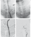
Salvage of the carotid artery with covered stent after perforation with dialysis sheath. A case report
- PMID: 23217633
- PMCID: PMC3520552
- DOI: 10.1177/159101991201800404
Salvage of the carotid artery with covered stent after perforation with dialysis sheath. A case report
Abstract
We present a rare case of carotid tear caused by iatrogenic erroneous insertion of a dialysis sheath into the common carotid artery (CCA). This was treated by placement of a covered stent-graft in the CCA over the puncture site. This treatment achieved hemostasis while preserving the carotid artery with good outcome. The technical details are presented and the relevant literature regarding treatment of carotid blowout syndrome is discussed. This case suggests that placement of a covered stent-graft is a good option not only for the "usual" blowout syndrome due to head and neck tumors, but also for treatment of iatrogenic injury to the carotid artery.
Figures


Similar articles
-
Dialysis access pseudoaneurysm: endovascular treatment with a covered stent.BMJ Case Rep. 2010 Oct 31;2010:bcr0720080477. doi: 10.1136/bcr.07.2008.0477. BMJ Case Rep. 2010. PMID: 22778110 Free PMC article.
-
Safety and effectiveness of endovascular embolization or stent-graft reconstruction for treatment of acute carotid blowout syndrome in patients with head and neck cancer: Case series and systematic review of observational studies.Head Neck. 2018 Apr;40(4):846-854. doi: 10.1002/hed.25018. Epub 2017 Nov 20. Head Neck. 2018. PMID: 29155470
-
[Case of a giant cervical carotid artery aneurysm with contralateral severe stenosis treated using a covered stent].No Shinkei Geka. 2012 Mar;40(3):255-60. No Shinkei Geka. 2012. PMID: 22392755 Japanese.
-
Endovascular Treatment of Carotid Blowout Syndrome.J Stroke Cerebrovasc Dis. 2021 Aug;30(8):105818. doi: 10.1016/j.jstrokecerebrovasdis.2021.105818. Epub 2021 May 25. J Stroke Cerebrovasc Dis. 2021. PMID: 34049016
-
Endovascular management of iatrogenic cervical internal carotid artery pseudoaneurysm in a 9-year-old child: Case report and literature review.Int J Pediatr Otorhinolaryngol. 2017 Apr;95:29-33. doi: 10.1016/j.ijporl.2017.01.031. Epub 2017 Jan 30. Int J Pediatr Otorhinolaryngol. 2017. PMID: 28576528 Review.
Cited by
-
Covered stents for exclusion of iatrogenic common carotid artery-internal jugular vein fistula and brachiocephalic artery pseudoaneurysm.BMJ Case Rep. 2015 Jun 23;2015:bcr2015011760. doi: 10.1136/bcr-2015-011760. BMJ Case Rep. 2015. PMID: 26106173 Free PMC article.
-
Iatrogenic Arterial Perforation During Endovascular Interventions.Cureus. 2020 Aug 25;12(8):e10018. doi: 10.7759/cureus.10018. Cureus. 2020. PMID: 32983713 Free PMC article. Review.
-
Neurovascular stents in pediatric population.Childs Nerv Syst. 2016 Mar;32(3):505-9. doi: 10.1007/s00381-015-2992-z. Epub 2015 Dec 29. Childs Nerv Syst. 2016. PMID: 26715300
-
MicroNET-covered stent use to seal carotid artery perforation.Postepy Kardiol Interwencyjnej. 2023 Sep;19(3):284-288. doi: 10.5114/aic.2023.131483. Epub 2023 Sep 27. Postepy Kardiol Interwencyjnej. 2023. PMID: 37854974 Free PMC article. No abstract available.
-
Endovascular treatment of post-pharyngitis internal carotid artery pseudoaneurysm with a covered stent in a child: a case report.Childs Nerv Syst. 2013 Aug;29(8):1369-73. doi: 10.1007/s00381-013-2083-y. Epub 2013 Mar 27. Childs Nerv Syst. 2013. PMID: 23532343
References
-
- McGettigan B, Parkes W, Gonsalves C, et al. The use of a covered stent in carotid blowout syndrome. Ear Nose Throat J. 2011;90:E17. - PubMed
-
- Wan WS, Lai V, Lau HY, et al. Endovascular treatment paradigm of carotid blowout syndrome: Review of 8-years experience. Eur J Radiol. 2011 Feb 8 Epub ahead of print. - PubMed
Publication types
MeSH terms
LinkOut - more resources
Full Text Sources
Medical

