. 2012 Dec 7;151(6):1283-95.
doi: 10.1016/j.cell.2012.10.041.

TMHS is an integral component of the mechanotransduction machinery of cochlear hair cells

Affiliations

TMHS is an integral component of the mechanotransduction machinery of cochlear hair cells

Wei Xiong et al. Cell. .

Abstract

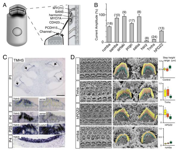
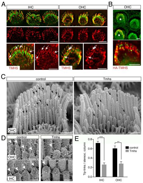
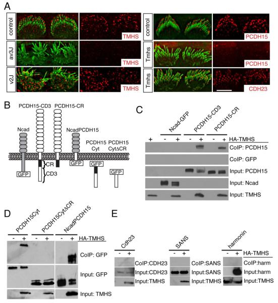
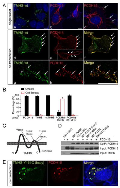
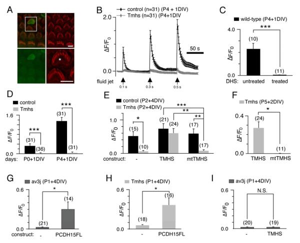
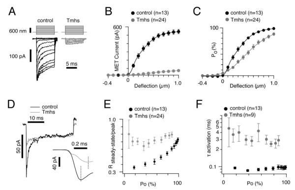
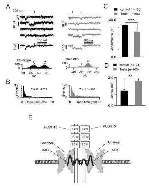
Hair cells are mechanosensors for the perception of sound, acceleration, and fluid motion. Mechanotransduction channels in hair cells are gated by tip links, which connect the stereocilia of a hair cell in the direction of their mechanical sensitivity. The molecular constituents of the mechanotransduction channels of hair cells are not known. Here, we show that mechanotransduction is impaired in mice lacking the tetraspan TMHS. TMHS binds to the tip-link component PCDH15 and regulates tip-link assembly, a process that is disrupted by deafness-causing Tmhs mutations. TMHS also regulates transducer channel conductance and is required for fast channel adaptation. TMHS therefore resembles other ion channel regulatory subunits such as the transmembrane alpha-amino-3-hydroxy-5-methyl-4-isoxazole propionic acid (AMPA) receptor regulatory proteins (TARPs) of AMPA receptors that facilitate channel transport and regulate the properties of pore-forming channel subunits. We conclude that TMHS is an integral component of the hair cell's mechanotransduction machinery that functionally couples PCDH15 to the transduction channel.


Figures

Figure 1
Figure 1. Mechanotransduction defects in TMHS-deficient mice
(A) Hair cell diagram showing on the right proteins that form tip links or are located in proximity to tip links. (B) Amplitude of mechanotransduction currents in mutant mouse lines. The values are expressed relative to the values in wild-type. The number of hair cells analyzed is indicated. Values are mean ± SEM. (C) In situ hybridization with TMHS antisense, sense control probes, and a Loxhd1 probe that reveals hair cells. The lowest panel shows vestibular hair cells, the magnified images hair cells at the apical-medial turn of the cochlea. Arrows point to hair cells. (D) SEM analysis of hair bundles from the mid-apical cochlea. On the right, OHCs are shown. The different rows of stereocilia have been colored. Whisker plots on the right show height differences between the first (longest) and second row of stereocilia (yellow); the second and third row (orange); the third row and surface of hair cells or lowest row (green) (number of evaluated hair bundles: control, n=16; Tmhs-/-, n=21; hPDZ, n=21; rumba, n=12; from 2 animals each). Scale bars: (C) black bar: 200 μm; white bar, 50 μm; gray bar, 20 μm; (D) left panel: 5 μm; right panel 2 μm. See also Figure S1.
Figure 2
Figure 2. Tip-link defects in hair cells from TMHS-/- mice
(A) Hair cells from wild-type mice at P5 were stained with antibodies to TMHS (red) and with phalloidin (green). Arrows point to TMHS in the tip-link region. (B) OHCs were electroporated to express HA-TMHS (electroporated cells labeled with an asterisk). Cells were stained for HA (red in upper panel, green in lower panel) and with phalloidin (green in upper panel, red in lower panel). (C) SEM analysis of OHCs at P7. Arrows point to tip links. (D) High resolution images showing tip links (arrows) in OHCs and IHCs. (E) Quantification of tip-link numbers at P7. Values are mean ± SD (**, p<0.01; ***, p<0.001). Scale bar: (A, B) 3 μm; (C) 0.5 μm; (D) 0.25 μm.
Figure 3
Figure 3. Interactions between TMHS and PCDH15
(A) IHCs from P1 wild-type mice, PCDH15 deficient Ames waltzerav3J/av3J mice, CDH23 deficient waltzerv2J/v2J mice, and Tmhs-/- mice were stained with antibodies to TMHS, PCDH15 and CDH23 (red) and phalloidin (green). Note the reduced staining of TMHS in PCDH15-muants and of PCDH15 in TMHS mutants. (B) Diagram of the constructs used for biochemical experiments. (C-E) HEK293 cells were transfected with the constructs indicated on top of each panel. Immunoprecipitations were carried out with HA antibodies that recognize HA-TMHS, followed by western blotting to detect PCDH15 constructs (C, D), CDH23, SANS, and harmonin (E). GFP-tagged PCDH15 and N-cadherin proteins were detected with an antibody to GFP, the remaining proteins with antibodies specific to the individual proteins. The lower rows show input protein, the upper rows co-immunoprecipitation (Co-IP) results. Note the specific interaction between TMHS and PCDH15. Scale bars: (A) 4 μm.
Figure 4
Figure 4. Wild-type but not mutant TMHS facilitates PCDH15 transport to the plasma membrane
(A) RPMI-2650 cells were transfected with expression vectors for TMHS and/or PCDH15 and analyzed for protein expression and localization one day after transfection (a-f) or four days after transfection (g-i). TMHS is shown in green, PCDH15 in red. Nuclei (a-c, blue) and actin (b and c, blue) were stained with DAPI and phalloidin, respectively. Note the low levels of PCDH15 at the cell surface in the absence of TMHS (c, arrows), and extensive co-localization of PCDH15 and TMHS at the surface of double-transfected cells (d-f, arrows). The insert in panel e show staining of non-permeabilized cells with an antibody to the PCDH15 extracellular domain. Panels g-i show co-localization of TMHS and PCDH15 in small aggregates in cell protrusions (arrows) after four days in culture. (B) Quantification of expression of proteins in the cytosol/at the plasma membrane in RPMI-2650 cells transfected to express TMHS and/or PCDH15 (at least 100 cells were quantified in 3 independent experiments). Values are mean ± SEM. (C) Diagram of TMHS and location of mutations within the protein. (D) Co-immunoprecipitation experiments were carried out with extracts from transfected HEK293 cells. Transfected constructs are indicated on top. The lower rows show input protein, the upper rows co-immunoprecipitation (Co-IP) results. (E) RPMI-2650 cells were co-transfected with expression vectors for PCDH15 and TMHS carrying the hurry-scurry mutation. TMHS is in green, PCDH15 in red, and nuclei and actin in blue. Scale bars: (A) 7 μm; (B) 5 μm.
Figure 5
Figure 5. Rescue of mechanotransduction in Tmhs-/- mice by acute expression of TMHS and PCDH15
(A) Example of G-CaMP3 expression (green) in transfected OHC. Hair bundles of transfected cells are morphologically intact (Phalloidin staining, red), which can be seen in detail (asterisk) in the bottom-right panel. (B) Representative example demonstrating fluid-jet induced Ca2+ response in control and Tmhs-/- OHCs. OHCs were transfected at P4 and cultured for 1 day in vitro (DIV). Fluid-jet pulse duration was increased from 0.1 sec, to 0.3 sec, to 0.5 sec. For quantitative analysis (panels C-I), the amplitude of the 2nd Ca2+ response peak was measured. (C) Ca2+ response of wild type OHCs with/without dihydrostreptomycin (DHS). (D) Quantification of peak amplitude in OHCs from control and Tmhs-/- mice transfected at P0 or P4 and cultured for 1 DIV. (E) Response of OHCs from control and Tmhs-/- mice transfected to express G-CaMP3 only (-), wild-type TMHS (TMHS), or TMHS carrying the hurry-scurry mutation (mtTMHS). (F) OHCs from Tmhs-/- mice at P5 were transfected with wild-type and mutant TMHS and analyzed after 2 DIV for mechanically evoked Ca2+ currents. (G) Ca2+ responses in OHCs from PCDH15-deficient Ames-waltzerav3J/av3J mice electroporated to express PCDH15-CD3. (H) Ca2+ responses in OHCs from Tmhs-/- mice electroporated to express PCDH15-CD3. (I) Ca2+ responses in OHCs from PCDH15-deficient Ames-waltzerav3J/av3J mice electroporated to express TMHS. Please note that the changes in fluorescence intensity vary in magnitude between panels since we used for the experiments hair cells of different ages. All values are mean ± SEM (*, p<0.05; **, p<0.01; ***, p<0.001). Scale bars: (A) 6 μm. See also Figure S2.
Figure 6
Figure 6. Macroscopic mechanotransduction currents in OHCs from Tmhs-/- mice
(A) Examples of transduction currents in OHCs from wild-type and Tmhs-/- mice in response to a set of 10 msec hair bundle deflections ranging from -400 nm to 1000 nm (100 nm steps). (B) Current displacement plots obtained from similar data as shown in (A). The number of analyzed hair cells is indicated. (C) Open probability (Po) displacement plots calculated from similar data as shown in (A). The number of analyzed hair cells is indicated. (D) Representative current traces to show adaptation in OHCs from control and Tmhs-/- mice. Traces were chosen from controls and mutants based on similar steady state currents and similar rate of slow adaptation. Deflection was for 30 msec. Inset showed magnified traces for the activation phase of mechanically induced currents. (E) Ratio of steady-state current versus peak current plotted against the channel open probability (Po). (F) Activation time constant (τactivation) plotted against Po. All values are mean ± SEM (*, p<0.05; **, p<0.01; ***, p<0.001). See also Figure S3.
Figure 7
Figure 7. Single channel responses in OHCs from Tmhs-/- mice
(A) Upper panels: representative single channel recordings from a control (black) and Tmhs-/- (grey) OHC. Close (C) and open state (O) induced by a 300 nm deflection are shown. Middle panels: ensemble averages from 25 responses in controls and from 52 responses in mutants. The adaptation phase was only evident in ensemble averages from control OHC. Bottom panels: amplitude histograms generated with the data from the second traces in upper panels. Gaussian fits of the 2 peaks in the histograms determine a single channel conductance of 6.8 pA in control OHC and 5.2 pA in Tmhs-/- OHC. (B) Open-time histograms of single channel events from all recorded cells (control: 1691 events from 24 cells; mutant: 1663 events from 18 cells). The histograms were fitted with exponential curves with time constants of 0.94 ms in control (black) OHCs and 1.57 ms in Tmhs-/- (grey) OHCs. The first bin was not included for fitting as described (Ricci et al., 2003). (C) Amplitude of single channel currents from all recorded OHCs. Single channel conductance in OHCs from controls were at 86.8 ± 1.6 pS, and at 68.1 ± 1.7pS in OHCs from Tmhs-/- mice (controls: 150 traces from 19 cells; mutants: 96 traces from 8 cells). (D) Latency between the onset of deflection and first event from all recorded OHCs. Latency was at 1.3 ± 0.1 msec in control OHCs and at 2.2 ± 0.1 msec in Tmhs-/- OHCs (controls: 171 traces from 5 cells; mutants: 263 traces from 10 cells). All values are mean ± SEM (*, p<0.05; **, p<0.01; ***, p<0.001). (E) Model of the function of TMHS in hair cells. According to the model, TMHS is a regulatory subunit of the mechanotransduction channel that connects the pore forming subunits of the channel to PCDH15 and might also affect membrane properties.

References

    1. Adato A, Michel V, Kikkawa Y, Reiners J, Alagramam KN, Weil D, Yonekawa H, Wolfrum U, El-Amraoui A, Petit C. Interactions in the network of Usher syndrome type 1 proteins. Hum Mol Genet. 2005;14:347–356. - PubMed
    1. Ahmed ZM, Goodyear R, Riazuddin S, Lagziel A, Legan PK, Behra M, Burgess SM, Lilley KS, Wilcox ER, Riazuddin S, et al. The tip-link antigen, a protein associated with the transduction complex of sensory hair cells, is protocadherin-15. J Neurosci. 2006;26:7022–7034. - PMC - PubMed
    1. Alagramam KN, Murcia CL, Kwon HY, Pawlowski KS, Wright CG, Woychik RP. The mouse Ames waltzer hearing-loss mutant is caused by mutation of Pcdh15, a novel protocadherin gene. Nat Genet. 2001;27:99–102. - PubMed
    1. Bahloul A, Michel V, Hardelin JP, Nouaille S, Hoos S, Houdusse A, England P, Petit C. Cadherin-23, myosin VIIa and harmonin, encoded by Usher syndrome type I genes, form a ternary complex and interact with membrane phospholipids. Hum Mol Genet. 2010;19:3557–3565. - PMC - PubMed
    1. Beurg M, Evans MG, Hackney CM, Fettiplace R. A large-conductance calcium-selective mechanotransducer channel in mammalian cochlear hair cells. J Neurosci. 2006;26:10992–11000. - PMC - PubMed

Publication types