. 2012 Dec 7;151(6):1296-307.
doi: 10.1016/j.cell.2012.11.002.

An interdomain energetic tug-of-war creates the allosterically active state in Hsp70 molecular chaperones

Affiliations

An interdomain energetic tug-of-war creates the allosterically active state in Hsp70 molecular chaperones

Anastasia Zhuravleva et al. Cell. .

Abstract

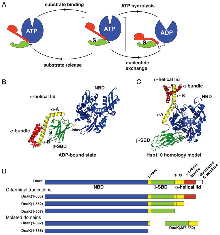
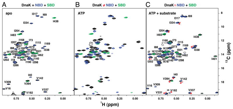
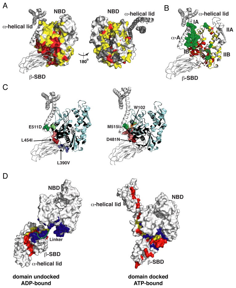
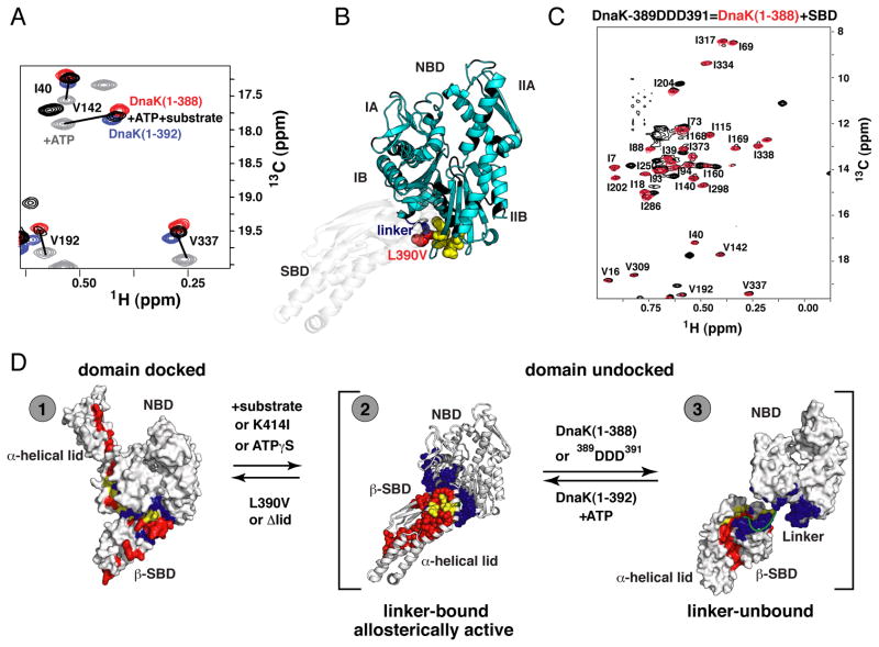
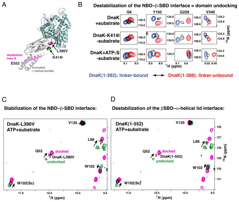
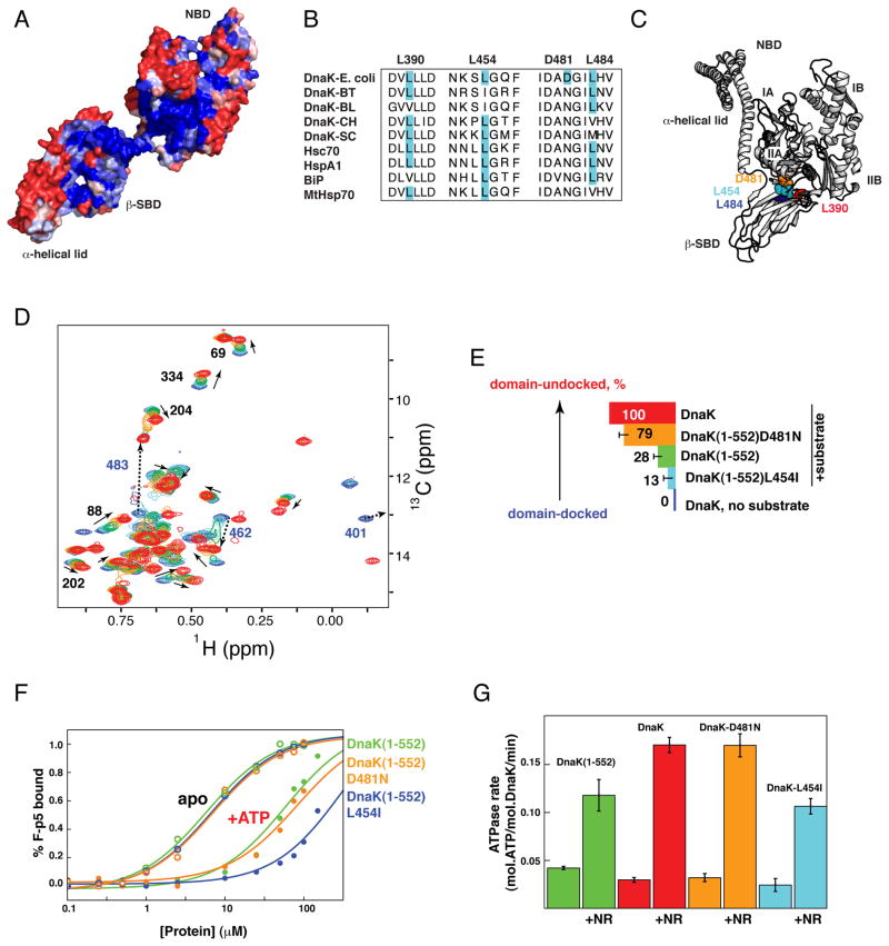
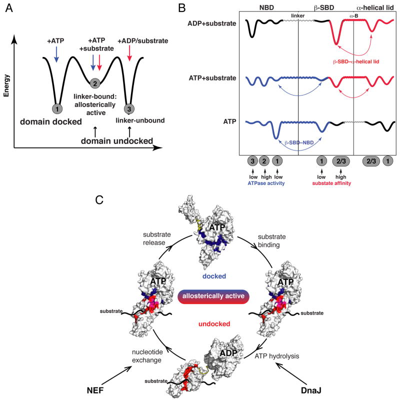
The allosteric mechanism of Hsp70 molecular chaperones enables ATP binding to the N-terminal nucleotide-binding domain (NBD) to alter substrate affinity to the C-terminal substrate-binding domain (SBD) and substrate binding to enhance ATP hydrolysis. Cycling between ATP-bound and ADP/substrate-bound states requires Hsp70s to visit a state with high ATPase activity and fast on/off kinetics of substrate binding. We have trapped this "allosterically active" state for the E. coli Hsp70, DnaK, and identified how interactions among the NBD, the β subdomain of the SBD, the SBD α-helical lid, and the conserved hydrophobic interdomain linker enable allosteric signal transmission between ligand-binding sites. Allostery in Hsp70s results from an energetic tug-of-war between domain conformations and formation of two orthogonal interfaces: between the NBD and SBD, and between the helical lid and the β subdomain of the SBD. The resulting energetic tension underlies Hsp70 functional properties and enables them to be modulated by ligands and cochaperones and "tuned" through evolution.


Figures

Figure 1
Figure 1. Conformational Insights into the Hsp70 Allosteric Cycle
(A) The Hsp70 allosteric cycle. (B) and (C) Ribbon representation of the structures of the two ‘end-point’ states of DnaK: the ADP-bound state (PDB ID code 2kho) and the Hsp110-based homology model of the ATP-bound state (Smock et al., 2010; coordinates available upon request); structural elements colored as in (D). (See also Figure S1.) (D) DnaK constructs used for NMR studies.
Figure 2
Figure 2. NMR Fingerprints of DnaK Reveal Three Different Ligand-Bound States in Its Allosteric Cycle
The isoleucine region of methyl-TROSY spectra of the two-domain DnaK constructs [black; DnaK(1-552) for (A, B) and full-length DnaK for (C), see Figure S2 for the full-length DnaK and ADP-bound state] are overlaid with the spectra of corresponding nucleotide and substrate-bound states of the individual domains, NBD, DnaK(1-388) (blue) and SBD, DnaK(387-552) (green): nucleotide-free (A), ATP-bound (B), and ATP-/substrate-bound (C). On (C), red arrows point to small but significant chemical shift differences between NBD resonances of the full-length ATP-/substrate-bound DnaK and those of its isolated NBD.
Figure 3
Figure 3. Experimental Validation of the Hsp110-based Homology Model for the ATP-bound (Domain-Docked) State of DnaK
(A, B) Mapping of the NBD residues significantly affected by interaction with the β-SBD onto the structure of the Hsp110-based homology model. Residues with significant chemical shift differences (see text) between DnaK(1-552) and either DnaK(1-392) or DnaK(1-507) are shown in yellow and green, respectively; residues showing enhanced μs-ms dynamics upon ATP binding, i.e., whose assignments were obtained for the isolated NBD but not for DnaK(1-552), are shown in red (see Supplemental Experimental Procedures); the unassigned residues in either constructs are shown in dark gray, the rest are shown in light gray (see also Figure S3A). (C) Ribbon representation of the Hsp110-based homology model showing effects of ‘soft’ mutations on backbone (L454I, D481N, L390V, and E511D) and methyl (M515I) chemical shifts of ATP-bound (docked) DnaK(1-552). A site of mutation and the residues affected (see Supplemental Experimental Procedures) are shown as dark and light colored spheres, respectively (See also Figure S3B and C); Trp102, which becomes solvent-exposed in the ATP-bound conformation of DnaK (Buchberger et al., 1995), is shown in orange sticks on right; NBD residues without backbone assignments are shown in black. (D) Interfaces between the NBD, β-SBD and α-helical lid are shown mapped onto the two endpoint Hsp70 states, domain-undocked/linker-unbound (PDB ID code 2kho) and domain-docked [Hsp110-based homology model (see Experimental Procedures)]: residues at the interface between the β-SBD and α-helical lid, which is stabilized by substrate binding are in red, and residues at the interface between the NBD and either the β-SBD or the α-helical lid, which is stabilized upon cooperative ATP and linker binding, are in blue; residues participating in both interfaces are in yellow.
Figure 4
Figure 4. The Allosterically Active DnaK intermediate
(A) Ligand-driven changes in the DnaK conformational ensemble: Blow-up of a representative region of methyl-TROSY spectra of ATP-bound DnaK in the absence of substrate (gray, the domain-docked state) and with substrate (black, the domain-undocked ensemble of linker-bound and –unbound conformations), overlaid on spectra of the isolated NBD, with linker [DnaK(1-392)] (blue, the linker-bound conformation) or without linker [DnaK(1-388)] (red, the linker-unbound, domain undocked conformation). Similar results were obtained for wild-type DnaK without the T199A mutation (see Figure S4). (B) Linker binding site on the NBD: Residues with significant chemical-shift differences (more than 0.03 and 0.3 ppm for 1HN and 15N atoms, respectively; shown as yellow spheres) in the NBD of ATPγS-bound DnaK(1-552) [reflecting the ensemble of two domain-undocked conformations, linker-bound and linker unbound (see text)] between DnaK(1-552) and its ‘soft’ mutant L390V mapped onto the modeled structure of the allosterically active conformation [to schematically model this conformation the SBD of Hsp110-based homology model was replaced by the SBD from the ADP-bound DnaK (PBD ID code 2kho)] (see also Figure S4). (C) The isoleucine region of the methyl-TROSY spectrum of the two-domain allosterically defective 389DDD391 DnaK(1-605) bound to ATP and substrate (black) showing near-perfect overlap with spectra of the individual, non-linker-bound NBD, DnaK(1-388) (red). (D) Schematic illustration of two coupled conformational transitions in DnaK: (i) between the domain-docked conformation and the domain-undocked ensemble [corresponding to a transition between gray (ATP-bound) and black (ATP-/substrate-bound) peaks on left] and (ii) between linker-bound and –unbound conformations [corresponding to a transition between NBD+linker (blue) and NBD only (red) peaks (A); note in full-length DnaK that this transition is fast on the NMR time scale, and that black peaks on the left correspond to the dynamic domain-undocked ensemble of these two conformations]. Interdomain interfaces are colored as in Figure 3D.
Figure 5
Figure 5. The Impact of Competition Between the NBD–β-SBD and β-SBD–α-Helical Lid Interfaces on the Hsp70 Allosteric Landscape
(A) DnaK sequence modifications that result in perturbations in its conformational ensemble are mapped onto the modeled structure of the allosterically active conformation (modeled as for Figure 4): L390V and C-terminal truncations, which favor domain docking, are shown in magenta, K414I, which favors domain undocking, is shown in green. (B) Destabilization of the NBD–β-SBD interface results in domain undocking even in the absence of substrate: Blow-up of the amide-TROSY spectra of DnaK(1-392) (blue, representing the NBD linker-bound state) and DnaK(1-388) (red, representing the NBD linker-unbound state) overlaid on the spectra of two-domain DnaK under conditions that stabilize domain undocking, either upon substrate binding to the ATP-bound DnaK(1-605) (top panels) or upon perturbation of the NBD–β-SBD interface, viz. ATP-bound DnaK(1-552)K414I (middle panels), or ATPγS-bound DnaK(1-552) (bottom panels). Resonances shown (Gly6, Tyr193, Gly229, and V340) report on long-range conformational changes in the nucleotide-binding site upon linker binding to the NBD (see Supplemental Experimental Procedures). The spectra of the isolated NBD constructs are with the corresponding nucleotide bound (ATP, top and middle panels) or ATPγS-bound (bottom panels). (C) Stabilization of the NBD–β-SBD interface in DnaK(1-605)L390V or (D) destabilization of the β-SBD–α-helical lid interaction in DnaK(1-552) favors the domain-docked conformation even in the presence of substrate. A representative region of the amide-TROSY spectra of the ATP-bound state of DnaK(1-552) (magenta; the domain-docked conformation) and ATP-/substrate-bound DnaK(1-605) (green; the domain-undocked ensemble of linker-bound and linker-unbound conformations) overlaid with spectra of DnaK(1-605)L390V (C) and DnaK(1-552) (D), shown in black. Consistent with previous biochemical studies (Kumar et al., 2011; Swain et al., 2007) neither the L390V mutation on the NBD–β-SBD interface nor disruption of the β-SBD–α-helical lid interface in DnaK(1-552) affects the ATP-bound conformation in the absence of substrate.
Figure 6
Figure 6. Evolutionary Variation at the NBD–β-SBD and β-SBD–α-Helical Lid Interfaces Modulates the Hsp70 Allosteric Landscape
(A–C) Sequence conservation and diversity at the NBD–β-SBD interdomain interface in the Hsp70 family. (A) Sequence conservation between different Hsp70 family members colored from blue for fully conserved residues to red for residues with no conservations (see Experimental Procedures) shown on the structure of ADP-bound DnaK state (PDB ID code 2kho). (B) Multiple sequence alignment (ClustalW) of DnaKs from E. coli, Bacteroides thetaiotaomicron VPI-5482 (BT), Bifidobacterium longum NCC2705 (BL), Cytophaga hutchinsonii (CH), and Streptomyces coelicolor A3(2) (SC), and four human Hsp70s (Hsc70, HspA1, endoplasmic reticulum-BiP, and mitochondrial-MtHsp70). (C) Observed amino acid substitutions identified in B are shown on the Hsp110-based homology model. (D) The isoleucine region of methyl-TROSY spectra of ATP-bound full-length DnaK in the absence of substrate (blue, corresponds to the docked state) and with substrate (red, corresponds to the domain-undocked ensemble: the interconverting mixture of linker-bound and linker-unbound conformations) overlaid with ATP-/substrate-bound DnaK(1-552) (green) and its variants L454I (light blue; the DnaK(1-552)L484I shows very similar results, see Table S1) and D481N (orange). All experiments were performed at saturating substrate concentrations (2mM of NR peptide) (See also Figure S5). (E) Histogram showing the degree of substrate-induced domain undocking for individual constructs, colored as in B and D. For each DnaK variant, the degree of domain undocking was estimated as described in the Experimental Procedures. (F and G) Tuning of DnaK functionality. (F) Equilibrium binding of a fluorescently labeled peptide substrate to DnaK(1-552) (green) and its variants L454I (cyan) and D481N (orange) in the presence of ADP (open circles) and ATP (filled circles); KD values are 5.7±1.0 (60±20), 6.9±1.0 (80±20), and 6.8±1.0 (300±70) μM for the ADP-(ATP-)bound state of DnaK(1-552), DnaK(1-552)-D481N, and DnaK(1-552)-L454I, respectively (see Supplemental Experimental Procedures). (C) Stimulation of the ATPase activity of wild-type DnaK (red) and its variants DnaK-L454I (cyan), DnaK-D481N (orange), and DnaK(1-552) (green) by 200 μM NR peptide. (See Figure S5I and Supplemental Experimental Procedures).
Figure 7
Figure 7. Mechanism of Hsp70 Allostery
(A,B) Schematic illustration of Hsp70 allosteric landscapes showing how the allosterically active state serves as an intermediate between the two ‘end-point’ states (A) and (B) how thermodynamic coupling of Hsp70 domains determines the conformations the protein populates along its allosteric cycle (see also Figures 4D and S6): Binding of ADP and substrate favors interactions between the β-SBD and α-helical lid (red, in the domain-undocked conformation), ATP-induced linker binding to the NBD favors NBD–SBD docking (blue, in the domain-docked conformation). In the presence of both ATP and substrate, an interdomain energetic ‘tug-of-war’ results in a highly dynamic and tunable conformational ensemble. The interdomain linker and helix B provide flexible and ligand-adjustable connections between the NBD, the β-SBD, and the α-helical lid. (C) Illustration of the roles of energetic interdomain coupling and ‘tunability’ in the allosteric cycle. The interfaces between the NBD and the β-SBD (blue) and between the β-SBD and the α-helical lid (red; magenta–residues participate in both interfaces) define the thermodynamics and kinetics of the allosteric cycle and can be modulated by either intrinsic (sequence changes) or extrinsic (binding to nucleotide, substrates, co-chaperones) factors.

Similar articles

Cited by

References

    1. Ahmad A, Bhattacharya A, McDonald RA, Cordes M, Ellington B, Bertelsen EB, Zuiderweg ERP. Heat shock protein 70 kDa chaperone/DnaJ cochaperone complex employs an unusual dynamic interface. Proc Natl Acad Sci USA. 2011;108:18966–18971. - PMC - PubMed
    1. Bertelsen EB, Chang L, Gestwicki JE, Zuiderweg ERP. Solution conformation of wild-type E. coli Hsp70 (DnaK) chaperone complexed with ADP and substrate. Proc Natl Acad Sci USA. 2009;106:8471–8476. - PMC - PubMed
    1. Bertelsen EB, Zhou HJ, Lowry DF, Flynn GC, Dahlquist FW. Topology and dynamics of the 10 kDa C-terminal domain of DnaK in solution. Prot Sci. 1999;8:343–354. - PMC - PubMed
    1. Bhattacharya A, Kurochkin AV, Yip GNB, Zhang Y, Bertelsen EB, Zuiderweg ERP. Allostery in Hsp70 chaperones is transduced by subdomain rotations. J Mol Biol. 2009;388:475–490. - PMC - PubMed
    1. Buchberger A, Theyssen H, Schroder H, McCarty JS, Virgallita G, Milkereit P, Reinstein J, Bukau B. Nucleotide-induced conformational-changes in the ATPase and substrate-binding domains of the DnaK chaperone provide evidence for interdomain communication. J Biol Chem. 1995;270:16903–16910. - PubMed

Publication types

MeSH terms