Review
. 2012 Dec 6;76(5):871-85.
doi: 10.1016/j.neuron.2012.11.020.

Apolipoprotein e sets the stage: response to injury triggers neuropathology

Affiliations
Review

Apolipoprotein e sets the stage: response to injury triggers neuropathology

Robert W Mahley et al. Neuron. .

Abstract

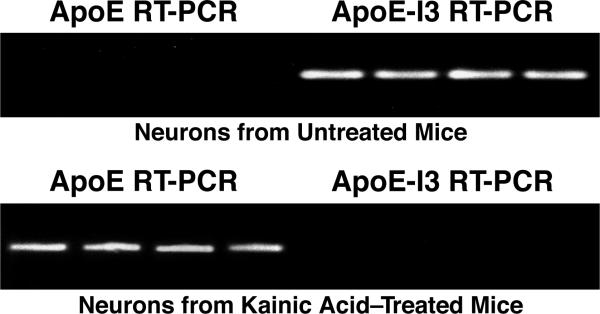
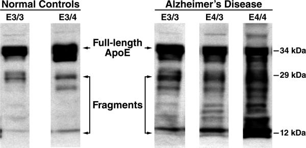
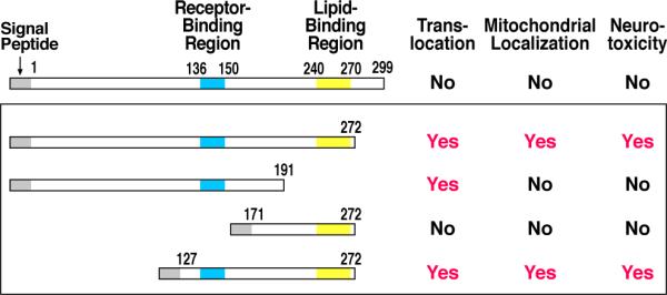
Apolipoprotein (apo) E4 is the major genetic risk factor for Alzheimer's disease and is associated with poor clinical outcome following traumatic brain injury and other neuropathological disorders. Protein instability and an isoform-specific apoE property called domain interaction are responsible for these neuropathological effects. ApoE4 is the most neurotoxic isoform and can induce neuropathology through various cellular pathways. Neuronal damage or stress induces apoE synthesis as part of the repair response; however, when apoE4 is expressed in neurons, its unique conformation makes it susceptible to proteolysis, resulting in the generation of neurotoxic fragments. These fragments cause pathological mitochondrial dysfunction and cytoskeletal alterations. Here, we review data supporting the hypothesis that apoE4 (> apoE3 > apoE2) has direct neurotoxic effects and highlight studies showing that blocking domain interaction reverses these detrimental effects.


Figures

Figure 1
Figure 1. Injurious Agents That Stress or Damage Neurons Induce ApoE Synthesis and Initiate Neuropathology
ApoE4, because of domain interaction, displays impaired trafficking through the ER and Golgi apparatus compared with apoE3. As a result, it is targeted to a neuron-specific protease that initially cleaves off the C-terminal 27–30 amino acids, generating neurotoxic fragments. These fragments escape the secretory pathway and enter the cytosol, where they cause mitochondrial dysfunction and cytoskeletal alterations, enhance tau phosphorylation, and form NFT-like structures (Mahley et al., 2006). Figure reprinted with permission from Mahley and Huang, Small-molecule structure correctors target abnormal protein structure and function: The structure corrector rescue of apolipoprotein E4–associated neuropathology. J. Med. Chem. 55: 8997–9008, 2012. Copyright 2012 American Chemical Society.
Figure 2
Figure 2. EGFP Inserted into One Allele of the ApoE Gene Acts as a Marker for ApoE Expression in Vivo
Top, schematic of the EGFP-apoE reporter cassette. Bottom, representative images of hippocampal sections taken from EGFPapoE-reporter knockin mice. In the uninjured mouse hippocampus (before kainic acid), astrocytes display abundant apoE synthesis (bottom, left); however, after injury with kainic acid, neurons turn on the synthesis of apoE, as indicated by the colocalization of EGFP (green) and NeuN (red) (bottom, right) (Xu et al., 2006). Modified from Figure 6, Xu, Q., Bernardo, A., Walker, D., Kanegawa, T., Mahley, R.W., and Huang, Y. Profile and regulation of apolipoprotein E (apoE) expression in the CNS in mice with targeting of green fluorescent protein gene to the apoE locus. J. Neurosci. 26: 4985–4994, 2006.
Figure 3
Figure 3. Kainic Acid–Induced Injury Triggers Switch from Intron 3–Containing to Mature ApoE mRNA Synthesis
EGFPapoE-reporter knockin mice were treated or not with kainic acid, and CA1 hippocampal neurons were isolated by laser-capture microdissection for reverse transcription–polymerase chain reaction (RT-PCR). Neurons from a normal, uninjured mouse demonstrate the presence of apoE mRNA with intron 3 (apoE-I3), whereas after injury there is a dramatic switch to mature apoE mRNA lacking intron 3 (Xu et al., 2008). Modified from Figure 7, Xu, Q., Walker, D., Bernardo, A., Brodbeck, J., Balestra, M.E., and Huang, Y. Intron-3 retention/splicing controls neuronal expression of apolipoprotein E in the CNS. J. Neurosci. 28: 1452–1459, 2008.
Figure 4
Figure 4. Models of the Structures of ApoE3 and ApoE4
(A) ApoE4 displays a unique property called domain interaction caused by the ionic interaction between arginine-61 in the N-terminal domain with glutamic acid–255 in the C-terminal domain. ApoE3 is significantly less likely to undergo domain interaction than apoE4. (B) ApoE4 domain interaction can be blocked by a small-molecule apoE4 structure corrector that disrupts the ionic interaction between arginine-61 and glutamic acid–255. This converts apoE4 to an apoE3-like molecule both structurally and functionally (Huang, 2010; Mahley and Rall, 2000; Mahley et al., 2006, 2009). ApoE4SC, apoE4 structure corrector. Figure reprinted with permission from Mahley and Huang, Small-molecule structure correctors target abnormal protein structure and function: The structure corrector rescue of apolipoprotein E4–associated neuropathology. J. Med. Chem. 55: 8997–9008, 2012. Copyright 2012 American Chemical Society.
Figure 5
Figure 5. ApoE4 Displays Impaired Trafficking through the Secretory Pathway That Can Be Corrected by Blocking ApoE4 Domain Interaction
Fluorescence recovery after laser photobleaching experiments were performed on EGFP-apoE3- and EGFP-apoE4-expressing Neuro-2a cells. For each section, the left and center panels show representative recovery curves for the ER and Golgi apparatus, respectively, while the right-hand panel shows quantitative histograms of apoE mobility. (A) Recovery curves show that apoE4 trafficking is decreased compared with apoE3 in both the ER (left) and Golgi apparatus (center). Right, histogram showing that the percent of the immobile fraction was greater with apoE4 than apoE3. (B) The impaired trafficking of apoE4 was reversed by site-directed mutagenesis of arginine-61, which blocked domain interaction (apoE4-R61T). This mutation reversed the impairment in apoE4 trafficking and mobility in both the ER and Golgi apparatus. (C) Treatment of apoE4-expressing cells with a structure corrector (PH-002) restored the trafficking of apoE4 through the secretory pathway. The structure corrector did not affect the trafficking of apoE3 or apoE4-R61T (Brodbeck et al., 2011). Modified from Figures 3, 4, 5 originally published in Brodbeck, J., McGuire, J., Liu, Z., Meyer-Franke, A., Balestra, M.E., Jeong, D.-e., Pleiss, M., McComas, C., Hess, F., Witter, D., Peterson, S., Childers, M., Goulet, M., Liverton, N., Hargreaves, R., Freedman, S., Weisgraber, K., Mahley, R.W., Huang, Y. Structure-dependent impairment of intracellular apolipoprotein E4 trafficking and its detrimental effects are rescued by small-molecule structure correctors. J. Biol. Chem. 2011; 286:17217–17226. © the American Society for Biochemistry and Molecular Biology.
Figure 6
Figure 6. ApoE Fragmentation Pattern in Human Temporal Cortex in Non-Demented Controls and in Age-Matched AD Patients
Compared with full-length apoE (34 kDa), proteolytic cleavage generates an initial fragment with a molecular weight of ~29 kDa. Subsequent to this, fragments of ~12–20 kDa are generated. In AD patients, there is an apoE4 gene–dose effect on apoE fragmentation, whereby apoE4/3 subjects have more fragments than apoE3/3 subjects and the apoE4/4 subjects have the greatest amount of fragments.
Figure 7
Figure 7. Regions of ApoE Responsible for Fragment Translocation into the Cytosol, Mitochondrial Targeting, and Neurotoxicity
Fragments containing the receptor-binding region (residues 136–150) and the lipid-binding region (residues 240–270) represent the minimal structure required for translocation, mitochondrial localization, and neurotoxicity (Mahley et al., 2006). Modified from Figure 4, Mahley, R.W., Weisgraber, K.H., and Huang, Y. Apolipoprotein E4: A causative factor and therapeutic target in neuropathology, including Alzheimer's disease. Proc. Natl. Acad. Sci. USA 103: 5644–5651, 2006. © by the National Academy of Sciences.
Figure 8
Figure 8. ApoE4-Transfected Cells Demonstrate Intracellular FRET Signal
ApoE constructs were generated to express YFP and CFP at the N and C termini, respectively, to act as reporter FRET fluorophores. When expressed in Neuro-2a cells, YFP-apoE4-CFP displays significantly more FRET signal compared to YFP-apoE3-CFP-expressing cells. This reflects the closer interaction between the N- and C-terminal domains in apoE4 and demonstrates that the biophysical property of domain interaction occurs in cells (Xu et al., 2004). Modified from Figure 1, Xu, Q., Brecht, W.J., Weisgraber, K.H., Mahley, R.W., and Huang, Y. Apolipoprotein E4 domain interaction occurs in living neuronal cells as determined by fluorescence resonance energy transfer. J. Biol. Chem. 279: 25511–25516, 2004. © the American Society for Biochemistry and Molecular Biology.
Figure 9
Figure 9. FRET Assay Identifies ApoE4 Structure Correctors
(A) The FRET signal is generated between the donor and acceptor fluorophores conjugated to the N and C termini of apoE. ApoE4 gives a higher FRET signal because of the closeness of the N- and C-terminal domains. The apoE4 signal is reduced by the small-molecule apoE structure corrector (SC) by disrupting domain interaction (Chen et al., 2012). (B) Dose-response analysis reveals the relative potencies of three active and one inactive structure corrector (Chen et al., 2012). (C) Mitochondrial cytochrome c oxidase (COX1) is reduced in Neuro-2a cells expressing apoE4 compared with apoE3. A small-molecule structure corrector (CB9032258) corrects the COX1 deficiency in the apoE4-expressing cells, but does not significantly affect apoE3-expressing cells. (A) and (B). Modified from Figure 2, Chen, H.-K., Liu, Z., Meyer-Franke, A., Brodbeck, J., Miranda, R.D., McGuire, J.G., Pleiss, M.A., Ji, Z.-S., Balestra, M.E., Walker, D.W., Xu, Q., Jeong, D.-e., Budamagunta, M.S., Voss, J.C., Freedman, S.B., Weisgraber, K.H., Huang, Y., Mahley, R.W. Small molecule structure correctors abolish detrimental effects of apolipoprotein E4 in cultured neurons. J. Biol. Chem. 2012; 287:5253–5266. © the American Society for Biochemistry and Molecular Biology.
Figure 10
Figure 10. ApoE Sets the Stage and Response to Neuronal Injury Triggers Neuropathology
(1) Injury to neurons induces the synthesis of apoE. ApoE (apoE4 > apoE3) is susceptible to proteolytic cleavage in neurons, and the neurotoxic fragments that are generated escape the secretory pathway and cause mitochondrial dysfunction and cytoskeletal alterations. This is most likely to occur when apoE4 is expressed (apoE4 > apoE3) due to its abnormal protein conformation (instability and domain interaction). (2) Exogenous apoE, primarily from astrocytes, could cause neuronal injury and could generate neurotoxic fragments by being shunted to the ER/Golgi apparatus, where proteolysis could occur. In addition, exogenous apoE does impact Aβ clearance/deposition. (3) Aβ expression can be induced by injured/stressed neurons, and together with other injurious agents could perpetuate the toxic cycle of injury in neurons. This would include apoE synthesis followed by proteolytic cleavage, toxic fragment formation, and neuropathology.

References

    1. Adams RA, Bauer J, Flick MJ, Sikorski SL, Nuriel T, Lassmann H, Degen JL, Akassoglou K. The fibrin-derived γ377-395 peptide inhibits microglia activation and suppresses relapsing paralysis in central nervous system autoimmune disease. J. Exp. Med. 2007;204:571–582. - PMC - PubMed
    1. Agosta F, Vossel KA, Miller BL, Migliaccio R, Bonasera SJ, Filippi M, Boxer AL, Karydas A, Possin KL, Gorno-Tempini ML. Apolipoprotein E ε4 is associated with disease-specific effects on brain atrophy in Alzheimer's disease and frontotemporal dementia. Proc. Natl. Acad. Sci. USA. 2009;106:2018–2022. - PMC - PubMed
    1. Akassoglou K, Adams RA, Bauer J, Mercado P, Tseveleki V, Lassmann H, Probert L, Strickland S. Fibrin depletion decreases inflammation and delays the onset of demyelination in a tumor necrosis factor transgenic mouse model for multiple sclerosis. Proc. Natl. Acad. Sci. USA. 2004;101:6698–6703. - PMC - PubMed
    1. Alberts MJ, Graffagnino C, McClenny C, DeLong D, Strittmatter W, Saunders AM, Roses AD. ApoE genotype and survival from intracerebral haemorrhage. Lancet. 1995;346:575. - PubMed
    1. Andrews-Zwilling Y, Bien-Ly N, Xu Q, Li G, Bernardo A, Yoon SY, Zwilling D, Yan TX, Chen L, Huang Y. Apolipoprotein E4 causes age- and tau-dependent impairment of GABAergic interneurons, leading to learning and memory deficits in mice. J. Neurosci. 2010;30:13707–13717. - PMC - PubMed

Publication types

MeSH terms

Substances