. 2013 Feb 5;436(1):127-33.
doi: 10.1016/j.virol.2012.10.038. Epub 2012 Dec 4.

The role of proteolytic processing and the stable signal peptide in expression of the Old World arenavirus envelope glycoprotein ectodomain

Affiliations

The role of proteolytic processing and the stable signal peptide in expression of the Old World arenavirus envelope glycoprotein ectodomain

Dominique J Burri et al. Virology. .

Abstract

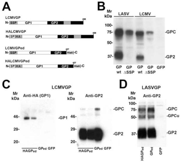
Maturation of the arenavirus GP precursor (GPC) involves proteolytic processing by cellular signal peptidase and the proprotein convertase subtilisin kexin isozyme 1 (SKI-1)/site 1 protease (S1P), yielding a tripartite complex comprised of a stable signal peptide (SSP), the receptor-binding GP1, and the fusion-active transmembrane GP2. Here we investigated the roles of SKI-1/S1P processing and SSP in the biosynthesis of the recombinant GP ectodomains of lymphocytic choriomeningitis virus (LCMV) and Lassa virus (LASV). When expressed in mammalian cells, the LCMV and LASV GP ectodomains underwent processing by SKI-1/S1P, followed by dissociation of GP1 from GP2. The GP2 ectodomain spontaneously formed trimers as revealed by chemical cross-linking. The endogenous SSP, known to be crucial for maturation and transport of full-length arenavirus GPC was dispensable for processing and secretion of the soluble GP ectodomain, suggesting a specific role of SSP in the stable prefusion conformation and transport of full-length GPC.


Figures

Figure 1
Figure 1. Expression and characterization of the soluble variant LCMV GPed
(A) Schematic representation of the recombinant LCMV GP variants: LCMV GP1 and GP2 are in white and black respectively. The trans-membrane domain of wild-type GP2 is indicated as a gray box. The stable signal peptide (SSP) and the C-terminal His-tag are indicated. (B) Expression of LCMV GPed and wild-type LCMV GP: LCMV GPed (LCMV GPed), wild-type LCMV GP (LCMV GP), and GFP were transiently expressed in HEK293H cells in serum-free medium (duplicates). After 48 h of expression in serum-free conditions, total protein was isolated from cell culture supernatant (supernatant) and cell lysates (cells). Proteins were separated by SDS-PAGE and transferred to nitrocellulose. Membranes were probed with mouse mAb 83.6 (anti-LCMV GP2) and a peroxidase-conjugated anti-mouse IgG using ECL for detection. Molecular masses and the expected positions of the precursors (GPC) and mature GP2 are indicated. The additional band at circa 45 kDa likely corresponds to an under-glycosylated form of the GPC (GPCu). (C) Deglycosylation with PNGaseF. LCMV GPed was expressed in HEK293H cells as in (B) and subjected to pull-down with NiNTA agarose. After washing, precipitated proteins were incubated with PNGaseF (+) or reaction buffer only. Proteins were separated by SDS-PAGE (long-run) and GP2 fragments detected in Western blot using an antibody to His6-tag. Apparent molecular masses are indicated. (D) Detection of GP1 and GP2 in supernatants by ELISA: LCMV GPed, and GFP were transiently expressed in HEK293H cells in serum-free medium. Conditioned supernatants were immobilized in microtiter plates and probed with monoclonal antibodies anti-GP1 (36.1 and 67.1) and anti-GP2 (33.1 and 83.6). Primary antibodies were detected with anti-mouse IgG coupled to peroxidase in a color reaction using ABTS substrate. OD (405) was measured using an ELISA reader (data are means ± SD, n = 3). (E) Expression of LASV GPed. LASV GPed (GPed) and wild-type LASV GP (GP), as well as GFP were expressed in HEK293H cells as in (B). The presence of LASV GP2 was detected in cell supernatants and total cell lysates with mAb 83.6 in Western blot as in (B). Molecular masses, GPC, and mature GP2 are indicated. As in (B), the band at circa 50 kDa likely corresponds to under-glycosylated GPC (GPCu).
Figure 2
Figure 2. LCMV GPed and LASV GPed are processed by SKI-1/S1P
Expression of LCMV GPed (A) and LASV GPed (B) in SRD12B and CHOK1 cells: SRD12B and CHOK1 cells were transfected with expression plasmids for LCMV GPed and LASV GPed using lipofectamine. After 48 hours of expression in serum-free conditions, total protein was isolated from conditioned cell culture medium (supernatant) and cell lysates (cells). Proteins were separated by SDS-PAGE and Western-blot analysis for LCMV GP2 performed as in Fig. 1. Molecular masses and the position of GPC, GP2, and GPCu are indicated.
Figure 3
Figure 3. The soluble GP2 domain forms trimers
Recombinant LCMV GPed was expressed in HEK293H cells in serum-free medium. Conditioned supernatants were dialyzed against cross-linking reaction buffer and subjected to chemical cross-linking with the indicated concentrations of S-DST. The reaction was quenched by the addition of 50 mM glycine, total protein isolated, separated by SDS-PAGE, and transferred to nitrocellulose. The blot was probed for GP2 with mAb 83.6. Molecular masses and the calculated apparent molecular weights of the cross-linked GP2 species are indicated.
Figure 4
Figure 4. Purified GP2 ectodomain forms trimers
(A-C) Purification of GP2 ectodomain from LCMV GPed: LCMV GPed was expressed in serum-free medium as described in Fig. 1 and purified from concentrated supernatants by NiNTA affinity chromatography. The matrix was washed under high-salt conditions (0.5 M NaCl) and bound GP2 eluted with EDTA. (A) Analysis of the indicated fractions by SDS-PAGE and silver staining (S: supernatant, W4, 5: = late wash fractions, E1-4: eluate fractions). Molecular masses are indicated. (B) Detection of GP2 by Western-blot: Supernatant (S) and eluate fractions E2 and E3 from (B) were subjected to Western blot analysis with mAb 83.6 (anti-LCMV GP2) as described in Fig. 1B. (C) Detection of GP1 and GP2 by ELISA: Supernatant (S), flow-through (FT), the wash fractions W1-W5, as well as the main eluate fractions E2 and E3 were immobilized in microtiter plates and GP1 and GP2 detected by ELISA using mAbs 36.1 (GP1) and 83.6 (GP2), respectively Note the high proportion of GP1 in the flow-through and the early wash fractions. (D) Chemical cross-linking of purified GP2: Eluate fractions E2 and E3 from (B) were pooled, dialyzed against reaction buffer, and chemical cross-linking performed with the indicated concentrations of S-DST as described in Fig. 3. After quenching of the reaction with glycine, total protein was isolated, separated by SDS-PAGE, and transferred to nitrocellulose. The blot was probed for GP2 with mAb 83.6. Molecular masses and the calculated apparent molecular weights of the cross-linked GP2 species are indicated.
Figure 5
Figure 5. The stable signal peptide (SSP) is required for transport and processing of full-length GPC, but not the GP ectodomain
(A) Schematic representation of the recombinant LCMV GP variants. The SSP and the HA-tag are indicated; SP corresponds to the signal peptide of Ig light chain. (B) The SSP is required for transport and processing of full-length LCMV and LASV GPC. Wild-type LCMV GP and LASV GP (GP wt) and HA-tagged LCMV GP and LASV GP containing the signal peptide of Ig light chain instead of the endogenous SSP (GP SSP) were transfected in HEK293T cells and protein processing detected in Western-blot with antibodies to GP2. (C) The SSP is dispensable for transport and processing of the soluble LCMV GP ectodomain. HALCMV GPed (HAGPed), wild-type LCMV GPed (GPed), and GFP were expressed in HEK293T cells. Recombinant proteins in the supernatant were detected by Western-blot using an antibody to HA (detection of GP1) and to GP2 as in (B). Apparent molecular masses and the positions of GP1, GPC, and GP2 are indicated. (D) Processing of LASV GPed containing the signal peptide of Ig light chain. LASV GPed containing the Ig light chain signal peptide (HAGPed), wild type LASV GP with the authentic SSP (GPed), and GFP were expressed in HEK293T cells. Processing of soluble LASV GP2 in the supernatant was detected by Western-blot as in (B). Molecular masses and the positions of GPC, GPCu and GP2 are indicated.

Similar articles

Cited by

References

    1. Agnihothram SS, York J, Nunberg JH. Role of the stable signal peptide and cytoplasmic domain of G2 in regulating intracellular transport of the Junin virus envelope glycoprotein complex. J Virol. 2006;80(11):5189–98. - PMC - PubMed
    1. Agnihothram SS, York J, Trahey M, Nunberg JH. Bitopic membrane topology of the stable signal peptide in the tripartite Junin virus GP-C envelope glycoprotein complex. J Virol. 2007;81(8):4331–7. Epub 2007 Jan 31. - PMC - PubMed
    1. Beyer WR, Popplau D, Garten W, von Laer D, Lenz O. Endoproteolytic processing of the lymphocytic choriomeningitis virus glycoprotein by the subtilase SKI-1/S1P. J Virol. 2003;77(5):2866–72. - PMC - PubMed
    1. Bonthius DJ, Nichols B, Harb H, Mahoney J, Karacay B. Lymphocytic choriomeningitis virus infection of the developing brain: critical role of host age. Ann Neurol. 2007;62(4):356–74. - PubMed
    1. Bonthius DJ, Perlman S. Congenital viral infections of the brain: lessons learned from lymphocytic choriomeningitis virus in the neonatal rat. PLoS Pathog. 2007;3(11):e149. - PMC - PubMed

Publication types

Substances

LinkOut - more resources