

Clinical phenotypes and prognostic full-field electroretinographic findings in Stargardt disease
- PMID: 23219216
- PMCID: PMC4494104
- DOI: 10.1016/j.ajo.2012.09.011
Clinical phenotypes and prognostic full-field electroretinographic findings in Stargardt disease
Abstract
Purpose: To investigate the relationships between clinical and full-field electroretinographic (ERG) findings and progressive loss of visual function in Stargardt disease.
Design: Retrospective cohort study.
Methods: We performed a retrospective review of data from 198 patients with Stargardt disease. Measures of visual function over time, including visual acuity, quantified Goldmann visual fields, and full-field ERG data were recorded. Data were analyzed using SAS statistical software. Subgroup analyses were performed on 148 patients with ERG phenotypic data, 46 patients with longitudinal visual field data, and 92 patients with identified ABCA4 mutations (46 with 1 mutation, and 47 with 2 or more mutations).
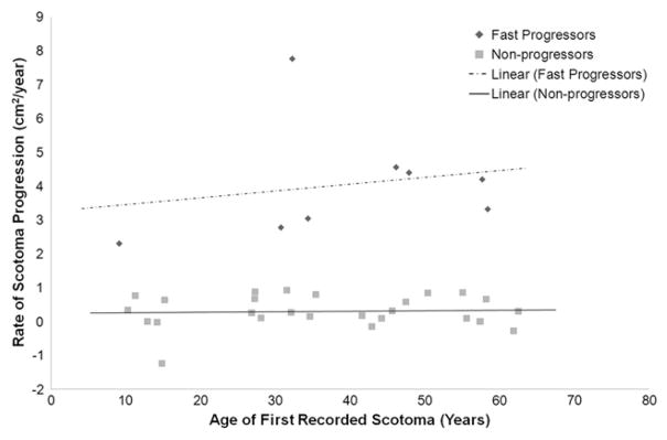
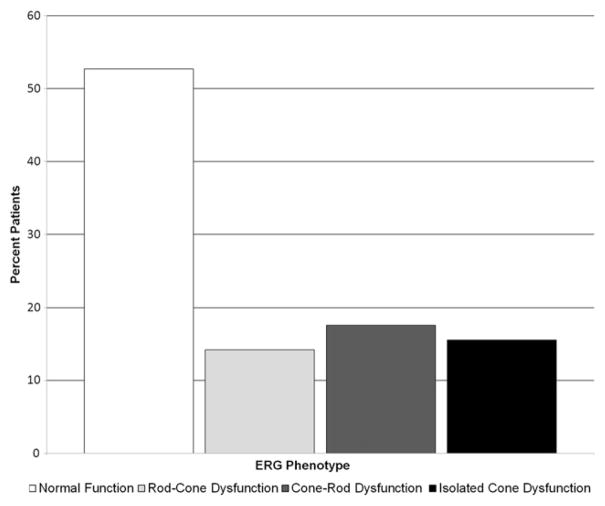
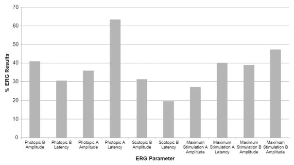
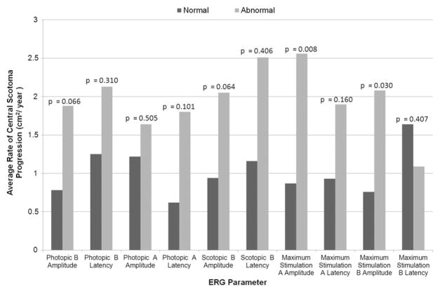
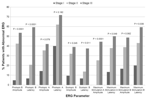
Results: Of 46 patients with longitudinal visual field data, 8 patients with faster central scotoma progression rates had significantly worse scotopic B-wave amplitudes at their initial assessment than 20 patients with stable scotomata (P = .014) and were more likely to have atrophy beyond the arcades (P = .047). Overall, 47.3% of patients exhibited abnormal ERG results, with rod-cone dysfunction in 14.2% of patients, cone-rod dysfunction in 17.6% of patients, and isolated cone dysfunction in 15.5% of patients. Abnormal values in certain ERG parameters were associated significantly with (maximum-stimulation A- and B-wave amplitudes) or tended toward (photopic and scotopic B-wave amplitudes) a higher mean rate of central scotoma progression compared with those patients with normal ERG values. Scotoma size and ERG parameters differed significantly between those with a single mutation versus those with multiple mutations.
Conclusions: Full-field ERG examination provides clinically relevant information regarding the severity of Stargardt disease, likelihood of central scotoma expansion, and visual acuity deterioration. Patients also may exhibit an isolated cone dystrophy on ERG examination.
Copyright © 2013 Elsevier Inc. All rights reserved.
Conflict of interest statement
ALL AUTHORS HAVE COMPLETED AND SUBMITTED THE ICMJE FORM FOR DISCLOSURE OF POTENTIAL CONFLICTS OF INTEREST and the following was reported. Dr Musch is a consultant for ReVision Therapeutics, Inc., for studies of fenretinide treatment for Stargardt disease. Dr Musch is a recipient of the Research to Prevent Blindness Lew R. Wasserman Merit Award. Involve din Design and conduct of study (K.T.J., K.B., J.R.H.); Collection, management, analysis, and interpretation of data (S.Z., K.T.J., W.R., N.K., L.M.N., D.C.M., J.R.H.); and Preparation, review, or approval of manuscript (S.Z., K.T.J., W.R., K.B., N.K., L.M.N., D.C.M., J.R.H.).
Figures
Similar articles
-
Full-field ERG as a predictor of the natural course of ABCA4-associated retinal degenerations.Mol Vis. 2018 Jan 4;24:1-16. eCollection 2018. Mol Vis. 2018. PMID: 29386879 Free PMC article.
-
Electroretinographic findings in patients with Stargardt disease and fundus flavimaculatus.Retina. 2004 Dec;24(6):920-8. doi: 10.1097/00006982-200412000-00013. Retina. 2004. PMID: 15579991
-
A longitudinal study of stargardt disease: clinical and electrophysiologic assessment, progression, and genotype correlations.Am J Ophthalmol. 2013 Jun;155(6):1075-1088.e13. doi: 10.1016/j.ajo.2013.01.018. Epub 2013 Mar 15. Am J Ophthalmol. 2013. PMID: 23499370
-
Vitamin A and fish oils for preventing the progression of retinitis pigmentosa.Cochrane Database Syst Rev. 2020 Jun 18;6(6):CD008428. doi: 10.1002/14651858.CD008428.pub3. Cochrane Database Syst Rev. 2020. PMID: 32573764 Free PMC article.
-
Insights into the Molecular Properties of ABCA4 and Its Role in the Visual Cycle and Stargardt Disease.Prog Mol Biol Transl Sci. 2015;134:415-31. doi: 10.1016/bs.pmbts.2015.06.008. Epub 2015 Jul 14. Prog Mol Biol Transl Sci. 2015. PMID: 26310168 Review.
Cited by
-
Clinical and electroretinographic profile of 27 patients with Stargardt disease treated at a hospital in Brazil.Arq Bras Oftalmol. 2021 Jul-Aug;84(4):367-373. doi: 10.5935/0004-2749.20210053. Arq Bras Oftalmol. 2021. PMID: 33567042 Free PMC article.
-
Comparison of Green Versus Blue Fundus Autofluorescence in ABCA4-Related Retinopathy.Transl Vis Sci Technol. 2018 Oct 1;7(5):13. doi: 10.1167/tvst.7.5.13. eCollection 2018 Sep. Transl Vis Sci Technol. 2018. PMID: 30279998 Free PMC article.
-
The Direct Healthcare Cost of Stargardt Disease: A Claims-Based Analysis.Ophthalmic Epidemiol. 2021 Dec;28(6):533-539. doi: 10.1080/09286586.2021.1883675. Epub 2021 Feb 21. Ophthalmic Epidemiol. 2021. PMID: 33615979 Free PMC article.
-
Complex inheritance of ABCA4 disease: four mutations in a family with multiple macular phenotypes.Hum Genet. 2016 Jan;135(1):9-19. doi: 10.1007/s00439-015-1605-y. Epub 2015 Nov 2. Hum Genet. 2016. PMID: 26527198 Free PMC article.
-
Prediction of Function in ABCA4-Related Retinopathy Using Ensemble Machine Learning.J Clin Med. 2020 Jul 29;9(8):2428. doi: 10.3390/jcm9082428. J Clin Med. 2020. PMID: 32751377 Free PMC article.
References
-
- Allikmets R, Singh N, Sun H, et al. A photoreceptor cell-specific ATP-binding transporter gene (ABCR) is mutated in recessive Stargardt macular dystrophy. Nat Genet. 1997;15(3):236–246. - PubMed
-
- Ayuso C, Garcia-Sandoval B, Najera C, Valverde D, Carballo M, Antinolo G. Retinitis pigmentosa in Spain. The Spanish Multicentric and Multidisciplinary Group for Research into Retinitis Pigmentosa. Clin Genet. 1995;48(3):120–122. - PubMed
-
- Blacharski P. Fundus flavimaculatus. In: Newsome DA, editor. Retinal Dystrophies and Degenerations. New York: Raven Press; 1988. pp. 135–159.
-
- Martinez-Mir A, Bayes M, Vilageliu L, et al. A new locus for autosomal recessive retinitis pigmentosa (RP19) maps to 1p13–1p21. Genomics. 1997;40(1):142–146. - PubMed
-
- Martinez-Mir A, Paloma E, Allikmets R, et al. Retinitis pigmentosa caused by a homozygous mutation in the Stargardt disease gene ABCR. Nat Genet. 1998;18(1):11–12. - PubMed
Publication types
MeSH terms
Substances
Grants and funding
LinkOut - more resources
Full Text Sources
Other Literature Sources
Medical
