. 2013 Feb;97(2):161-8.
doi: 10.1016/j.antiviral.2012.11.005. Epub 2012 Nov 28.

Potent inhibition of feline coronaviruses with peptidyl compounds targeting coronavirus 3C-like protease

Affiliations

Potent inhibition of feline coronaviruses with peptidyl compounds targeting coronavirus 3C-like protease

Yunjeong Kim et al. Antiviral Res. 2013 Feb.

Abstract

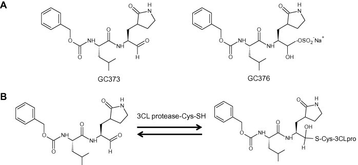
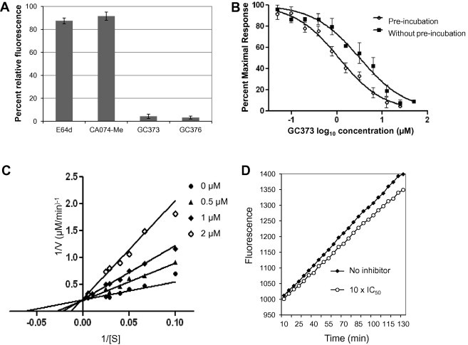
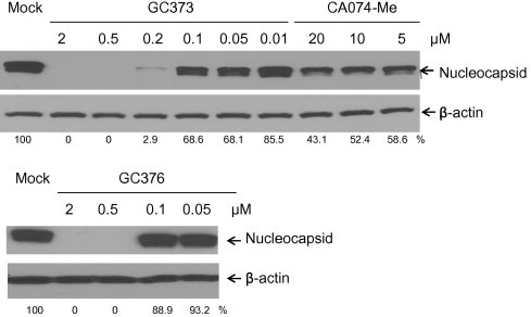
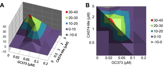
Feline coronavirus infection is common among domestic and exotic felid species and usually associated with mild or asymptomatic enteritis; however, feline infectious peritonitis (FIP) is a fatal disease of cats that is caused by systemic infection with a feline infectious peritonitis virus (FIPV), a variant of feline enteric coronavirus (FECV). Currently, there is no specific treatment approved for FIP despite the importance of FIP as the leading infectious cause of death in young cats. During the replication process, coronavirus produces viral polyproteins that are processed into mature proteins by viral proteases, the main protease (3C-like [3CL] protease) and the papain-like protease. Since the cleavages of viral polyproteins are an essential step for virus replication, blockage of viral protease is an attractive target for therapeutic intervention. Previously, we reported the generation of broad-spectrum peptidyl inhibitors against viruses that possess a 3C or 3CL protease. In this study, we further evaluated the antiviral effects of the peptidyl inhibitors against feline coronaviruses, and investigated the interaction between our protease inhibitor and a cathepsin B inhibitor, an entry blocker, against a feline coronavirus in cell culture. Herein we report that our compounds behave as reversible, competitive inhibitors of 3CL protease, potently inhibited the replication of feline coronaviruses (EC(50) in a nanomolar range) and, furthermore, combination of cathepsin B and 3CL protease inhibitors led to a strong synergistic interaction against feline coronaviruses in a cell culture system.


Figures

Fig. 1
Fig. 1
(A) Dipeptidyl protease inhibitors, GC373 and GC376. (B) Proposed mechanism of transition state inhibition of 3CL protease by GC373.
Fig. 2
Fig. 2
The sequence alignment of the 3CL protease of TGEV (Miller strain) and feline coronaviruses (WSU-1146 and DF2 strains). Asterisks mark the position of the conserved cysteine (144) and histidine (41) residues in the active site.
Fig. 3
Fig. 3
FRET-based protease assay. (A) The effects of the 3CL protease inhibitors, GC373 and GC376, a cathepsin B inhibitor CA074-Me, and a pan-cysteine cathepsin inhibitor E64d on the activity of TGEV 3CL protease in the FRET-protease assay. TGEV 3CL protease was incubated with each compound at 50 μM for 20 min before the substrate was added to the mixture. Each bar represents the percent relative fluorescence (mean ± standard error of the mean [SEM]). (B) A plot of log10 GC373 concentration versus percent maximal response. TGEV 3CL protease was incubated with GC373 for 20 min at increasing concentrations prior to addition of substrate (open circles), or GC373 was added at the same time with the substrate (filled squares). The fluorescence signals were detected by a spectrophotometer, and the data are then plotted as percent maximal response against the log concentrations of the compound. The data points represent the percent maximal response (mean ± SEM). (C) Lineweaver–Burk plots of kinetic data of the TGEV 3CL protease incubated with various concentrations of GC373. The enzymatic activities were measured using 10–200 μM substrate in the absence (filled circles) or presence of 0.5 (filled triangles), 1 (filled diamonds), and 2 (open diamonds) μM GC373. (D) Progress curves for the recovery of enzymatic activities of TGEV 3CL protease after 3CL protease-GC373 complex was rapidly diluted to the normal assay solution. TGEV 3CL protease was incubated with DMSO (filled diamonds) or 10 μM (open circles) GC373 for 20 min, then rapidly diluted 100-fold in reaction buffer prior to substrate addition, and assayed for enzymatic activity.
Fig. 4
Fig. 4
Western blot analysis of the effects of GC373, GC376, and CA074-Me on the accumulation of coronavirus nucleocapsid proteins in CRFK cells infected with WSU-1146. CRFK cells were treated with 0.1% DMSO, GC373, GC376, or CA074-Me for 2 h, followed by virus infection at an MOI of 5, and further incubated for 12 h. Cell extracts were analyzed by Western blot for expression of coronavirus nucleocapsid protein and β-actin was loaded as an internal control. Numbers below each lane indicate the values of coronavirus nucleocapsid proteins normalized to β-actin obtained with densitometric scanning using TotalLab Quant software (TotalLab Ltd.).
Fig. 5
Fig. 5
Three-dimensional plots showing the interaction of GC373 and CA074-Me on the replication of WSU-1146. (A and B) CRFK cells were incubated with CA074-Me (0.5–5 μM), GC373 (0.02–0.2 μM) or combinations of CA074-Me and GC373 for 2 h before the virus was inoculated in the cells at an MOI of 0.05. The cells were further incubated in the presence of each compound for 24 h, and virus replication was measured by the TCID50 method. Drug–drug interactions were analyzed by the three-dimensional model of Prichard and Shipman, using the MacSynergy II software at a 95% confidence interval. Surface above the plane of 0% synergy in the plot indicate synergy. (B) Contour plots (two-dimensional representations of the data) for easier identification of the concentration ranges where statistically significant synergistic or antagonistic effects occurred.

References

    1. Anand K., Palm G.J., Mesters J.R., Siddell S.G., Ziebuhr J., Hilgenfeld R. Structure of coronavirus main proteinase reveals combination of a chymotrypsin fold with an extra alpha-helical domain. EMBO J. 2002;21:3213–3224. - PMC - PubMed
    1. Barlough J.E., Shacklett B.L. Antiviral studies of feline infectious peritonitis virus in vitro. Vet. Rec. 1994;135:177–179. - PubMed
    1. Benetka V., Kubber-Heiss A., Kolodziejek J., Nowotny N., Hofmann-Parisot M., Mostl K. Prevalence of feline coronavirus types I and II in cats with histopathologically verified feline infectious peritonitis. Vet. Microbiol. 2004;99:31–42. - PMC - PubMed
    1. Chang H.W., de Groot R.J., Egberink H.F., Rottier J.M. Feline infectious peritonitis: insights into feline coronavirus pathobiogenesis and epidemiology based on genetic analysis of the viral 3c gene. J. Gen. Virology. 2010;91:415–420. - PubMed
    1. Chang H.W., Egberink H.F., Rottier P.J. Sequence analysis of feline coronaviruses and the circulating virulent/avirulent theory. Emerg. Infect. Dis. 2011;17:744–746. - PMC - PubMed

Publication types