

Macular telangiectasia type 2
- PMID: 23219692
- PMCID: PMC3638089
- DOI: 10.1016/j.preteyeres.2012.11.002
Macular telangiectasia type 2
Abstract
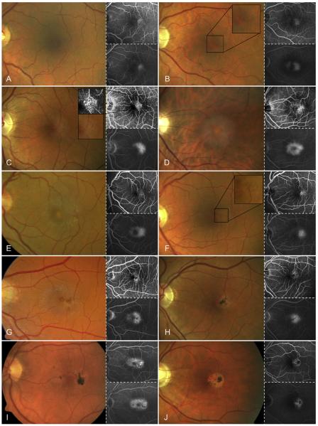
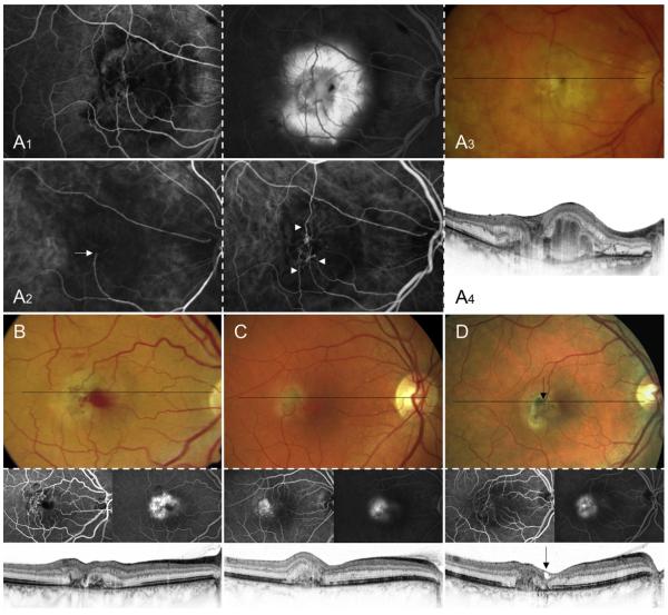
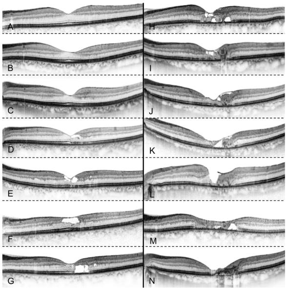
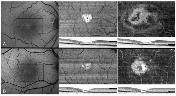
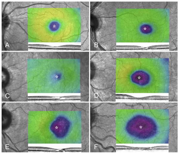
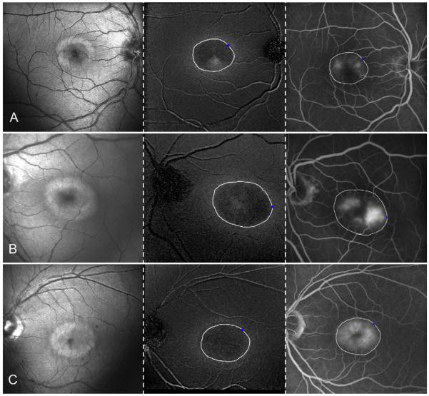
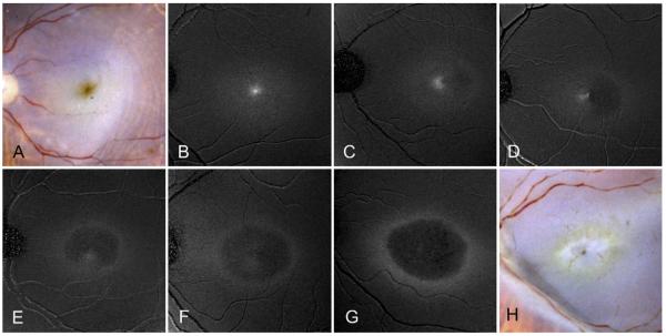
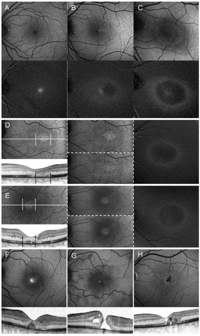
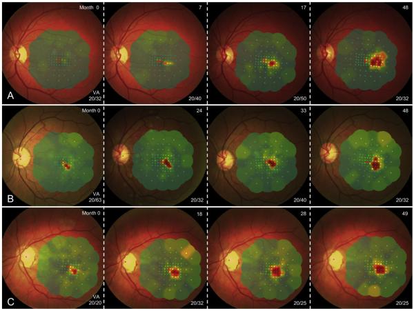
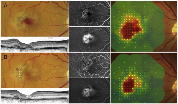
Macular telangiectasia type 2 is a bilateral disease of unknown cause with characteristic alterations of the macular capillary network and neurosensory atrophy. Its prevalence may be underestimated and has recently been shown to be as high as 0.1% in persons 40 years and older. Biomicroscopy may show reduced retinal transparency, crystalline deposits, mildly ectatic capillaries, blunted venules, retinal pigment plaques, foveal atrophy, and neovascular complexes. Fluorescein angiography shows telangiectatic capillaries predominantly temporal to the foveola in the early phase and a diffuse hyperfluorescence in the late phase. High-resolution optical coherence tomography (OCT) may reveal disruption of the photoreceptor inner segment-outer segment border, hyporeflective cavities at the level of the inner or outer retina, and atrophy of the retina in later stages. Macular telangiectasia type 2 shows a unique depletion of the macular pigment in the central retina and recent therapeutic trials showed that such depleted areas cannot re-accumulate lutein and zeaxanthin after oral supplementation. There have been various therapeutic approaches with limited or no efficacy. Recent clinical trials with compounds that block vascular endothelial growth factor (VEGF) have established the role of VEGF in the pathophysiology of the disease, but have not shown significant efficacy, at least for the non-neovascular disease stages. Recent progress in structure-function correlation may help to develop surrogate outcome measures for future clinical trials. In this review article, we summarize the current knowledge on macular telangiectasia type 2, including the epidemiology, the genetics, the clinical findings, the staging and the differential diagnosis of the disease. Findings using retinal imaging are discussed, including fluorescein angiography, OCT, adaptive optics imaging, confocal scanning laser ophthalmoscopy, and fundus autofluorescence, as are the findings using visual function testing including visual acuity and fundus-controlled microperimetry. We provide an overview of the therapeutic approaches for both non-neovascular and neovascular disease stages and provide a perspective of future directions including animal models and potential therapeutic approaches.
2012 Elsevier Ltd. All rights reserved
Figures
References
-
- Abujamra S, Bonanomi MT, Cresta FB, Machado CG, Pimentel SL, Caramelli CB. Idiopathic juxtafoveolar retinal telangiectasis: clinical pattern in 19 cases. Ophthalmologica. 2000;214:406–411. - PubMed
-
- Albini TA, Benz MS, Coffee RE, Westfall AC, Lakhanpal RR, McPherson AR, Holz ER. Optical coherence tomography of idiopathic juxtafoveolar telangiectasia. Ophthalmic Surg. Lasers Imaging. 2006;37:120–128. - PubMed
-
- Alldredge CD, Garretson BR. Intravitreal triamcinolone for the treatment of idiopathic juxtafoveal telangiectasis. Retina. 2003;23:113–116. - PubMed
-
- Arevalo JF, Sanchez JG, Garcia RA, Wu L, Berrocal MH, Rodriguez FJ, Rodriguez A, Novoa LA, Garcia-Amaris R. Indocyanine-green-mediated photothrombosis (IMP) with intravitreal triamcinolone acetonide for macular edema secondary to group 2A idiopathic parafoveal telangiectasis without choroidal neovascularization: a pilot study. Graefes Arch. Clin. Exp. Ophthalmol. 2007;245:1673–1680. - PubMed
Publication types
MeSH terms
Grants and funding
LinkOut - more resources
Full Text Sources
Other Literature Sources
Medical
