Review
. 2013 Nov;31(7):657-66.
doi: 10.1016/j.ijdevneu.2012.11.010. Epub 2012 Dec 5.

First-generation neuronal precursors in the crayfish brain are not self-renewing

Affiliations
Review

First-generation neuronal precursors in the crayfish brain are not self-renewing

Jeanne L Benton et al. Int J Dev Neurosci. 2013 Nov.

Abstract

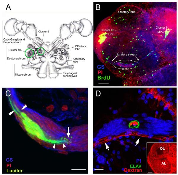
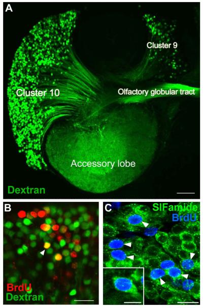
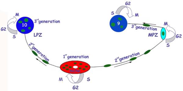
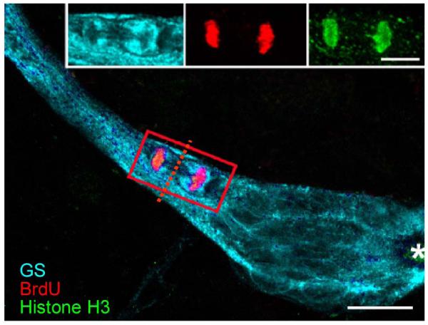
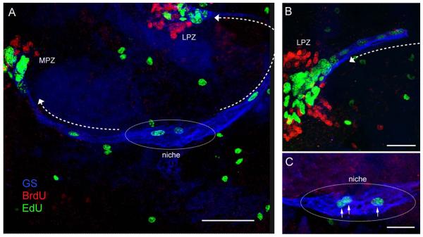
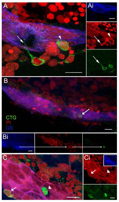
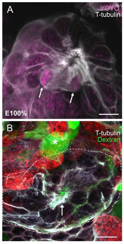
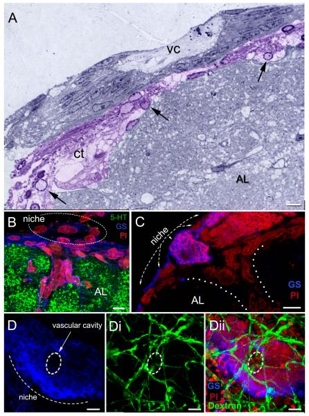
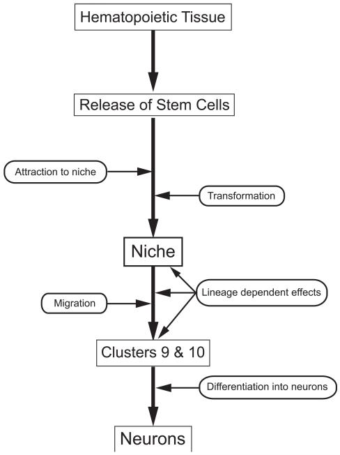
Adult-born neurons in crayfish (Procambarus clarkii) are the progeny of 1st-generation precursor cells (functionally analogous to neuronal stem cells in vertebrates) that are located in a neurogenic niche on the ventral surface of the brain. The daughters of these precursor cells migrate along the processes of bipolar niche cells to proliferation zones in the cell clusters where the somata of the olfactory interneurons reside. Here they divide again, producing offspring that differentiate into olfactory local and projection neurons. The features of this neuronal assembly line, and the fact that it continues to function when the brain is isolated and perfused or maintained in organotypic culture, provide opportunities unavailable in other organisms to explore the sequence of cellular and molecular events leading to the production of new neurons in adult brains. Further, we have determined that the 1st-generation precursor cells are not a self-renewing population, and that the niche is, nevertheless, not depleted as the animals grow and age. We conclude, therefore, that the niche is not a closed system and that there must be an extrinsic source of neuronal stem cells. Based on in vitro studies demonstrating that cells extracted from the hemolymph are attracted to the niche, as well as the intimate relationship between the niche and vasculature, we hypothesize that the hematopoietic system is a likely source of these cells.

Keywords: 5-HT; 5-bromo-2′-deoxyuridine; 5-ethynyl-2′deoxyuridine; AL; APC; BrdU; CTG; CellTracker™ Green CMFDA; EdU; GS; HPT; Hematopoietic system; Hemocytes; LPS; LPZ; MMS; MPZ; MSC; Neurogenic niche; OGT; OL; Olfactory pathway; ROS; Serotonin; Stem cell; accessory lobe; anterior proliferation center; glutamine synthetase; hematopoietic tissue; lateral proliferation zone; lipopolysaccharide; medial proliferation zone; mesenchymal stem cell; methiothepin mesylate salt; olfactory globular tract; olfactory lobe; reactive oxygen species; serotonin.


Figures

Figure 1
Figure 1
(A) Diagram of the eureptantian (crayfish, lobster) brain including the optic ganglia, and showing the locations of the proto-, trito- and deutocerebral neuropils. The soma clusters 9 and 10 (circles), locations of neurogenesis in the adult brain, flank two prominent neuropil regions of the deutocerebrum, the olfactory and accessory lobes. (Names of brain areas are according to Sandeman et al., 1992.) (B) Left side of the brain of Procambarus clarkii labeled immunocytochemically for the S-phase marker BrdU (green). Labeled cells are found in the lateral proliferation zone (LPZ) contiguous with Cluster 10 and in the medial proliferation zone (MPZ) near Cluster 9. The two zones are linked by a chain of cells in the migratory stream, labeled immunocytochemically for glutamine synthetase (GS; blue). These streams originate in the oval region ‘niche’ (dotted circle) containing cells labeled with the nuclear marker propidium iodide (PI, red). (C) Several niche cells are labeled by intracellular injection of Lucifer yellow. Each of these has a short process (arrowheads) projecting to the vascular cavity (arrow) and longer fibers (double arrowheads) that fasciculate to form the tracts projecting to the LPZ and MPZ, along which the daughters of the niche cells (2nd-generation neuronal precursors) migrate (the ‘streams’). Blue, GS; red, propidium iodide (PI). (D) The vascular connection of the cavity in the center of the glial soma cluster was demonstrated by injecting a dextran dye into the dorsal artery. The cavity, outlined in green by its reactivity to an antibody to ELAV, contains the dextran dye (red), which is also contained within a larger blood vessel that runs along beneath the niche. PI (blue) labeling of nuclei in the niche cells is also shown. Inset: dextran-filled vasculature in the olfactory (OL) and accessory (AL) lobes on the left side of the brain. Scale bars: B, 100 μm; C and D, 20 μm; inset in D, 100 μm. [C and D from Sullivan et al., 2007a].
Figure 2
Figure 2
A. The left side of a brain of P. clarkii in which dextran was applied to the accessory lobe using the technique of Utting et al. (2000). The dextran (green) enters neurons that have their terminals in the accessory lobe and labels the corresponding cell bodies and axons. From this it is clear that both projection neurons in Cluster 10, and local interneurons in Cluster 9, have their terminals in the accessory lobes and the axons from the projection neurons lie in the olfactory globular tract. B. Cluster 10 cell bodies from an animal that was exposed to BrdU for 12 days and sacrificed 4 months later, at which time dextran fluorescein 3000 MW was applied to the accessory lobe. Cells labeled red indicate that they passed through the cell cycle in the presence of BrdU. Cells labeled green indicate that they have terminals in the accessory lobe but did not pass through a cell cycle in the presence of BrdU. Double-labeled cells (orange) are cells that passed through a cell cycle in the presence of BrdU and have differentiated into neurons with their terminals in the accessory lobe. C. Cluster 10 cell bodies with BrdU (blue) and crustacean-SIFamide (green) labeled six months after being exposed to BrdU. Arrowheads point to double-labeled cells, green (cytoplasm) and blue (nuclei). Crustacean-SIFamide immunoreactivity is known to be expressed in olfactory interneurons in P. clarkii (Yasuda-Kamatani and Yasuda, 2006) and the presence of double labeling indicates that these cells were born in the adult animal and have differentiated into olfactory interneurons. Scale bars: A, 10 μm; B, C, 20 μm; C insert, 10 μm. [From Sullivan et al., 2007b; based on the experiments published in Sullivan and Beltz, 2005]
Figure 3
Figure 3
A model summarizing the events leading to the production of new olfactory interneurons in adult crayfish. Neuronal precursor (1st generation) cells reside within a neurogenic niche where they divide symmetrically. Their daughters (2nd generation precursors) migrate along tracts created by the fibers of the niche cells, towards either the LPZ or the MPZ. At least one more division will occur in the LPZ and MPZ before the progeny (3rd and subsequent generations) differentiate into neurons. [From Beltz et al., 2011]
Figure 4
Figure 4
Triple-labeled M-phase cell near the emergence of the streams immunolabeled for glutamine synthetase (cyan), phosphohistone-H3 (green) and BrdU (red). The cleavage planes of niche cells are always oriented perpendicular to the track of the stream. Cytoplasmic labeling of the dividing cell for GS confirms that GS-labeled niche cells are the precursors of the neuronal lineage. Outlines of other niche cells also are GS-labeled. Asterisk marks vascular cavity. Scale bar 20 μm. [From Zhang et al., 2009]
Figure 5
Figure 5
Double-nucleoside labeling of the niche, streams and proliferation zones in the crayfish brain. Crayfish were incubated in BrdU (red) for 6 hrs and then maintained in fresh pond water for 7 days. Just before sacrifice, they were treated with EdU (green) for 6 hrs. Fixed brains were labeled for glutamine synthetase (blue) to reveal the niche and streams. (A) Only EdU labels S phase cells in the niche and streams, demonstrating that migration is uni-directional (away from the niche [in the direction of the arrows]) and that the earlier-labeled BrdU cells do not remain in the niche, suggesting that niche cell divisions are not self-renewing. (B) Higher magnification image of the LPZ. BrdU-labeled cells (labeled first in the sequence) are found only in the proliferation zones (MPZ, LPZ) and not in the niche or streams. Arrow indicates direction of migration. (C) Higher magnification image of a neurogenic niche reveals that cells in S-phase labeled with EdU also labeled for glutamine synthatase (arrows), as do the other cells residing in the niche. Scale bars: A, 100 μm; B, 50 μm. C, 25 μm.
Figure 6
Figure 6
Cells circulating in the hemolymph are attracted to the niche in vitro. CellTracker™ Green (CTG)-labeled hemocytes are found in the vascular cavity and in, and on, the neurogenic niche. (A) Niche on a desheathed brain co-cultured with CTG-labeled cells extracted from the hemolymph: merged confocal fluorescent channels of stacked images. Several CTG-labeled cells reside just outside the niche, one with a long process extending into the niche (arrow), and another that is inserting on the outer margin of the niche (arrowhead). (Ai) Separate channels: GS outlining the niche (top); PI revealing cell nuclei, arrow/arrowhead pointing to the respective CTG-labeled cells in A (middle); and CTG-labeled cells with arrow pointing to the fine process from the CTG-labeled cell (bottom). (B) Projection of stacked images from a more dorsal region of the neurogenic niche than in (A) reveals a CTG-labeled cell just below the surface of the niche. (Bi) Separate confocal channels of the region in (B) with arrows pointing to the same CTG-filled cell also labeled for GS (left); PI, revealing cell nucleus (middle); and CTG-labeling (right). (C) In another example, a CTG-labeled cell (arrowhead) resides in the cavity and a second CTG-labeled cell (arrow) is embedded in the outer edge of the niche. (Ci) PI labeling of cell nuclei with arrowhead and arrow pointing to the corresponding nuclei with CTG-labeling in C. Insert, GS labeling of the niche. Bottom, separate channel, CTG-labeled cells. Scale bars: A, 20 μm; Ai, 10 μm; B, Bi, C and Ci, 20 μm.
Figure 7
Figure 7
(A) The protoniche at hatching (embryonic stage 100%; E100%) in the marbled crayfish, Procambarus fallax, labeled immunocytochemically for tyrosinated tubulin (white) and the nuclear stain YOYO (magenta). Dividing cells in the protoniche (arrows) are close to a central fibrous area that demarcates the emerging vascular cavity. (B) P. clarkii embryos at E95-100% and postembryonic stage 1 (POII) were micro-injected into the dorsal sinus with fluorescently-labeled dextran (green), which fills the brain vasculature (for technical details see Sintoni et al., 2012). Embryos were subsequently labeled immunocytochemically for tyrosinated tubulin (white), which delineates the protoniche (outlined with a broken white line, with an arrow pointing to the central pore). Dextran-filled fine vascular elements (green) run throughout the protoniche. A fine capillary with an expanded blind ending (lacuna; Sandeman, 1967) near the protoniche is noted with an asterisk. BrdU (red) labels cells associated with the deutocerebral proliferative system. Scale bars: A, 10 μm; B, 20 μm. [A from Sintoni et al., 2012]
Figure 8
Figure 8
The relationship between the neurogenic niche and vascular tissues. (A) Semi-thin section stained with toluidine blue, showing the niche and central vascular cavity (vc). The connective tissue (ct) below the niche (colorized purple) has many cells (arrows) with features resembling hemocytes. (B, C) Sagittal sections through the brain. These images show the niche lying on the ventral surface of the accessory lobe (AL), which is labeled immunocytochemically for serotonin (5-HT; green) in B. A blood vessel (dotted lines in C) emerges from the accessory lobe, Some cells within the blood vessel and others forming a layer between the niche and accessory lobe (B) are immunoreactive for glutamine synthetase, as are the niche cells. (D) The dorsal sinus in adult crayfish (15-20 carapace length) was injected with fluorescently-labeled dextran, which rapidly filled the brain vasculature. Fine blood vessels (dextran, green) associated with the niche are revealed on the ventral surface of the niche (Di, Dii), with some of these infiltrating the edge of the vascular cavity (broken line circle). Scale bars: A and C,10 μm; B, 6 μm; D, 20 μm. [A from Chaves da Silva et al., 2012]
Figure 9
Figure 9
Our current model of the sequence of events involved in the production of adult born neurons, beginning with hematopoietic tissue, the release of stem cells, their attraction to the niche and transformation into 1st-generation neuronal precursor cells. These aspects of the model are hypothetical. These precursor cells in the niche label for glutamine synthetase and produce daughters that migrate along processes of the niche cells towards Cluster 9 or 10. As these cells migrate, lineage-dependent changes in their physiological status are apparent, for example that they begin to express specific serotonin receptor subtypes as they approach the proliferation zones (Zhang et al., 2011). These 2nd-generation precursors divide at least once more in the proliferation zones in Clusters 9 and 10, and then differentiate into neurons (Sullivan and Beltz, 2005). These aspects of the model are supported by published data (Sullivan et al., 2007a, b; Beltz et al., 2011).

Similar articles

Cited by

References

    1. Beltz BS, Sandeman DC. Regulation of life-long neurogenesis in the decapod crustacean brain. Arthropod. Struct. Dev. 2003;32:39–60. - PubMed
    1. Beltz BS, Zhang Y, Benton JL, Sandeman DC. Adult neurogenesis in the decapod crustacean brain: a hematopoietic connection? Eur. J. Neurosci. 2011;34:870–883. - PMC - PubMed
    1. Benton JL, Goergen EM, Rogan SC, Beltz BS. Hormonal and synaptic influences of serotonin on adult neurogenesis. Gen. Comp. Endocrin. 2008;158:183–190. - PMC - PubMed
    1. Benton JL, Zhang Y, Kirkhart CR, Sandeman DC, Beltz BS. Primary neuronal precursors in adult crayfish brain: replenishment from a non-neuronal source. BMC Neurosci. 2011;12:53. - PMC - PubMed
    1. Blondheim NR, Levy YS, Ben-zur T, Burshtein A, Cherlow T, Kan I, Barzilai R, Bahat-Stromza M, Barhum Y, Bulvik S, Melamed E, Offen D. Human mesenchymal stem cells express neural genes, suggesting a neural predisposition. Stem Cells Dev. 2006;15:141. - PubMed

Publication types