. 2013 Mar;1828(3):956-66.
doi: 10.1016/j.bbamem.2012.11.030. Epub 2012 Dec 3.

Stomatin interacts with GLUT1/SLC2A1, band 3/SLC4A1, and aquaporin-1 in human erythrocyte membrane domains

Affiliations

Stomatin interacts with GLUT1/SLC2A1, band 3/SLC4A1, and aquaporin-1 in human erythrocyte membrane domains

Stefanie Rungaldier et al. Biochim Biophys Acta. 2013 Mar.

Abstract

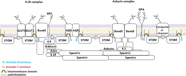
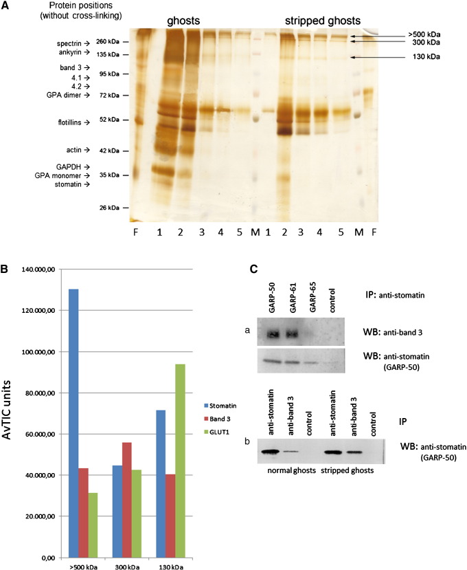
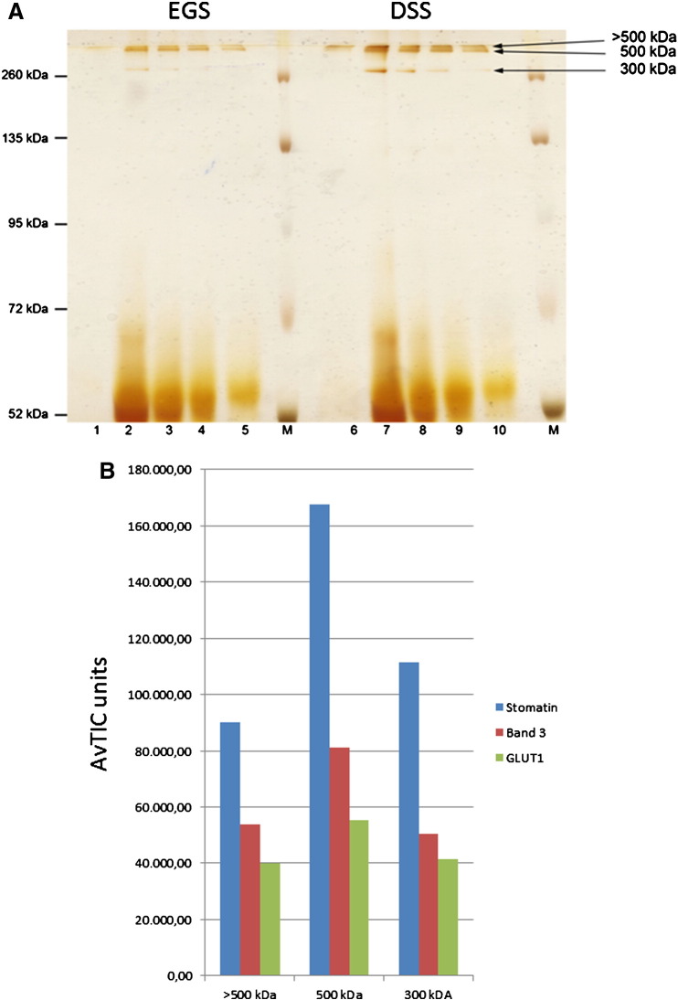
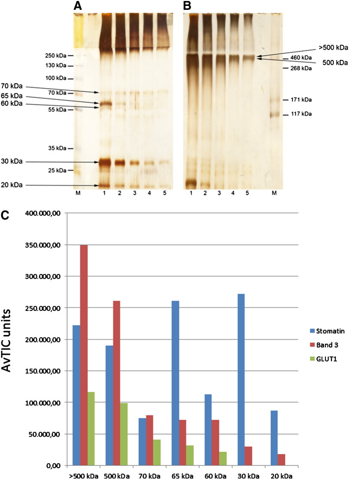
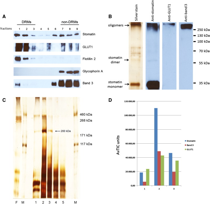
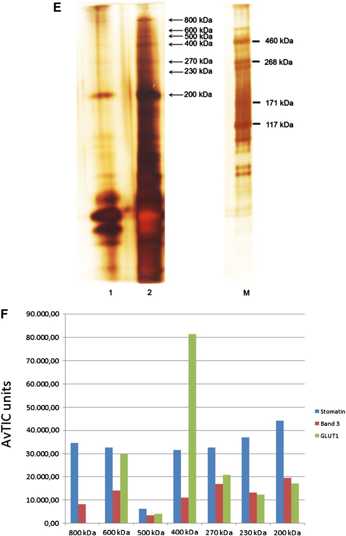
The widely expressed, homo-oligomeric, lipid raft-associated, monotopic integral membrane protein stomatin and its homologues are known to interact with and modulate various ion channels and transporters. Stomatin is a major protein of the human erythrocyte membrane, where it associates with and modifies the glucose transporter GLUT1; however, previous attempts to purify hetero-oligomeric stomatin complexes for biochemical analysis have failed. Because lateral interactions of membrane proteins may be short-lived and unstable, we have used in situ chemical cross-linking of erythrocyte membranes to fix the stomatin complexes for subsequent purification by immunoaffinity chromatography. To further enrich stomatin, we prepared detergent-resistant membranes either before or after cross-linking. Mass spectrometry of the isolated, high molecular, cross-linked stomatin complexes revealed the major interaction partners as glucose transporter-1 (GLUT1), anion exchanger (band 3), and water channel (aquaporin-1). Moreover, ferroportin-1 (SLC40A1), urea transporter-1 (SLC14A1), nucleoside transporter (SLC29A1), the calcium-pump (Ca-ATPase-4), CD47, and flotillins were identified as stomatin-interacting proteins. These findings are in line with the hypothesis that stomatin plays a role as membrane-bound scaffolding protein modulating transport proteins.


Figures

None
Graphical abstract
Fig. 1
Fig. 1
Isolation and analysis of EGS-cross-linked stomatin complexes and immunochemical verification of stomatin-band 3 interaction. (A) Erythrocyte membranes (“ghosts”), either untreated (left side) or depleted of cytoskeleton (“stripped ghosts”, right side), were cross-linked with 0.8 mM EGS, quenched, and solubilised; stomatin-complexes were isolated by immunoaffinity chromatography. Elution fractions were analysed by 10% SDS-PAGE/silver staining, excision of bands, as indicated, and mass spectrometry. Positions of un-cross-linked erythrocyte membrane proteins are indicated. Flow-through and elution fractions are shown. (B) Three stomatin-complexes isolated from stripped ghosts, > 500 kDa, about 300 kDa, and 130 kDa, contain stomatin, band 3, and GLUT1 as major components. Semi-quantitative results are shown as Average Total Ion Current (AvTIC) units. (C) Verification of the stomatin-band 3 interaction. (a) IP/WB analysis with monoclonal anti-stomatin antibodies against N- (GARP-50) and C-terminal (GARP-61, -65) epitopes and monoclonal anti-band 3, as indicated (GARP-65 has weak affinity). Control is an irrelevant antibody. (b) IP/WB analysis using normal or stripped ghosts. Co-IP works better with stripped ghosts. M, marker; F, flow-through; 1–5, respective elution fractions 1–5; GPA, glycophorin A; IP, immunoprecipitation; WB, Western blotting.
Fig. 2
Fig. 2
Isolation and analysis of DSS-cross-linked stomatin complexes. (A) Stripped ghosts were cross-linked with either 0.8 mM EGS (left side) or DSS (right side), and stomatin-complexes were isolated as before (see Fig. 1). Elution fractions were analysed by 10% SDS-PAGE (prolonged running time 24 h at 70 V), silver staining, and mass spectrometry of excised bands, as indicated ( > 500 kDa, 500 kDa, and 300 kDa). (B) Major components of DSS-cross-linked stomatin-complexes are band 3 and GLUT1. Semi-quantitative data are given in AvTIC units. M, marker; 1–5 and 6–10, respective elution fractions 1–5.
Fig. 3
Fig. 3
Analysis of stomatin-complexes after cross-linking with DSS and flotation. Stripped ghosts were cross-linked with (A) 0.8 mM EGS or (B) 0.8 mM DSS. Respective membranes were treated with cold Triton X-100, subjected to flotation, and cross-linked stomatin was identified by Western blotting of density gradient fractions. Note that cross-linked stomatin floats to low density. The dimer (about 65 kDa) is visible as major product. The monomer is not seen due to prolonged electrophoresis (24 h at 70 V). (C) In a different experiment, gradient fractions were analysed for cross-linked stomatin and GLUT1 by Western blotting, as indicated. Note that both cross-linked proteins float to low density. (D) DRM-fractions 3 and 4 from DSS-cross-linked membranes were pooled, solubilised, and stomatin-complexes were isolated by immunoaffinity chromatography. Elution fractions were analysed by SDS-PAGE/silver staining and MS of excised bands, as indicated. (E) Semi-quantitative data of the 500 kDa and 300 kDa complexes are given in AvTIC units. Note the major amount of aquaporin-1 (AQP1) associated with the 300 kDa protein complex. M, marker; 1–5, respective elution fractions.
Fig. 4
Fig. 4
Analysis of EGS-cross-linked stomatin-complexes in DRMs. Stripped ghosts were treated with cold Triton X-100 and subjected to flotation. DRMs were isolated and cross-linked with 16 μM EGS. After quenching and solubilisation with SDS, the mixture was adjusted to 1% Triton X-100 and stomatin-complexes were immunopurified. Elution fractions were analysed by (A) 10% or (B) 7% SDS-PAGE/silver staining. Stained bands were excised, as indicated, and analysed by MS. (C) Semi-quantitative results (AvTIC units) show the size-distribution of stomatin, band 3, and GLUT1. Stomatin peaks are clearly seen at the size of oligomers ( ≥ 500 kDa), the homo-dimer (65 kDa), and monomer (30 kDa). The 20 kDa stomatin may represent an intramolecularly cross-linked species. Low-molecular band 3 may represent fragments or contamination. M, marker; 1–5, respective elution fractions.
Fig. 5
Fig. 5
Preparation and analysis of EGS-cross-linked high molecular stomatin-complexes in DRMs. (A) Normal ghosts were treated with cold Triton X-100, mixed with alkaline sucrose, and subjected to flotation. Gradient fractions were analysed by Western blotting as indicated. To prevent GLUT1 overstaining in the non-DRM dense fractions 6–9, these fractions were diluted 1:50. Glycophorin A was used as a non-raft marker. (B) Combined DRM fractions 1 + 2 were incubated with 8 μM EGS, quenched, solubilised with SDS, adjusted to 1% Triton X-100, and stomatin-complexes were immunopurified. Elution fraction 1 was analysed by mini-gel 10% SDS-PAGE/silver staining and Western blotting as indicated. High-molecular complexes are visible. Note the major band of non-cross-linked stomatin. (C) Flow-through and elution fractions 1–5 were analysed by 7% SDS-PAGE/silver staining and mass spectrometry of excised 200 kDa bands (fractions 1–3), as indicated. (D) Semi-quantitative MS-analysis shows the composition of the three 200 kDa complexes eluted in fractions 1–3, respectively, reflecting the mass distribution in the fractions. (E) High molecular stomatin-complexes were separated on a 4–12% gradient gel, excised, as indicated, and analysed by MS. (F) Semi-quantitative MS-analysis shows that the region between 200 and 800 kDa contains stomatin-complexes of varying composition. Whereas stomatin is the major constituent of most complexes, the 400 kDa complex contains GLUT1 as major component. Data are compared by AvTIC units. F, flow-through; 1–5, respective elution fractions; M, marker.
Fig. 5
Fig. 5
Preparation and analysis of EGS-cross-linked high molecular stomatin-complexes in DRMs. (A) Normal ghosts were treated with cold Triton X-100, mixed with alkaline sucrose, and subjected to flotation. Gradient fractions were analysed by Western blotting as indicated. To prevent GLUT1 overstaining in the non-DRM dense fractions 6–9, these fractions were diluted 1:50. Glycophorin A was used as a non-raft marker. (B) Combined DRM fractions 1 + 2 were incubated with 8 μM EGS, quenched, solubilised with SDS, adjusted to 1% Triton X-100, and stomatin-complexes were immunopurified. Elution fraction 1 was analysed by mini-gel 10% SDS-PAGE/silver staining and Western blotting as indicated. High-molecular complexes are visible. Note the major band of non-cross-linked stomatin. (C) Flow-through and elution fractions 1–5 were analysed by 7% SDS-PAGE/silver staining and mass spectrometry of excised 200 kDa bands (fractions 1–3), as indicated. (D) Semi-quantitative MS-analysis shows the composition of the three 200 kDa complexes eluted in fractions 1–3, respectively, reflecting the mass distribution in the fractions. (E) High molecular stomatin-complexes were separated on a 4–12% gradient gel, excised, as indicated, and analysed by MS. (F) Semi-quantitative MS-analysis shows that the region between 200 and 800 kDa contains stomatin-complexes of varying composition. Whereas stomatin is the major constituent of most complexes, the 400 kDa complex contains GLUT1 as major component. Data are compared by AvTIC units. F, flow-through; 1–5, respective elution fractions; M, marker.

Similar articles

Cited by

References

    1. Hiebl-Dirschmied C.M., Adolf G.R., Prohaska R. Isolation and partial characterization of the human erythrocyte band 7 integral membrane protein. Biochim. Biophys. Acta. 1991;1065:195–202. - PubMed
    1. Hiebl-Dirschmied C.M., Entler B., Glotzmann C., Maurer-Fogy I., Stratowa C., Prohaska R. Cloning and nucleotide sequence of cDNA encoding human erythrocyte band 7 integral membrane protein. Biochim. Biophys. Acta. 1991;1090:123–124. - PubMed
    1. Stewart G.W., Hepworth-Jones B.E., Keen J.N., Dash B.C., Argent A.C., Casimir C.M. Isolation of cDNA coding for an ubiquitous membrane protein deficient in high Na+, low K + stomatocytic erythrocytes. Blood. 1992;79:1593–1601. - PubMed
    1. Salzer U., Mairhofer M., Prohaska R. Stomatin: a new paradigm of membrane organization emerges. Dyn. Cell Biol. 2007;1:20–33.
    1. Green J.B., Young J.P. Slipins: ancient origin, duplication and diversification of the stomatin protein family. BMC Evol. Biol. 2008;8:44. - PMC - PubMed

Publication types

MeSH terms