. 2013 Feb 1;85(3):404-16.
doi: 10.1016/j.bcp.2012.11.015. Epub 2012 Dec 3.

3-(1H-indol-3-yl)-2-[3-(4-nitrophenyl)ureido]propanamide enantiomers with human formyl-peptide receptor agonist activity: molecular modeling of chiral recognition by FPR2

Affiliations

3-(1H-indol-3-yl)-2-[3-(4-nitrophenyl)ureido]propanamide enantiomers with human formyl-peptide receptor agonist activity: molecular modeling of chiral recognition by FPR2

Igor A Schepetkin et al. Biochem Pharmacol. .

Abstract

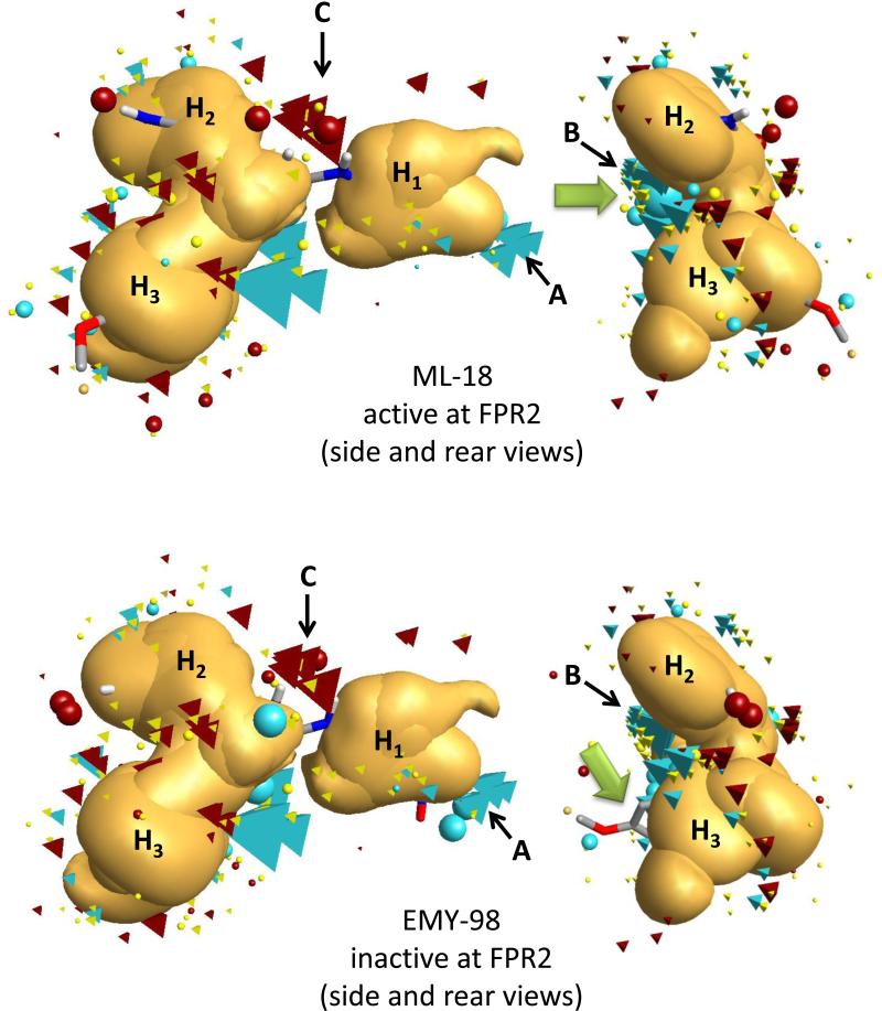
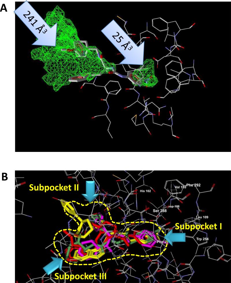
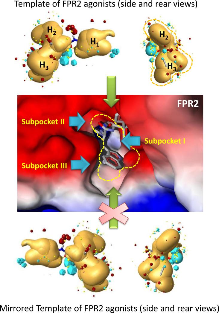
N-formyl peptide receptors (FPRs) are G protein-coupled receptors (GPCRs) that play critical roles in inflammatory reactions, and FPR-specific interactions can possibly be used to facilitate the resolution of pathological inflammatory reactions. Recent studies indicated that FPRs have stereo-selective preference for chiral ligands. Here, we investigated the structure-activity relationship of 24 chiral ureidopropanamides, including previously reported compounds PD168368/PD176252 and their close analogs, and used molecular modeling to define chiral recognition by FPR2. Unlike previously reported 6-methyl-2,4-disubstituted pyridazin-3(2H)-ones, whose R-forms preferentially activated FPR1/FPR2, we found that four S-enantiomers in the seven ureidopropanamide pairs tested preferentially activated intracellular Ca(2+) flux in FPR2-transfected cells, while the R-counterpart was more active in two enantiomer pairs. Thus, active enantiomers of FPR2 agonists can be in either R- or S-configurations, depending on the molecular scaffold and specific substituents at the chiral center. Using molecular modeling approaches, including field point methodology, homology modeling, and docking studies, we propose a model that can explain stereoselective activity of chiral FPR2 agonists. Importantly, our docking studies of FPR2 chiral agonists correlated well with the FPR2 pharmacophore model derived previously. We conclude that the ability of FPR2 to discriminate between the enantiomers is the consequence of the arrangement of the three asymmetric hydrophobic subpockets at the main orthosteric FPR2 binding site with specific orientation of charged regions in the subpockets.


Figures

Figure 1
Figure 1
Effect of the FPR2 antagonist WRW4 on Ca2+ mobilization induced by EMY-96. FPR2 HL60 cells were pretreated for 30 min with DMSO (control) or WRW4 (2 μM), followed by the addition of 10 μM compound EMY-96, and Ca2+ flux was monitored, as described under Materials and Methods. Control samples were treated with 1 nM of WKYMVM. The data are presented as % of response induced by WKYMVM and are the mean ± S.D. of triplicate samples from one experiment that is representative of three independent experiments.
Figure 2
Figure 2
Analysis of β-arrestin recruitment in cells treated with PD176252 and PD-362. FPR1-CHO-K1 (○,□) and FPR2-CHO-K1 (■,▲) cells were incubated with the indicated concentrations of PD176252 (○,■) or PD-362 (□,▲) and analyzed, as described under Materials and Methods. Representative of three independent experiments.
Figure 3
Figure 3
Analysis of Ca2+ mobilization in FPR2 transfected HL-60 cells treated with S- (ML-8) and R- (EMY-87) enantiomers. FPR2 HL-60 cells were loaded with Fluo-4 AM dye, and Ca2+ flux in response to the indicated concentrations of compounds or control WKYMVM peptide (5 nM) was analyzed, as described under Materials and Methods. The data are presented as % of response induced by WKYMVM and are the mean ± S.D. of triplicate samples from one experiment that is representative of three independent experiments.
Figure 4
Figure 4
Desensitization of formyl peptide-induced Ca2+ mobilization in human neutrophils by EMY-96. Neutrophils were pretreated with the indicated concentrations of EMY-96 for 30 min, and Ca2+ mobilization was monitored after addition of fMLF (10 nM), WKYMVm (1 nM), or WKYMVM (10 nM). Representative of three independent experiments.
Figure 5
Figure 5
Overlay of molecular conformations of enantiomer pair (S)-ML-18/(R)-EMY-98 with the best fit to the geometry of the FPR2 template. Superimpositions of the conformations to the template were refined by the simplex optimization algorithm incorporated in FieldAlign. Field points are colored as follows: blue, electron-rich (negative); red, electron-deficient (positive); yellow, van der Waals attractive (steric). All field points belonging to FPR templates are tetrahedral shaped, and field points, belonging to ML-18/EMY-98 are spherical. The hydrophobic field surface is colored in orange. The main groups of negative field points are marked as “A” and “B”, the main group of positive field points is marked as “C”, and the three hydrophobic surfaces are marked as H1, H2, and H3 in accordance with our previously reported FPR2 pharmacophore model [12]. The green arrows point to positions of the large negative field points (blue spheres) corresponding to both carbonyl groups of compound ML-18 and an incomplete geometric overlap of EMY-98 with the template.
Figure 6
Figure 6
Homology model of FPR2 agonist docking. Panel A. A PDB file of the homology model for FPR2, based on bovine rhodopsin template, was loaded into MVD software and the “Detect cavity” feature was applied with probe size 1.2 Å to identify potential areas of the protein where ligands could be docked. Two cavities were found with volumes 241 Å3 and 25 Å3 (indicated by arrows). The docking pose of PD176252 in the FPR2 lignd-binding site is shown. Panel B. Overlapping docking poses of FPR2 agonists S-(-)-5e (red), AG-10/5 (magenta), AG-10/8 (green), and EMY-96 (yellow) in the FPR2 ligand-binding site with schematic representation of the three receptor subpockets (yellow dashed line) described previously [12].
Figure 7
Figure 7
Model of chiral compound docking to FPR2. Geometry of the hydrophobic field surface of the pharmacophore model, but not its mirrored (chiral) template matches to the binding site geometry of FPR2. An FPR2 agonist can approach the FPR2 binding site from the top (“mouth”) of the cavity, shown by dashed yellow line around the agonist template (hydrophobic regions H2 and H3) and around the cavity mouth, which includes subpockets II and III. Field points are colored as follows: blue, electron-rich (negative); red, electron-deficient (positive); yellow, van der Waals attractive (steric). Hydrophobic region H1 (usually associated with 4-nitrophenyl or 4-bromophenyl groups in FPR2 agonists) should properly fit into subpocket I of the FPR2 ligand-binding site. The cavity of the FPR2 binding site shows the position of side chain tails of EMY-96 in subpockets II and III. Surface coloring was made according to electrostatic properties, whereby negatively and positively charged areas are shown in red and blue, respectively. It should be noted, that blue (positively charged) surface areas of the receptor correspond to blue field points obtained with positive probe atom and red (negatively charged) surface areas of the receptor correspond to red field points obtained with negative probe atom. Areas of subpockets are indicated with light-blue arrows. Numeration of subpockets and the hydrophobic surface of the FPR2 pharmacophore model are as described previously [12].

Similar articles

Cited by

References

    1. Ye RD, Boulay F, Wang JM, Dahlgren C, Gerard C, Parmentier M, Serhan CN, Murphy PM. International Union of Basic and Clinical Pharmacology. LXXIII. Nomenclature for the formyl peptide receptor (FPR) Family. Pharmacol Rev. 2009;61:119–61. - PMC - PubMed
    1. Schiffmann E, Corcoran BA, Wahl SM. N-formylmethionyl peptides as chemoattractants for leucocytes. Proc Natl Acad Sci USA. 1975;72:1059–62. - PMC - PubMed
    1. Gao JL, Murphy PM. Species and subtype variants of the N-formyl peptide chemotactic receptor reveal multiple important functional domains. J Biol Chem. 1993;268:25395–401. - PubMed
    1. Migeotte I, Communi D, Parmentier M. Formyl peptide receptors: a promiscuous subfamily of G protein-coupled receptors controlling immune responses. Cytokine Growth Factor Rev. 2006;17:501–19. - PubMed
    1. Gavins FN. Are formyl peptide receptors novel targets for therapeutic intervention in ischaemia-reperfusion injury? Trends Pharmacol Sci. 2010;31:266–76. - PMC - PubMed

Publication types

MeSH terms