. 2013 Apr:57:47-58.
doi: 10.1016/j.yjmcc.2012.11.013. Epub 2012 Dec 7.

In vivo and in vitro cardiac responses to beta-adrenergic stimulation in volume-overload heart failure

Affiliations

In vivo and in vitro cardiac responses to beta-adrenergic stimulation in volume-overload heart failure

Anuradha Guggilam et al. J Mol Cell Cardiol. 2013 Apr.

Abstract

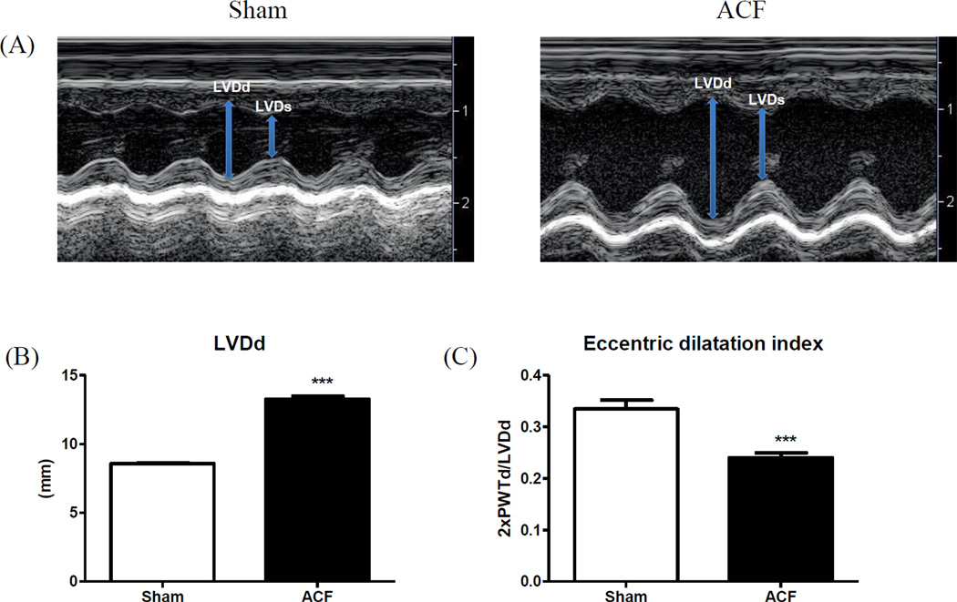
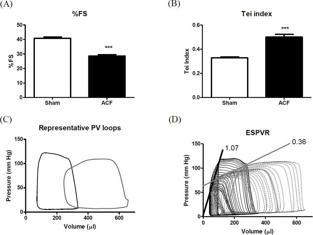
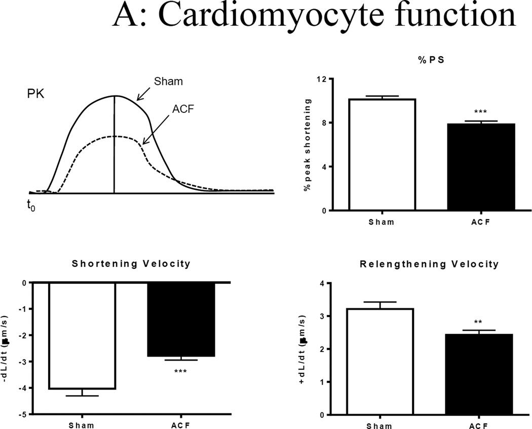
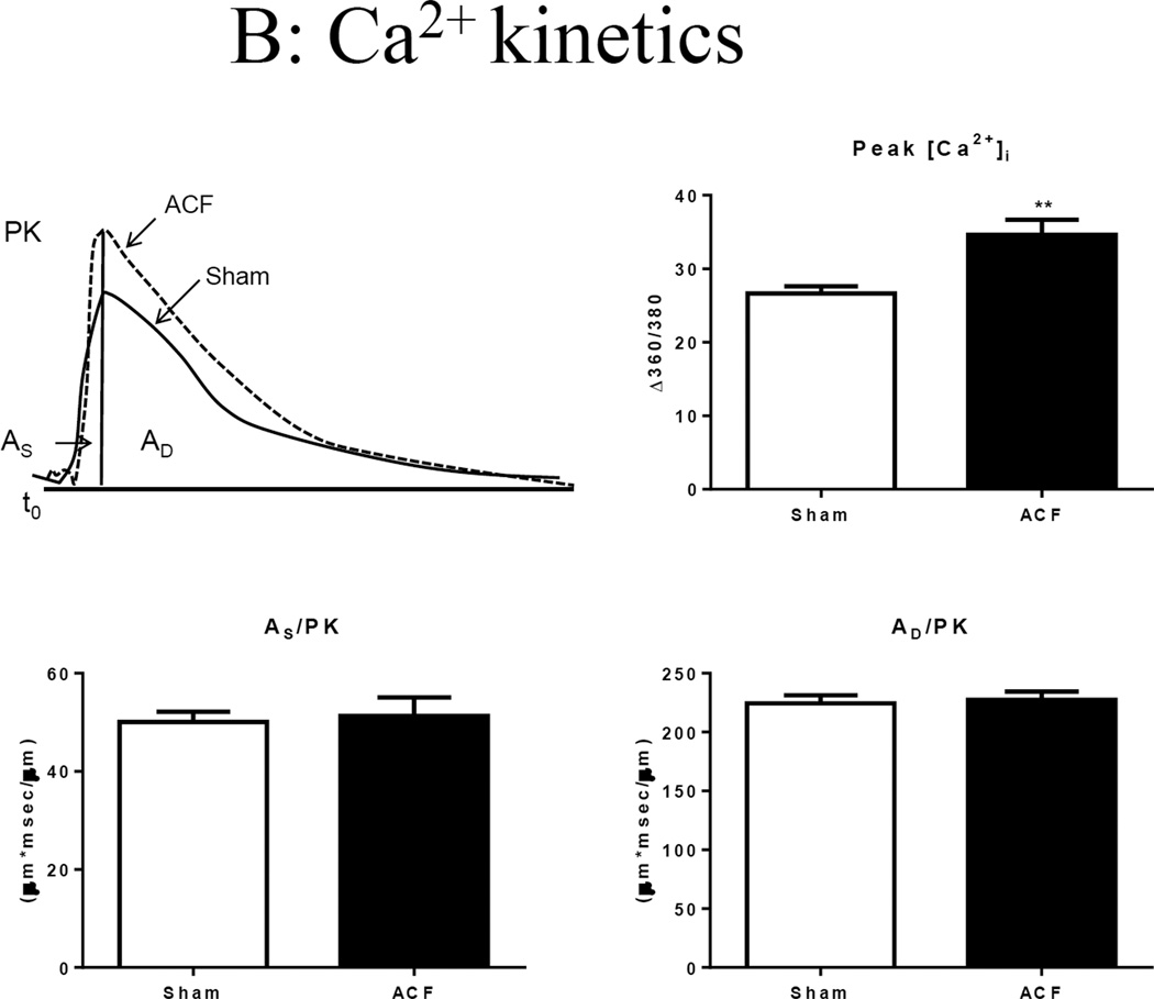
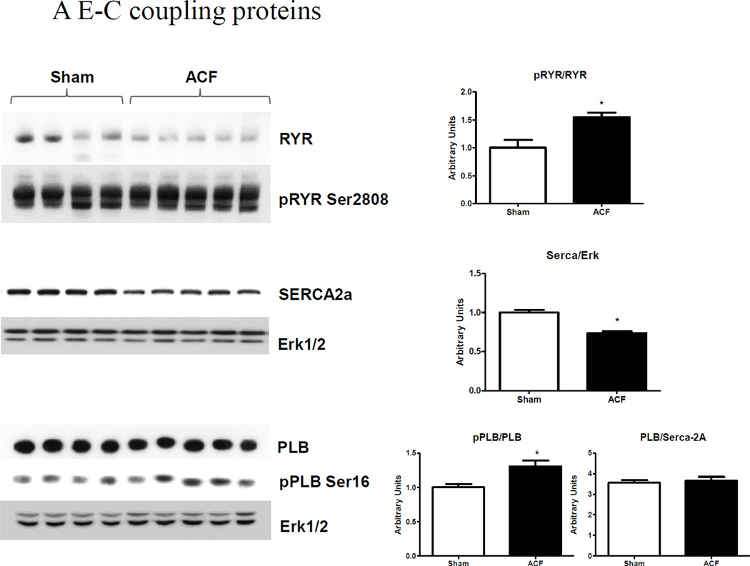
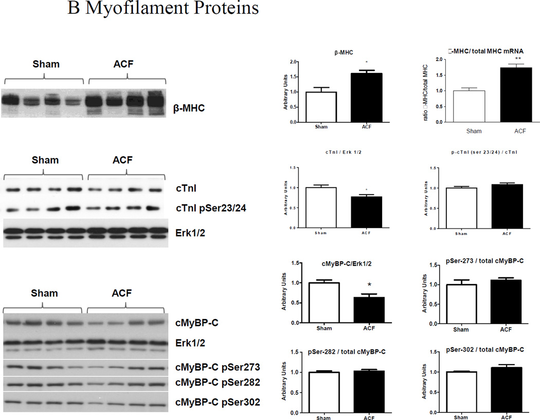
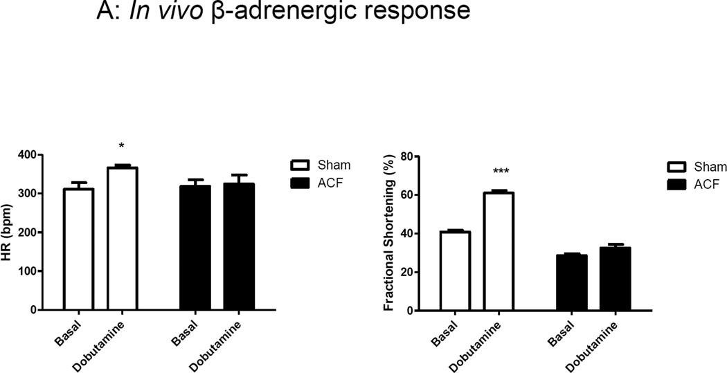
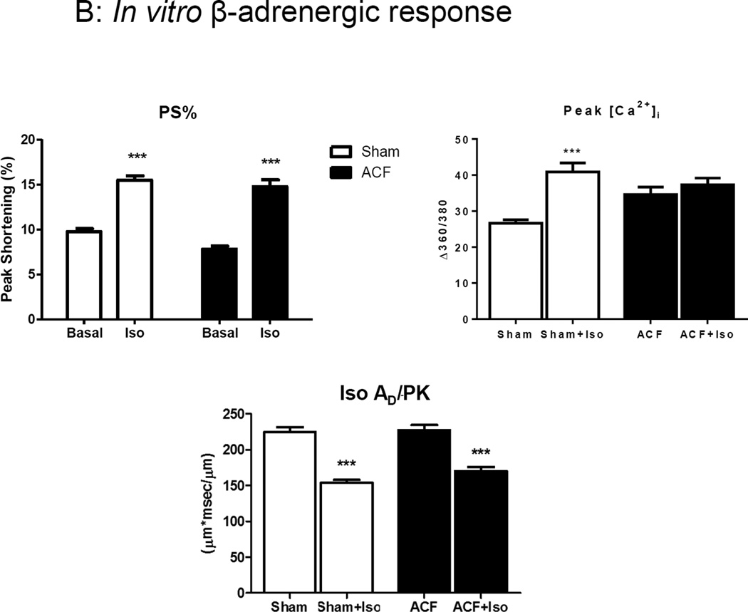
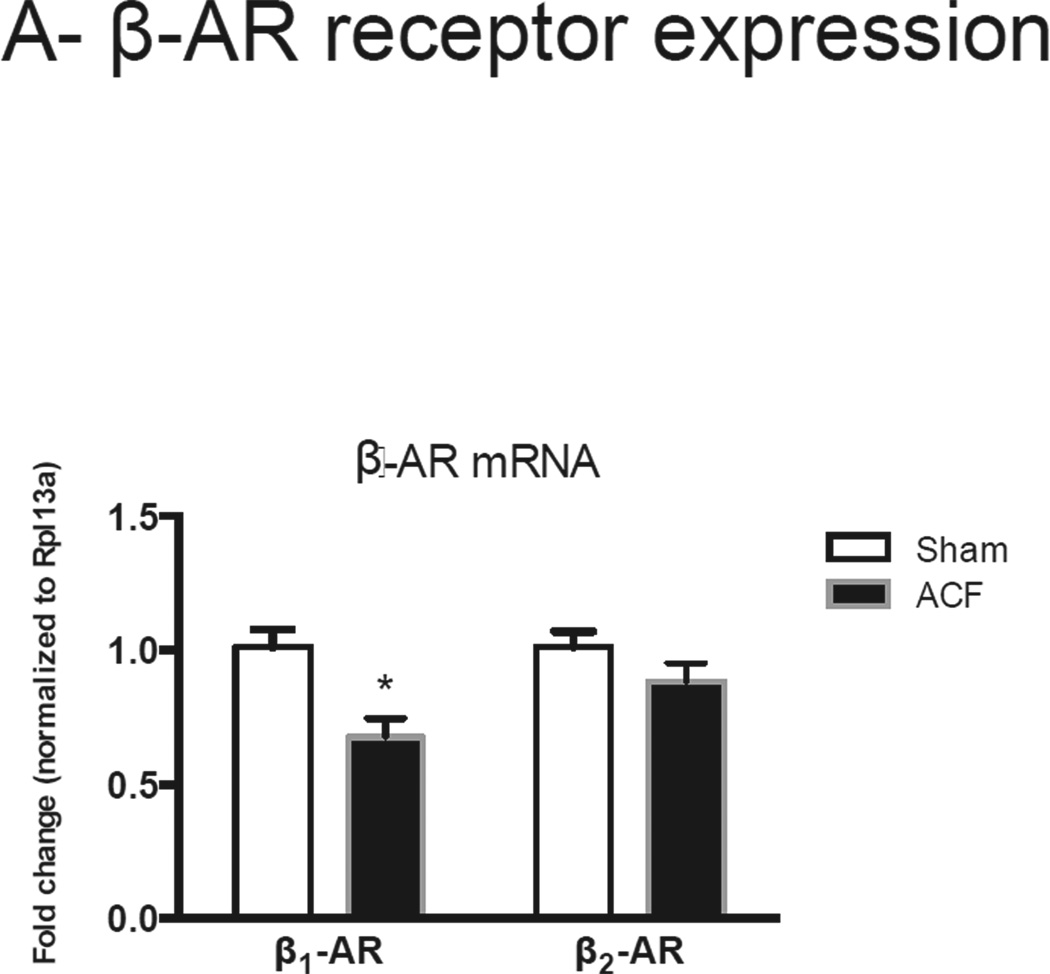
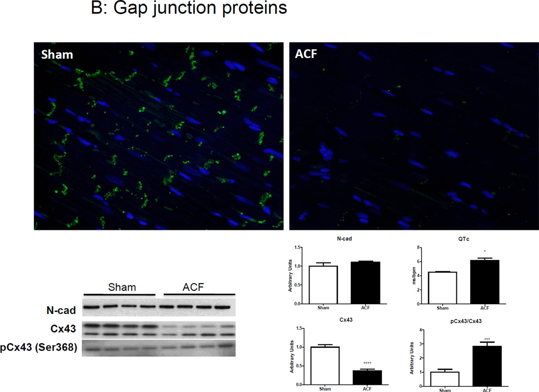
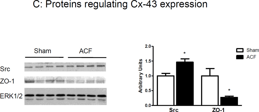
Hearts in volume overload (VO) undergo progressive ventricular hypertrophy resulting in chronic heart failure that is unresponsive to β-adrenergic agonists. This study compared left ventricular (LV) and isolated cardiomyocyte contractility and β-adrenergic responsiveness in rats with end-stage VO heart failure (HF). Adult male Sprague-Dawley rats were studied 21 weeks after aortocaval fistula (ACF) or sham surgery. Echocardiography revealed decreased fractional shortening accompanied by increased LV chamber diameter and decreased eccentric dilatation index at end-stage ACF compared to sham. Hemodynamic measurements showed a decrease in the slope of end-systolic pressure-volume relationship, indicating systolic dysfunction. Isolated LV myocytes from ACF exhibited decreased peak sarcomere shortening and kinetics. Both Ca2+ transient amplitude and kinetics were increased in ACF myocytes, with no change under the integrated Ca2+ curves relating to contraction and relaxation phases. Increases in ryanodine receptor and phospholamban phosphorylation, along with a decrease in SERCA2 levels, were observed in ACF. These changes were associated with decreased expression of β-myosin heavy chain, cardiac troponin I and cardiac myosin binding protein-C. In vivo inotropic responses to β-adrenergic stimulation were attenuated in ACF. Interestingly, ACF myocytes exhibited a similar peak shortening to those of sham in response to a β-adrenergic agonist. The protein expression of the gap junction protein connexin-43 was decreased, although its phosphorylation at Ser-368 increased. These changes were associated with alterations in Src and ZO-1. In summary, these data suggest that the disconnect in β-adrenergic responsiveness between in vivo and in vitro conditions may be associated with altered myofilament Ca2+ sensitivity and connexin-43 degradation.


Conflict of interest statement

Disclosures: The authors declare no conflict of interest.

Figures

Figure 1
Figure 1
LV structural remodeling. (A) Representative M-mode images and (B) cumulative data for LV end-systolic and end diastolic diameters (LVDs and LVDd, respectively). (C) Eccentric dilatation index (2xPWTd/LVDd). ***p<0.001.
Figure 2
Figure 2
LV function. (A) %Fractional shortening; (B) Tei index; (C) Representative pressure-volume loops and (D) slope of ESPVR. ***p<0.001.
Figure 3
Figure 3
Single cell shortening and Ca2+ transients in 21-wk Sham and ACF myocytes. (A) Top panel: Representative sarcomere shortening in sham and ACF on the left, bar graph to the right. Bottom panel: Velocity of shortening (+dL/dt) and relengthening (-dL/dt). ***p<0.001. (B) Top panel: Representative Ca2+ transients from sham and ACF myocytes (left) and intracellular calcium amplitude (peak [Cai2+]) in response to electrical stimulation (right). **p<0.01. Bottom panel: Ca2+ transient, area under the curver for systolic phase (AS/PK) and area under the curve for cytosolic Ca2+ decay (AD/PK) normalized to peak. Data represent mean±SEM from 20–30 cells from 4–5 rats/group.
Figure 3
Figure 3
Single cell shortening and Ca2+ transients in 21-wk Sham and ACF myocytes. (A) Top panel: Representative sarcomere shortening in sham and ACF on the left, bar graph to the right. Bottom panel: Velocity of shortening (+dL/dt) and relengthening (-dL/dt). ***p<0.001. (B) Top panel: Representative Ca2+ transients from sham and ACF myocytes (left) and intracellular calcium amplitude (peak [Cai2+]) in response to electrical stimulation (right). **p<0.01. Bottom panel: Ca2+ transient, area under the curver for systolic phase (AS/PK) and area under the curve for cytosolic Ca2+ decay (AD/PK) normalized to peak. Data represent mean±SEM from 20–30 cells from 4–5 rats/group.
Figure 4
Figure 4
LV protein analyses. (A) E-C coupling proteins. Representative immunoblots (left) and cumulative data (right) of RyR, phospho-RyR2808, SERCA-2a, PLB and phospho-PLB Ser16 expression. (B) Myofilament proteins. Ratio of β-MHC to total MHC mRNA and representative blots (left) and cumulative data (right) of β-MHC, TnI and phospho-TnI Ser23/24, cMyBP-C, phospho-cMyBP-C (pSer273, p282 and p302) protein expression. ERK1/2 was used as a lane loading control. (n=4–5 rats/group) *p<0.05, **P<0.01.
Figure 4
Figure 4
LV protein analyses. (A) E-C coupling proteins. Representative immunoblots (left) and cumulative data (right) of RyR, phospho-RyR2808, SERCA-2a, PLB and phospho-PLB Ser16 expression. (B) Myofilament proteins. Ratio of β-MHC to total MHC mRNA and representative blots (left) and cumulative data (right) of β-MHC, TnI and phospho-TnI Ser23/24, cMyBP-C, phospho-cMyBP-C (pSer273, p282 and p302) protein expression. ERK1/2 was used as a lane loading control. (n=4–5 rats/group) *p<0.05, **P<0.01.
Figure 5
Figure 5
In vivo and in vitro β-adrenergic responsiveness. (A) In vivo response to dobutamine in ACF versus sham. (B) In vitro responses Iso. Iso had decreased the area under the curve for cytosolic Ca2+ decay phase (AD/PK) in both Sham and ACF groups. ***p<0.001.
Figure 5
Figure 5
In vivo and in vitro β-adrenergic responsiveness. (A) In vivo response to dobutamine in ACF versus sham. (B) In vitro responses Iso. Iso had decreased the area under the curve for cytosolic Ca2+ decay phase (AD/PK) in both Sham and ACF groups. ***p<0.001.
Figure 6
Figure 6
(A) β1 and β2-adrenoceptor mRNA expression. (B) Top Panel: Representative photomicrographs of connexin-43 and N-cadherin staining. Bottom panel: Representative immunoblots (left) and histograms (right) showing quantification of N-cad, Cx-43 and phosphor-Cx43 Ser368. Note elongated QT interval in ACF animals. (B) Representative immunoblots (left) and cumulative data (right) for Src and ZO-1, normalized to ERK1/2 expression. (n=4–5 rats/group) *p<0.05.
Figure 6
Figure 6
(A) β1 and β2-adrenoceptor mRNA expression. (B) Top Panel: Representative photomicrographs of connexin-43 and N-cadherin staining. Bottom panel: Representative immunoblots (left) and histograms (right) showing quantification of N-cad, Cx-43 and phosphor-Cx43 Ser368. Note elongated QT interval in ACF animals. (B) Representative immunoblots (left) and cumulative data (right) for Src and ZO-1, normalized to ERK1/2 expression. (n=4–5 rats/group) *p<0.05.

Similar articles

Cited by

References

    1. Carabello BA. Models of volume overload hypertrophy. J Card Fail. 1996;2:55–64. - PubMed
    1. Ryan TD, Rothstein EC, Aban I, Tallaj JA, Husain A, Lucchesi PA, et al. Left ventricular eccentric remodeling and matrix loss are mediated by bradykinin and precede cardiomyocyte elongation in rats with volume overload. J Am Coll Cardiol. 2007;49:811–821. - PubMed
    1. Ding YF, Brower GL, Zhong Q, Murray D, Holland M, Janicki JS, et al. Defective intracellular Ca2+ homeostasis contributes to myocyte dysfunction during ventricular remodelling induced by chronic volume overload in rats. Clin Exp Pharmacol Physiol. 2008;35:827–835. - PubMed
    1. Juric D, Yao X, Thandapilly S, Louis X, Cantor E, Chaze B, et al. Defects in ryanodine receptor function are associated with systolic dysfunction in rats subjected to volume overload. Exp Physiol. 2010;95:869–879. - PubMed
    1. Takahashi T, Allen PD, Lacro RV, Marks AR, Dennis AR, Schoen FJ, et al. Expression of dihydropyridine receptor (Ca2+ channel) and calsequestrin genes in the myocardium of patients with end-stage heart failure. J Clin Invest. 1992;90:927–935. - PMC - PubMed

Publication types

MeSH terms