Review
. 2013 May:58:67-76.
doi: 10.1016/j.yjmcc.2012.11.018. Epub 2012 Dec 6.

CaV1.2 sparklets in heart and vascular smooth muscle

Affiliations
Review

CaV1.2 sparklets in heart and vascular smooth muscle

Manuel F Navedo et al. J Mol Cell Cardiol. 2013 May.

Abstract

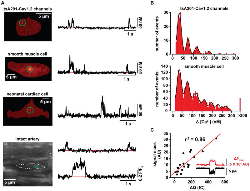
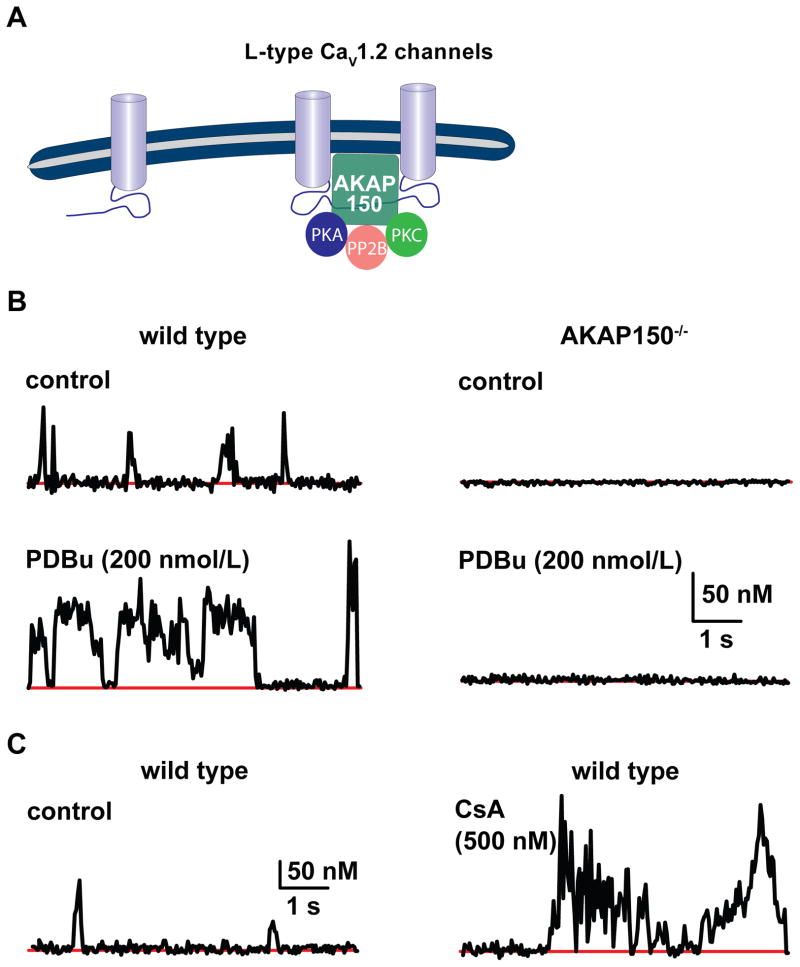
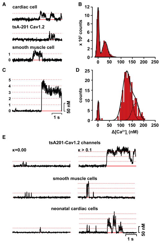
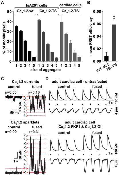
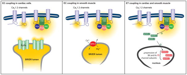
CaV1.2 sparklets are local elevations in intracellular Ca(2+) ([Ca(2+)]i) resulting from the opening of a single or small cluster of voltage-gated, dihydropyridine-sensitive CaV1.2 channels. Activation of CaV1.2 sparklets is an early event in the signaling cascade that couples membrane depolarization to contraction (i.e., excitation-contraction coupling) in cardiac and arterial smooth muscle. Here, we review recent work on the molecular and biophysical mechanisms that regulate CaV1.2 sparklet activity in these cells. CaV1.2 sparklet activity is tightly regulated by a cohort of protein kinases and phosphatases that are targeted to specific regions of the sarcolemma by the anchoring protein AKAP150. We discuss a model for the local control of Ca(2+) influx via CaV1.2 channels in which a signaling complex formed by AKAP79/150, protein kinase C, protein kinase A, and calcineurin regulates the activity of individual CaV1.2 channels and also facilitates the coordinated activation of small clusters of these channels. This results in amplification of Ca(2+) influx, which strengthens excitation-contraction coupling in cardiac and vascular smooth muscle.


Figures

Figure 1
Figure 1. CaV1.2 sparklets
A) Total internal reflection fluorescence and swept-field confocal images of CaV1.2 sparklets recorded from representative tsA201 cells expressing CaV1.2 channels, isolated vascular smooth muscle, neonatal cardiac cells or smooth muscle cells in an intact artery. The traces to the right of each image show the time-course of [Ca2+]i in the sites highlighted by the green circle (square) in the images. B) Amplitude histograms of CaV1.2 sparklets recorded from tsA201 cells expressing CaV1.2 channels or vascular smooth muscle cells. The solid black lines in the histograms are the best fit to the data following a multicomponent Gaussian function with a quantal unit of Ca2+ influx of 38 nM Ca2+. C) Plot of the relationship between signal mass as a function of Ca2+ flux in vascular smooth muscle. Parts of this figure have been adapted from Navedo et al [10, 27], and Amberg et al [8].
Figure 2
Figure 2. Mechanisms of heterogeneous CaV1.2 sparklet activity
A) Schematic diagram of the interaction of AKAP150 with a subpopulation of CaV1.2 channels. B) Representative time-course of [Ca2+]i traces illustrating that activation of PKC with PDBu increases CaV1.2 sparklet activity in vascular smooth muscle. Right panel shows fluorescence traces indicating that AKAP150 is required for increased CaV1.2 sparklet activity. C) Time-course of [Ca2+]i demonstrating that inhibition of calcineurin with cyclosporine A increases CaV1.2 sparklet activity. Parts of this figure have been adapted from Navedo et al [20, 27].
Figure 3
Figure 3. Coupled gating of CaV1.2 channels
A) Representative optical recordings of single CaV1.2 channels in neonatal cardiac cells (top), tsA201 cells (middle), and vascular smooth muscle cells (bottom). B) All-points histogram of CaV1.2 sparklet traces in A. C) Fluorescence time-course of a CaV1.2 sparklet site with coupled channels. D) All-points histogram of CaV1.2 sparklet traces in D. E) Representative fluorescence traces of stochastic (left panel) and coupled CaV1.2 sparklets in tsA201 cells, vascular smooth muscle and cardiac cells. Parts of this figure have been adapted from Navedo et al [12].
Figure 4
Figure 4. Ca2+ signaling amplification by oligomerization of CaV1.2 channels
A) Bar plot of the number & brightness analysis showing the relative percentage of mobile pixels in tsA201 cells or cardiac cells expressing wild type (wt) or Timothy syndrome (TS) CaV1.2 channels tagged with tRFP. B) Bar plot of the mean FRET efficiency between wt CaV1.2 channels or TS CaV1.2 channels. C) Representative recordings of CaV1.2 currents (upper panel) and sparklets (lower panel) obtained from tsA201 cells transfected with CaV1.2 channels tagged with a light-activated fusion system [58] before and after induction of channels fusion. D) Evoked Ca2+ transients and simultaneous cell length changes in un-transfected cardiac cells (upper panel) and cells expressing CaV1.2 channels tagged with the light-activated fusion system (lower panel) before and after channels fusion. Adapted from Dixon et al [11].
Figure 5
Figure 5. Proposed model for the role of CaV1.2 sparklet activity and coupled gating events on EC and ET coupling in cardiac and vascular smooth muscle
See main text for details.

Similar articles

Cited by

References

    1. Guatimosim S, Dilly K, Santana LF, Saleet Jafri M, Sobie E, Lederer W. Local Ca2+ Signaling and EC Coupling in Heart: Ca2+ Sparks and the Regulation of the [Ca2+]i Transient. Journal of molecular and cellular cardiology. 2002;34:941. - PubMed
    1. Vega AL, Yuan C, Votaw VS, Santana LF. Dynamic changes in sarcoplasmic reticulum structure in ventricular myocytes. J Biomed Biotechnol. 2011;2011:382586. - PMC - PubMed
    1. Gollasch M, Lohn M, Furstenau M, Nelson MT, Luft FC, Haller H. Ca2+ channels, ‘quantized’ Ca2+ release, and differentiation of myocytes in the cardiovascular system. J Hypertens. 2000;18:989–98. - PubMed
    1. Wang SQ, Song LS, Lakatta EG, Cheng H. Ca2+ signalling between single L-type Ca2+ channels and ryanodine receptors in heart cells. Nature. 2001;410:592–6. - PubMed
    1. Rubart M, Patlak JB, Nelson MT. Ca2+ currents in cerebral artery smooth muscle cells of rat at physiological Ca2+ concentrations. J Gen Physiol. 1996;107:459–72. - PMC - PubMed

Publication types

MeSH terms