. 2013 Mar;45(3):604-11.
doi: 10.1016/j.biocel.2012.11.022. Epub 2012 Dec 7.

Overexpression of human selenoprotein H in neuronal cells enhances mitochondrial biogenesis and function through activation of protein kinase A, protein kinase B, and cyclic adenosine monophosphate response element-binding protein pathway

Affiliations

Overexpression of human selenoprotein H in neuronal cells enhances mitochondrial biogenesis and function through activation of protein kinase A, protein kinase B, and cyclic adenosine monophosphate response element-binding protein pathway

Suresh L Mehta et al. Int J Biochem Cell Biol. 2013 Mar.

Abstract

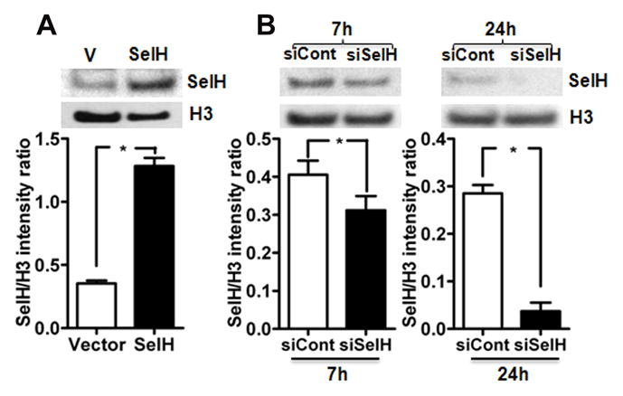
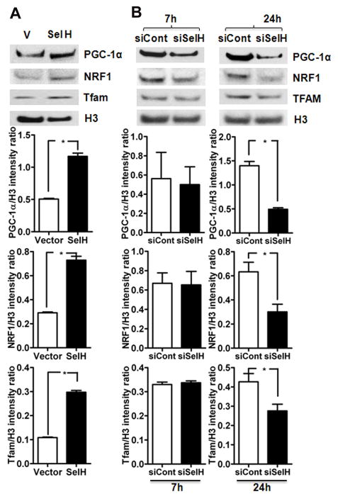
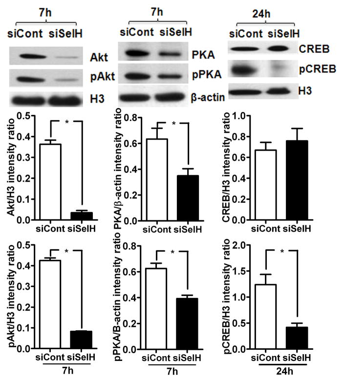
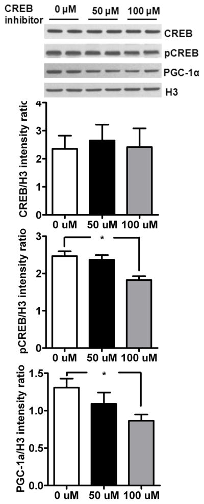
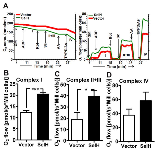
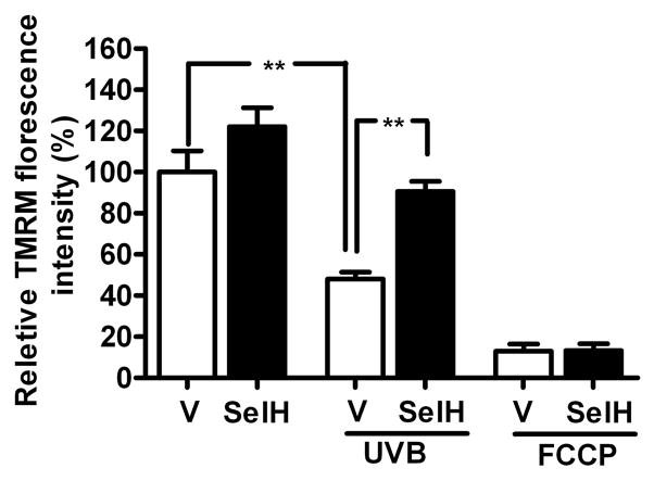
Mitochondrial biogenesis is activated by nuclear encoded transcription co-activator peroxisome proliferator-activated receptor γ coactivator-1α (PGC-1α), which is regulated by several upstream factors including protein kinase A and Akt/protein kinase B. We have previously shown that selenoprotein H enhances the levels of nuclear regulators for mitochondrial biogenesis, increases mitochondrial mass and improves mitochondrial respiratory rate, under physiological condition. Furthermore, overexpression of selenoprotein H protects neuronal HT22 cells from ultraviolet B irradiation-induced cell damage by lowering reactive oxygen species production, and inhibiting activation of caspase-3 and -9, as well as p53. The objective of this study is to identify the cell signaling pathways by which selenoprotein H initiates mitochondrial biogenesis. We first confirmed our previous observation that selenoprotein H transfected HT22 cells increased the protein levels of nuclear-encoded mitochondrial biogenesis factors, peroxisome proliferator-activated receptor γ coactivator-1α, nuclear respiratory factor 1 and mitochondrial transcription factor A. We then observed that total and phosphorylation of protein kinase A, Akt/protein kinase B and cyclic adenosine monophosphate response element-binding protein (CREB) were significantly increased in selenoprotein H transfected cells compared to vector transfected HT22 cells. To verify whether the observed stimulating effects on mitochondrial biogenesis pathways are caused by selenoprotein H and mediated through CREB, we knocked down selenoprotein H mRNA level using siRNA and inhibited CREB with napthol AS-E phosphate in selenoprotein H transfected cells and repeated the measurements of the aforementioned biomarkers. Our results revealed that silencing of selenoprotein H not only decreased the protein levels of PGC-1α, nuclear respiratory factor 1 and mitochondrial transcription factor A, but also decreased the total and phosphorylation levels of protein kinase A, protein kinase B, and CREB. Similarly, CREB inhibition reduced CREB activation and PGC-1α protein levels in selenoprotein H transfected cells. Moreover, selenoprotein H transfection increased the activity of mitochondrial complexes and prevented the ultraviolet B induced fall of mitochondrial membrane potential. We conclude that the effects of selenoprotein H on mitochondrial biogenesis and mitochondrial function are probably mediated through protein kinase A-CREB-PGC-1α and Akt/protein kinase B-CREB-PGC-1α pathways.


Conflict of interest statement

Disclosures

No conflict of interest are declared by the authors

Figures

Fig. 1
Fig. 1
Effect of transfection and silencing of hSelH gene in HT22 cells. A) Protein level and quantitative analysis of SelH in nuclear fraction of vector and SelH-transfected cells. Transfection of SelH increased its protein level as compared to vector control. B) SelH gene silencing decreased the protein level of SelH in HT22 cells. Quantitative analysis of silencing of SelH gene on its protein level at 7 and 24 hours post- transfection. Protein level of SelH decreased after 7 and 24 hours post siRNA transfection. The decrease was more pronounced after 24 hours of post-siRNA silencing. The data are collected from three independent experiments and presented as mean±SD. *P<0.05 vs. respective control. V, vector transfected cells; SelH, SelH-transfected cells; H3, histone H3; siCont, control siRNA; siSelH, SelH siRNA; 7h, 7 hours; 24h, 24 hours.
Fig. 2
Fig. 2
Effect of SelH transfection and SelH silencing on the protein levels of mitochondrial biogenesis regulators. A) Protein levels of mitochondrial biogenesis regulators, PGC-1α, NRF1 and Tfam increased after SelH transfection whereas B) silencing of SelH reduced the protein levels of PGC-1α, NRF1 and Tfam as compared to control in nuclear fraction. The data are collected from three independent experiments and presented as mean±SD. *P<0.05 vs. respective control. V, vector transfected cells; SelH, SelH transfected cells; H3, histone H3; siCont, control siRNA; siSelH, SelH siRNA; 7h, 7 hours; 24h, 24 hours.
Fig. 3
Fig. 3
SelH regulate mitobiogenesis by Akt, PKA and CREB. A) Protein and phosphorylation levels of Akt, PKA and CREB in vector and SelH transfected cells. Both, total protein and phospho- product of Akt (nuclear), PKA (cytosolic) and CREB (nuclear) increased in SelH cells as compared to vector transfected cells. B) Quantitative western analysis of Akt, PKA and CREB revealed that SelH transfection not only increased the protein levels of Akt, PKA and CREB but also significantly elevated the phosphorylation of these proteins. The data are collected from three independent experiments and presented as mean±SD. *P<0.05 vs. respective control. V, vector transfected cells; SelH, SelH-transfected cells; H3, histone H3.
Fig. 4
Fig. 4
Effect of SelH siRNA silencing on total and phospho levels of Akt, PKA and CREB. SelH silencing decreased the total protein and phospho product of Akt, PKA and CREB in SelH transfected cells. Quantitative analysis revealed that the silencing has significant effect on protein and phosphorylation of Akt, PKA and CREB in SelH transfected cells. The data are collected from three independent experiments and presented as mean±SD. *P<0.05 vs. respective control. SelH, SelH-transfected cells; H3, histone H3; siCont, control siRNA; siSelH, SelH siRNA; 7h, 7 hours; 24h, 24 hours.
Fig. 5
Fig. 5
Inhibition of CREB phosphorylation blocks mitochondrial biogenesis in SelH-transfected cells. Incubation of SelH-HT22 cells with 50 and 100 μM of CREB inhibitor for 24 hours decreased phosphor-CREB and PGC-1α protein levels. The data are collected from three independent experiments and presented as mean±SD. *P<0.05 and **p<0.01 vs. respective control. SelH, SelH transfected cells; H3, histone H3.
Fig. 6
Fig. 6
Effect of SelH transfection on mitochondrial respiratory rate at different complexes. A) Typical oxygraph curve of digitonin permeabilized cells from vector and SelH transfected cells. Oxygen consumption hence respiratory activity (pmol/s/Mill cells) increased with the addition of specific substrates and decreased with the addition of inhibitors. Arrow indicates the addition of substrates/inhibitor to activate or inhibit specific mitochondrial complex. The activity of each complex was calculated from the difference in oxygen flow in the presence of specific substrate(s) and specific inhibitor. B–D) Activities of mitochondrial complex I (B), II+III (C) and IV (D). Note the increase in complex activities in SelH as compared to vector transfected cells. The data are collected from four independent experiments and presented as mean±SD. *P<0.05, ***P<0.001 vs. respective complex. AA, antimycin A; As, ascorbate; GM, glutamate and malate; Rot, rotenone; Sc, succinate; TMPD, N,N m,N′,N′-tetramethyl-p-phenylenediamine dihydrochloride.
Fig. 7
Fig. 7
Mitochondrial membrane potential (Δψm) in vector and SelH transfected HT22 cells challenged to UVB. UVB- irradiation significantly lowered Δψ in vector transfected cells. In contrast, SelH transfection preserved mitochondrial membrane potential in UVB challenged SelH transfected cells as compared to vector cells. Data are presented as means ± SD. The data are collected from three independent experiments and presented as mean±SD. **p<0.01 vs. vector. V, vector-transfected cells and SelH, SelH-transfected cells.

Similar articles

Cited by

References

    1. Alvarez-Guardia D, Palomer X, Coll T, Davidson MM, Chan TO, Feldman AM, Laguna JC, Vazquez-Carrera M. The p65 subunit of NF-kB binds to PGC-1a, linking inflammation and metabolic disturbances in cardiac cells. Cardiovascular Research. 2010;87:449–58. - PubMed
    1. Arteel GE, Mostert V, Oubrahim H, Briviba K, Abel J, Sies H. Protection by selenoprotein P in human plasma against peroxynitrite-mediated oxidation and nitration. Biological Chemistry. 1998;379:1201–5. - PubMed
    1. Ben Jilani KE, Panee J, He Q, Berry MJ, Li PA. Overexpression of selenoprotein H reduces Ht22 neuronal cell death after UVB irradiation by preventing superoxide formation. International Journal Biological Sciences. 2007;3:198–204. - PMC - PubMed
    1. Chen J, Hattori Y, Nakajima K, Eizawa T, Ehara T, Koyama M, Hirai T, Fukuda Y, Kinoshita M, Sugiyama A, Hayashi J, Onaya T, Kobayashi T, Tawata M. Mitochondrial complex I activity is significantly decreased in a patient with maternally inherited type 2 diabetes mellitus and hypertrophic cardiomyopathy associated with mitochondrial DNA C3310T mutation: a cybrid study. Diabetes Research and Clinical Practice. 2006;74:148–53. - PubMed
    1. Chowanadisai W, Bauerly KA, Tchaparian E, Wong A, Cortopassi GA, Rucker RB. Pyrroloquinoline quinone stimulates mitochondrial biogenesis through cAMP response element-binding protein phosphorylation and increased PGC-1alpha expression. Journal of Biological Chemistry. 2010;285:142–52. - PMC - PubMed

Publication types

MeSH terms

Substances