. 2013 Jul;1832(7):962-71.
doi: 10.1016/j.bbadis.2012.11.015. Epub 2012 Dec 4.

Resident mesenchymal cells and fibrosis

Affiliations

Resident mesenchymal cells and fibrosis

Nicol Hutchison et al. Biochim Biophys Acta. 2013 Jul.

Abstract

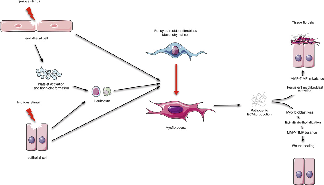
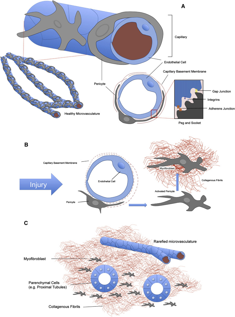
Fibrosis is a major clinical problem associated with as many as 45% of all natural deaths in developed nations. It can affect all organs and accumulating evidence indicates that fibrogenesis is not merely a bystander product of injury, but is a central pathological problem directly contributing to loss of organ function. In the majority of clinical cases, fibrogenesis is strongly associated with the recruitment of leukocytes, even in the absence of infection. Although chronic infections are a significant cause of fibrogenesis, in most cases fibrotic disease occurs in the context of sterile injury, such as microvascular disease, toxic epithelial injury or diabetes mellitus. Fibrogenesis is a direct consequence of the activation of extensive, and previously poorly appreciated, populations of mesenchymal cells in our organs which are either wrapped around capillaries and known as 'pericytes', or embedded in interstitial spaces between cell structures and known as resident 'fibroblasts'. Recent fate-mapping and complementary studies in several organs indicate that these cells are the precursors of the scar-forming myofibroblasts that appear in our organs in response to injury. Here we will review the literature supporting a central role for these cells in fibrogenesis, and highlight some of the critical cell to cell interactions that are necessary for the initiation and continuation of the fibrogenic process. This article is part of a Special Issue entitled: Fibrosis: Translation of basic research to human disease.


Conflict of interest statement

Conflict of interest

JSD is on the Scientific Advisory Board of Promedior Inc., and Regulus Therapeutics. He is the founder of Muregen LLC. He has consulted with Takeda, Boehringer Ingelheim, Bristol Mayer Squibb, GlaxoSmithKline, Biogen Idec and Gilead Pharmaceutical Companies.

Figures

Fig. 1
Fig. 1
Overview of wound healing: tissue regeneration or fibrosis. Epithelial and/or endothelial damage caused by various insults release inflammatory mediators that initiate an anti-fibrinolytic-coagulation cascade, which triggers blood clot formation. This is followed by an inflammatory phase, during which leukocytes are recruited at the site of injury. Damaged epithelial and/or endothelial can also directly activate the pericyte–myofibroblast transition. Then, myofibroblasts derived from pericytes/resident fibroblasts/mesenchymal cells produce fibrillar ECM components. Collagen fibers become organized, blood vessels are restored to normal, scar tissue is eliminated, and epithelial and/or endothelial cells divide and migrate to regenerate the damaged tissue. However, persistent inflammation can lead to chronic myofibroblast activation and excessive accumulation of ECM components ultimately resulting in fibrosis.
Fig. 2
Fig. 2
Characterization of pericytes in normal human kidney biopsy sample and in Coll-GFPTg mouse kidney cortex. (A) Normal adult human kidney cortex, immunostained for CollagenIα(1) protein. Note that CollagenIα(1) protein is strongly expressed in glomerular podocytes (arrowheads), and in perivascular cells from capillaries (arrows). (B) Normal adult Coll-GFPTg kidney cortex, immunostained for GFP. Coll-GFPTg mice express GFP under regulation by the Coll1α1 promoter, so GFP protein expression indicates Coll1α1 transcription. Note that GFP-expressing cells (glomerular podocytes (arrowheads), perivascular cells (arrows)) in Coll-GFPTg mice are the same as Coll1α1-expressing cells in human kidney cortex. Therefore, Coll-GFPTg mice are faithful reporters of all cells that produce CollagenIα(1) protein in human kidney. (C) Four color-confocal image of the cortex of the Coll-GFPTg mouse kidney stained for the capillary basement membrane-specific protein Lamininα4 (red) and the endothelial marker CD31 (white). Note numerous GFP+ cells that lack endothelial, epithelial or leukocyte markers (not shown) that form extensive processes along capillaries. Note direct interactions with endothelial cell bodies (arrows) and note Coll-GFP+ processes passing through splits in capillary basement membrane (arrowheads).
Fig. 3
Fig. 3
Results of fate mapping of Foxd1 progenitors in normal adult and injured kidney using the Foxd1-Cre;Rosa26-tdTomatoR mouse. (A) Schema showing the cross of Foxd1-Cre recombinase allele with TdTomato reporter allele driven by the universal promoters at the Rosa26 locus. Bigenic mice recombine genomic DNA at the Rosa locus only in cells that have activated the Foxd1 transcription factor gene in nephrogenesis. (B) Combining the Foxd1-Cre and Rosa26-tdTomatoR alleles with Coll-GFPTg in a single mouse indicates that perivascular cells of the Foxd1 lineage in kidney overlap almost completely with Coll-GFP+ pericytes (arrowheads). Image shows kidney cortex and kidney medulla. Note that vascular smooth muscle cells in arteriole do not express Coll1α1 (arrows). (C) In kidney injury in Foxd1-Cre;Rosa26-tdTomatoR mice (shown here is UUO d7), perivascular fibroblast and pericyte populations expand. However, now all of the expanded population of interstitial Foxd1-progenitor derived cells co-express αSMA, the marker which defines these cells as myofibroblasts.
Fig. 4
Fig. 4
Pericyte localization in the microvasculature. (A) Schematic depicting pericyte–endothelial interaction in healthy tissue. Pericytes are mesenchymal cells that are found in the microvasculature where they partially cover capillary endothelial cells. Pericytes have long membranous processes that are partially or completely embedded in capillary basement membrane (CBM), which they share with endothelial cells. The CBM is frequently incomplete, allowing direct cell to cell contact with the endothelial cell. Here a number of connections can form, including cytoplasmic invaginations known as peg & socket processes. It is thought that the peg and sockets can help facilitate bilateral communication between endothelial cells and pericytes, including PDGF–VEGF signaling. (B) In response to injury or wounding, pericytes detach from capillaries, proliferate and begin to deposit pathological fibrillar matrix. In the process, CBM is degraded and endothelial cells lose the support of pericytes (both their mechanical support, and pericyte VEGF secretion and other trophic signals). (C) Pericyte detachment has two major consequences. Firstly, the injured organ is left with a rarefied microvasculature, with wide-spread capillary drop-out (indicated schematically by a reduction in their number). The resulting hypoxia in the organ can feed-back to produce more fibrogenic signals. Secondly, the deposition of collagens and laminins by pericytes/myofibroblasts distorts parenchymal tissue architecture (for example, nephrons in the kidney) and hardens the interstitial spaces between these functional tissues, impeding their normal functions.

References

    1. Wynn TA. Common and unique mechanisms regulate fibrosis in various fibroproliferative diseases. J. Clin. Invest. 2007;117:524–529. - PMC - PubMed
    1. Bellomo R, Kellum JA, Ronco C. Acute kidney injury. Lancet. 2012;380:756–766. - PubMed
    1. Selman M, Rojas M, Mora A, Pardo A. Aging and interstitial lung diseases: unraveling an old forgotten player in the pathogenesis of lung fibrosis. Semin. Respir. Crit. Care Med. 2010;31:607–617. - PubMed
    1. Monroe DM, Hoffman M. The clotting system – a major player in wound healing. Haemophilia. 2012;18:11–16. - PubMed
    1. Salloum S, Tai AW. Treating hepatitis C infection by targeting the host. Transl. Res. 2012;159:421–429. - PMC - PubMed