

Polychromatic in vivo imaging of multiple targets using visible and near infrared light
- PMID: 23220327
- PMCID: PMC3672391
- DOI: 10.1016/j.addr.2012.10.015
Polychromatic in vivo imaging of multiple targets using visible and near infrared light
Abstract
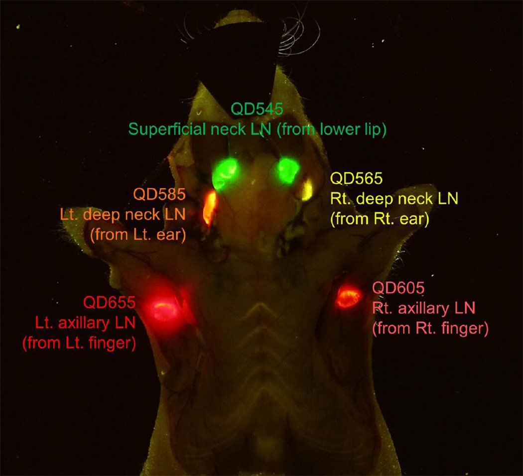
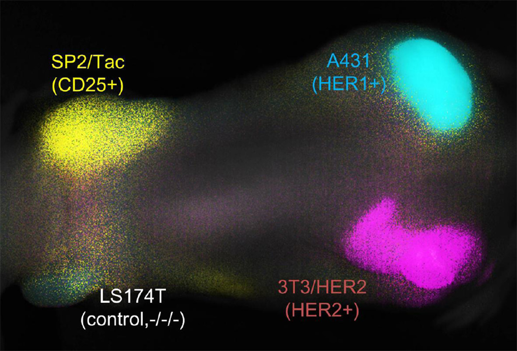
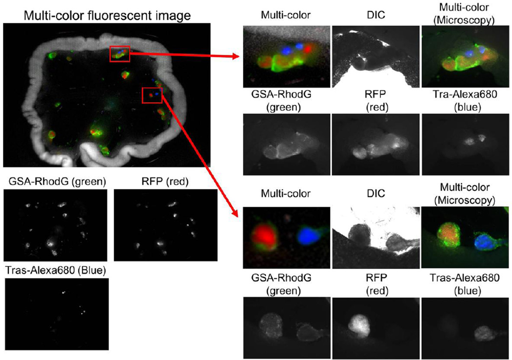
Conventional diagnostic imaging methods such as X-ray CT, MRI, and nuclear medicine are inherently monochromatic meaning that they can depict only one molecular target at a time. Optical imaging has the unique ability to be polychromatic and therefore multi-color imaging employing targeted agents conjugated to fluorophores of varying wavelength enables multiple simultaneous readouts thus providing greater multiplexed information. Numerous successful multicolor imaging techniques have recently been reported using optical imaging in in vivo animal disease models, thus adding to a growing body of research supporting the clinical viability and applicability of these technologies. Herein, we review multicolor optical imaging from the basic chemistry and physics perspective and then extend this to biological and medical applications.
Keywords: Cancer; Endoscope; Fluorescence; Fluorescence-guidance; Molecular imaging; Multi-color; Surgery.
Published by Elsevier B.V.
Figures
Similar articles
-
Second near-infrared window fluorescence nanoprobes for deep-tissue in vivo multiplexed bioimaging.Adv Drug Deliv Rev. 2023 Feb;193:114697. doi: 10.1016/j.addr.2023.114697. Epub 2023 Jan 11. Adv Drug Deliv Rev. 2023. PMID: 36641080 Review.
-
Characterizing short-wave infrared fluorescence of conventional near-infrared fluorophores.J Biomed Opt. 2019 Mar;24(3):1-5. doi: 10.1117/1.JBO.24.3.035004. J Biomed Opt. 2019. PMID: 30851014 Free PMC article.
-
Stable, Wavelength-Tunable Fluorescent Dyes in the NIR-II Region for In Vivo High-Contrast Bioimaging and Multiplexed Biosensing.Angew Chem Int Ed Engl. 2019 Jun 11;58(24):8166-8171. doi: 10.1002/anie.201904182. Epub 2019 May 8. Angew Chem Int Ed Engl. 2019. PMID: 31008552
-
Targeted multicolor in vivo imaging over 1,000 nm enabled by nonamethine cyanines.Nat Methods. 2022 Mar;19(3):353-358. doi: 10.1038/s41592-022-01394-6. Epub 2022 Feb 28. Nat Methods. 2022. PMID: 35228725
-
Quicker, deeper and stronger imaging: A review of tumor-targeted, near-infrared fluorescent dyes for fluorescence guided surgery in the preclinical and clinical stages.Eur J Pharm Biopharm. 2020 Jul;152:123-143. doi: 10.1016/j.ejpb.2020.05.002. Epub 2020 May 8. Eur J Pharm Biopharm. 2020. PMID: 32437752 Review.
Cited by
-
Immunogenic Cell Death Activates the Tumor Immune Microenvironment to Boost the Immunotherapy Efficiency.Adv Sci (Weinh). 2022 Aug;9(22):e2201734. doi: 10.1002/advs.202201734. Epub 2022 Jun 2. Adv Sci (Weinh). 2022. PMID: 35652198 Free PMC article. Review.
-
Efficient co-delivery of immiscible hydrophilic/hydrophobic chemotherapeutics by lipid emulsions for improved treatment of cancer.Int J Nanomedicine. 2017 Apr 7;12:2871-2886. doi: 10.2147/IJN.S129091. eCollection 2017. Int J Nanomedicine. 2017. PMID: 28435264 Free PMC article.
-
Endoscopic molecular imaging of cancer.Future Oncol. 2013 Oct;9(10):1501-13. doi: 10.2217/fon.13.123. Future Oncol. 2013. PMID: 24106901 Free PMC article. Review.
References
-
- Brancato R, Trabucchi G. Fluorescein and indocyanine green angiography in vascular chorioretinal diseases. Seminars in ophthalmology. 1998;13:189–198. - PubMed
-
- Flower RW, Hochheimer BF. Indocyanine green dye fluorescence and infrared absorption choroidal angiography performed simultaneously with fluorescein angiography. The Johns Hopkins medical journal. 1976;138:33–42. - PubMed
Publication types
MeSH terms
Substances
Grants and funding
LinkOut - more resources
Full Text Sources
Other Literature Sources
