. 2012 Dec 5;4(163):163ra158.
doi: 10.1126/scitranslmed.3004430.

Overexpression of methyl-CpG binding protein 2 impairs T(H)1 responses

Affiliations

Overexpression of methyl-CpG binding protein 2 impairs T(H)1 responses

Tianshu Yang et al. Sci Transl Med. .

Abstract

The DNA binding protein methyl-CpG binding protein 2 (MeCP2) critically influences neuronal and brain function by modulating gene expression, and children with overexpression of the MECP2 gene exhibit postnatal neurological syndromes. We demonstrate that some children with MECP2 duplication also display variable immunological abnormalities that include reductions in memory T and B cells and natural killer cells and immunoglobulin assay responses. Moreover, whereas mice with MeCP2 overexpression were unable to control infection with the intra-macrophage parasite Leishmania major and secrete interferon-γ (IFN-γ) from involved lymph nodes, they were able to control airway fungal infection by Aspergillus niger and mount protective T helper cell type 2 (T(H)2)-dependent allergic responses. Relative to normal T cells, T(H) cells from children and mice with MECP2 duplication displayed similar impairments in IFN-γ secretion and T(H)1 responses that were due to both MeCP2-dependent suppression of IFN-γ transcription and sequestration of the IFN-γ locus as assessed by chromatin immunoprecipitation assay. Thus, overexpressed MeCP2 aberrantly suppresses IFN-γ secretion from T(H) cells, potentially leading to a partially immunodeficient state. Our findings establish a rational basis for identifying, treating, and preventing infectious complications potentially affecting children with MECP2 duplication.


Figures

Fig. 1
Fig. 1
Children with MECP2 duplication syndrome have reduced numbers of memory T and B cells and NK cells. (A to F) Peripheral blood from 27 children with MECP2 duplication syndrome was analyzed for total leukocytes (A) and the leukocyte subgroups (B) mature neutrophils, (C) immature neutrophils, (D) eosinophils, (E) basophils, and (F) monocytes, expressed as percentages of total leukocytes. (G to M) Lymphocyte subgroups are indicated as the proportion of CD45+ cells that were (G) B cells (CD45+CD19+), (H) CD4+ T cells (CD45+CD3+CD4+), (I) CD8+ T cells (CD45+CD3+CD8+), (J) memory CD4+ T cells (CD45+CD4+CD45RO+), (K) naïve CD4+ T cells (CD45+CD4+CD45RA+), (L) memory B cells (CD45+CD19+CD27+), and (M) NK cells (CD45+CD56+) as determined by flow cytometry. Age-based normal ranges (TCH) for each parameter are indicated by gray shading. n = 27. *P < 0.05; **P < 0.01; ***P < 0.001 relative to normal ranges. Statistics was performed with the R program binominal distribution.
Fig. 2
Fig. 2
MeCP2 overexpression does not alter immune system development and homeostasis in mice. Splenocytes and thymocytes from wild-type (WT) and MeCP2 overexpression (MeCP2Tg3) mice were analyzed for the frequency of major lymphocyte subsets and memory T cells by flow cytometry by comparing the expression of the lineage markers CD4, CD8, CD19, and NK1.1. (A) Representative analysis of CD4 and CD8 expression on splenocytes (top panel) and thymocytes (bottom panel). (B) Absolute numbers of splenic CD4 and CD8 T cells, B cells, and NK cells (n = 4 per group). (C) Representative analysis of the activation and memory CD4+ T cell markers CD44 and CD62L from splenocytes. (D) Absolute number of CD44hiCD62lo CD4+ splenocytes (n = 5 per group).
Fig. 3
Fig. 3
Impaired TH1, but normal TH2, responses in MeCP2Tg3 mice. (A and B) Impaired control of L. major infection and reduced IFN-γ responses in MeCP2Tg3 mice. WT (n = 7) Fvb and syngeneic MECP2 transgenic mice (MeCP2Tg3, n = 6) were infected in footpads with stationary-phase L. major promastigotes. Footpad thickness was measured over 80 days (A), and total popliteal lymph node cells spontaneously secreting IFN-γ and IL-4 were quantified by enzyme-linked immunospot (ELISpot) (B). A third group of MeCP2Tg3 mice received intraperitoneally naïve WT CD4+ T cells 24 hours before infection (MeCP2Tg3 + CD4, n = 6). The footpad thickness data were analyzed with Student’s t test for each time point. Statistics refer to WT versus MeCP2Tg3. The ELISpot data were analyzed with Mann-Whitney U test. (C to E) Normal TH2-dependent allergic responses in MeCP2Tg3 mice. (C) Airway responsiveness to acetylcholine provocation was assessed in WT and MeCP2Tg3 mice challenged intranasally over 2 weeks with either phosphate-buffered saline (PBS) or the conidia of A. niger. (D) Total IL-4– and IFN-γ–secreting cells from whole lung of the same mice as determined by ELISpot assay. (E) Total numbers of eosinophils (Eos), monocytes (Mono), neutrophils (Neutro), and lymphocytes (Lym) in bronchoalveolar lavage (BAL) fluid of the same mice. n = 3 in PBS WT; n = 4 in PBS MeCP2Tg3; n = 7 in both fungus WT and fungus MeCP2Tg3 group. Data were analyzed with Student’s t test. (F and G) MeCP2 overexpression impairs TH1 development in vitro. Naïve splenic CD4+ and CD8+ T cells were activated under neutral (TH0) or TH1-differentiating conditions as indicated. Flow cytometry was used to determine intracytoplasmic production of IL-4 and IFN-γ. The aggregate percent of IFN-γ–secreting cells deriving from naïve CD4+ T cells using this protocol is shown (n = 4 per group; **P < 0.01, Student’s t test). (H) Impaired IFN-γ–dependent IgG isotype production in MeCP2Tg3 mice. WT and MeCP2Tg3 mice were immunized with OVA/alum weekly for 3 weeks, after which serum levels of OVA-specific IgG1 and IgG2a were determined by enzyme-linked immunosorbent assay (ELISA) (n = 6 per group; *P < 0.05, Students’ t test). Data are averages ± SEM. *P < 0.05, **P < 0.01.
Fig. 4
Fig. 4
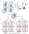
CD4+ T cells from PBMCs of children with MECP2 duplication syndrome exhibit impaired TH1 differentiation and dysregulated proliferation in vitro. (A) Representative flow cytometric analyses of intracytoplasmic IFN-γ production (y axis) and cell proliferation as assessed by dilution of the cytoplasmic dye carboxyfluorescein diacetate succinimidyl ester (CFSE) (x axis) of stimulated CD4+ T cells from a normal (control) volunteer and one with MECP2 duplication (MeCP2). (B) Aggregate analysis of the percent of IFN-γ–secreting cells under TH1 differentiation conditions comparing control and MECP2-overexpressing T cells from 10 independent experiments. Statistics was performed with paired t test. Bar represents mean. (C and D) Representative histograms depicting proliferation of IFN-γ–secreting and nonsecreting T cells as assessed by intracellular CFSE dilution under the indicated conditions.
Fig. 5
Fig. 5
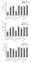
TLR signaling is unaffected in children with MECP2 duplication syndrome. Production of the indicated cytokines by PBMCs from children with MECP2 duplication syndrome (MECP2 dupl) after stimulation with the indicated TLR ligands was determined by multiplex analysis (control, n = 18; MECP2 duplication, n = 9). (A) IL-1β. (B) TNF-α. (C) IL-6.
Fig. 6
Fig. 6
MeCP2 overexpression suppresses T-bet–dependent IFN-γ transcription. (A and B) IFN-γ mRNA level is regulated at the transcriptional level in TH1 cells from both MeCP2Tg3 mice and children with MECP2 duplication. (A) CD4+ splenocytes from WT and MeCP2Tg3 mice were cultured as in Fig. 4, and IFN-γ mRNA was quantified by real-time quantitative polymerase chain reaction (qPCR) (n = 4 per group). (B) CD4+ T cells isolated from PBMCs of children with MECP2 duplication and controls were cultured under TH1-polarizing conditions for 3 days, and IFN-γ mRNA levels were quantified by real-time qPCR (n = 4 per group). (C) Intracellular T-bet was quantified by flow cytometry comparing TH1 cells from a control individual and one with MECP2 duplication. Accumulated data from six independent subjects in each group are shown. Statistics was performed with paired t test. Bar represents mean. (D) Endogenous IFN-γ transcription is suppressed when T-bet is overexpressed with MeCP2. Jurkat T cells were transfected with vectors expressing either TBET or TBET and MECP2, and the effect on IFN-γ mRNA expression was determined by real-time qPCR (*P < 0.05, n = 3 per group). (E) Impaired IFN-γ secretion is partially rescued by MECP2 siRNA knockdown in CD4+ T cells. CD4+ T cells from children with MECP2 duplication were transfected with MECP2-specific and irrelevant (control) siRNA, and CD4+ T cells from control individuals received control siRNA. IFN-γ and MeCP2 transcripts were then quantified by real-time qPCR (n = 3 per group). (F and G) The IFN-γ locus is sequestered in MeCP2Tg3 T cells. Naïve CD4+ T cells from WT and MeCP2Tg3 mice were cultured under TH1 biasing conditions. After 3 days, chromatin regions linked to acetylated histone protein 3 (H3Ac) were immunoprecipitated with a specific antibody, and the relative abundance of the IFN-γ promoter was assessed by qPCR. (G) The ratio of anti-H3Ac–associated PCR product to input-associated product from three independent experiments was determined by densitometric quantitation of the PCR bands. All statistical comparison were performed with Student’s t test (*P < 0.05).

Comment in

References

    1. Guy J, Cheval H, Selfridge J, Bird A. The role of MeCP2 in the brain. Annu. Rev. Cell Dev. Biol. 2011;27:631–652. - PubMed
    1. Lubs H, Abidi F, Bier JA, Abuelo D, Ouzts L, Voeller K, Fennell E, Stevenson RE, Schwartz CE, Arena F. XLMR syndrome characterized by multiple respiratory infections, hypertelorism, severe CNS deterioration and early death localizes to distal Xq28. Am. J. Med. Genet. 1999;85:243–248. - PubMed
    1. Ramocki MB, Tavyev YJ, Peters SU. The MECP2 duplication syndrome. Am. J. Med. Genet. A. 2010;152A:1079–1088. - PMC - PubMed
    1. Van Esch H, Bauters M, Ignatius J, Jansen M, Raynaud M, Hollanders K, Lugtenberg D, Bienvenu T, Jensen LR, Gecz J, Moraine C, Marynen P, Fryns JP, Froyen G. Duplication of the MECP2 region is a frequent cause of severe mental retardation and progressive neurological symptoms in males. Am. J. Hum. Genet. 2005;77:442–453. - PMC - PubMed
    1. Carvalho CM, Zhang F, Liu P, Patel A, Sahoo T, Bacino CA, Shaw C, Peacock S, Pursley A, Tavyev YJ, Ramocki MB, Nawara M, Obersztyn E, Vianna-Morgante AM, Stankiewicz P, Zoghbi HY, Cheung SW, Lupski JR. Complex rearrangements in patients with duplications of MECP2 can occur by fork stalling and template switching. Hum. Mol. Genet. 2009;18:2188–2203. - PMC - PubMed

Publication types

LinkOut - more resources