. 2013 Feb;3(2):224-37.
doi: 10.1158/2159-8290.CD-12-0349. Epub 2012 Dec 7.

Activating HER2 mutations in HER2 gene amplification negative breast cancer

Affiliations

Activating HER2 mutations in HER2 gene amplification negative breast cancer

Ron Bose et al. Cancer Discov. 2013 Feb.

Abstract

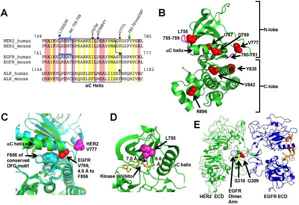
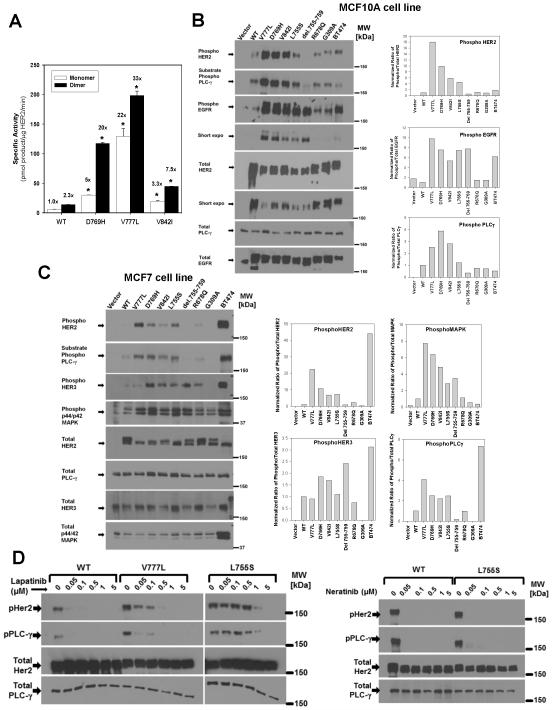
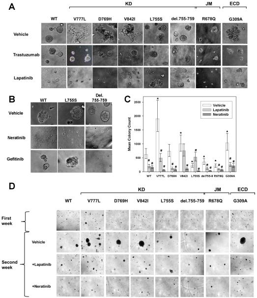
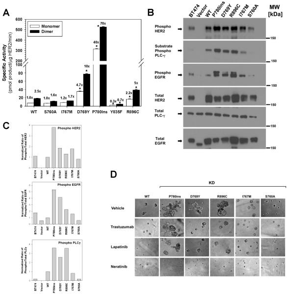
Data from 8 breast cancer genome-sequencing projects identified 25 patients with HER2 somatic mutations in cancers lacking HER2 gene amplification. To determine the phenotype of these mutations, we functionally characterized 13 HER2 mutations using in vitro kinase assays, protein structure analysis, cell culture, and xenograft experiments. Seven of these mutations are activating mutations, including G309A, D769H, D769Y, V777L, P780ins, V842I, and R896C. HER2 in-frame deletion 755-759, which is homologous to EGF receptor (EGFR) exon 19 in-frame deletions, had a neomorphic phenotype with increased phosphorylation of EGFR or HER3. L755S produced lapatinib resistance, but was not an activating mutation in our experimental systems. All of these mutations were sensitive to the irreversible kinase inhibitor, neratinib. These findings show that HER2 somatic mutation is an alternative mechanism to activate HER2 in breast cancer and they validate HER2 somatic mutations as drug targets for breast cancer treatment.

Significance: We show that the majority of HER2 somatic mutations in breast cancer patients are activating mutations that likely drive tumorigenesis. Several patients had mutations that are resistant to the reversible HER2 inhibitor lapatinib, but are sensitive to the irreversible HER2 inhibitor, neratinib. Our results suggest that patients with HER2 mutation–positive breast cancers could benefit from existing HER2-targeted drugs.


Figures

Figure 1
Figure 1
HER2 somatic mutations in breast cancer. A, Clinical information on patients with HER2 somatic mutations. ER = estrogen receptor, PR = progesterone receptor. B, HER2 somatic mutations observed in 25 patients are shown. Blue circles represent each case of the indicated mutation. Two patients had 2 HER2 somatic mutations each, resulting in a total of 27 mutations in 25 patients. del.755-759* indicates that 2 patients had del.755-759 and one patient had del.755-759 with a S760A change. ECD = Extracellular domain, TM = transmembrane region, JM = juxtamembrane region. C, HER2 gene copy number in patient 15687, who has V777L mutation. X-axis values represent the genomic position on chromosome 17, with numbering based on the March 2006 human reference sequence (NCBI Build 36.1/hg18). D, The exome sequencing-based copy number, as determined by VarScan 2 (45), for the ERBB2 gene and the level 3 gene expression values from the Agilent 244K Custom Gene Expression 4502A-07-03 platform are plotted. The red data points and the arrows to them indicate the patients with HER2 somatic mutations. E, Expression of mutant allele in tumors harboring HER2 mutations by Sanger sequencing of patient mRNA samples.
Figure 2
Figure 2
Protein structure visualizations of the HER2 somatic mutations. A, Multi-sequence alignment of HER2, EGFR, and ALK tyrosine kinases. Location of somatic mutations is marked with stars. Blue line and green arrow indicate del.755-759 and P780ins, respectively. Identical residues are shaded pink and similar residues yellow. B, HER2 kinase domain structure (PDB: 3PP0) showing the locations of point mutations (red spheres) and deletions/insertions (magenta). C, HER2 V777 and EGFR V769 are visualized in the active conformation of the EGFR kinase domain (cyan, PDB: 2GS2) and the inhibitor bound conformation of HER2 (green, PDB: 3PP0). D, Proximity of the HER2 L755 to the tyrosine kinase inhibitor SYR127063 is shown. SYR127063 has the same core chemical structure as lapatinib (23). E, Alignment of HER2-EGFR extracellular domain structures PDB: 1N8Y (HER2) and PDB: 1IVO (EGFR).
Figure 3
Figure 3
HER2 mutations differentially activate HER2 signaling. A, HER2 WT or mutant kinase domain constructs were recombinantly expressed and assayed in vitro. Fold change relative to HER2 WT monomer is indicated above each bar. * = p<0.01. B, MCF10A cells were retrovirally transduced with HER2 WT or the respective mutants and lysates were probed with the indicated antibodies. Bar graphs represents quantification of western blots bands, which was performed with Bio-Rad ChemiDoc XRS system or ImageJ software. C, MCF7 cells were retrovirally transduced with HER2 WT or the respective mutants and lysates were probed and quantified as in B. D, MCF10A cells expressing HER2 WT, V777L, or L755S were treated with lapatinib (left) or neratinib (right) at the indicated concentrations for 4 hours, and analyzed by western blot as above.
Figure 4
Figure 4
HER2 mutations V777L, D769H, V842I, G309A induce gain of function over HER2 WT in MCF10A mammary epithelial cells. A, HER2 WT or mutants were seeded on 3D Matrigel culture in the presence or absence of DMSO vehicle (0.5%), trastuzumab (100 μg/ml) or lapatinib (0.5 μM). Phase contrast images of acini or invasive structures were obtained at 200x magnification on day 8. KD = kinase domain, JM = juxtamembrane region, and ECD = extracellular domain. B, HER2 WT, L755S, and del.755-759 cells were grown in Matrigel in the presence of DMSO vehicle (0.5%), neratinib (0.5 μM) or gefitinib (0.5 μM). Phase contrast images were obtained as in A. C, MCF10A-HER2 WT or mutants were seeded in soft agar. After 7 days of growth, they were treated with DMSO vehicle (0.5%), lapatinib (0.5μM) or neratinib (0.5μM) for an additional week. Error bars represent 95% highest posterior density intervals. * indicates a significant difference between the HER2 mutant and HER2 WT and # indicates the effect of inhibitor treatment was significant (95% highest posterior density interval did not contain 0 for both). D, Photomicrographs of the colonies in soft agar on day 12, magnification 40x.
Figure 5
Figure 5
HER2 mutants V777L, G309A, D769H formed tumors more rapidly than HER2 WT. A, 0.5×10⁶ cells of NIH3T3 expressing either HER2 WT or mutant were injected in the flanks of nude mice. N=5 mice for each construct. Tumor size was measured every 2-3 days. A linear mixed effects model was fit to the logarithm of tumor volumes to statistically test whether HER2 mutants grew faster than HER2 WT. * = p<0.01. B, Photographs were taken on day 11. C, NIH3T3 cells expressing HER2 WT, G309A, or del.755-759 were injected into nude mice as in a. N=3 mice for G309A and WT and N=4 mice for del.755-759. * = p<0.01. D, Photographs taken on day 12.
Figure 6
Figure 6
HER2 kinase domain mutations differentially activate HER2 signaling, induce an invasive phenotype and respond to HER2-targeted drugs. A, Six additional HER2 kinase domain mutations were tested by in vitro kinase assay. Fold change relative to HER2 WT monomer is indicated above each bar. * = p<0.01. B, MCF10A cells were retrovirally transduced with HER2 WT or the respective mutants and lysates were probed with the indicated antibodies. C, Quantitation of western blots from panel B. D, HER2 WT or mutants were seeded on 3D Matrigel culture in the presence of DMSO vehicle (0.5%), trastuzumab (100 μg/ml), lapatinib (0.5 μM), or neratinib (0.5μM). Phase contrast images of acini or invasive structures were obtained at 200x magnification on day 6.

Comment in

Similar articles

Cited by

References

    1. Baselga J. Targeting tyrosine kinases in cancer: the second wave. Science. 2006;312:1175–8. - PubMed
    1. Chin L, Andersen JN, Futreal PA. Cancer genomics: from discovery science to personalized medicine. Nat Med. 2011;17:297–303. - PubMed
    1. Hynes NE, Lane HA. ERBB receptors and cancer: the complexity of targeted inhibitors. Nat Rev Cancer. 2005;5:341–54. - PubMed
    1. Awada A, Bozovic-Spasojevic I, Chow L. New therapies in HER2-positive breast cancer: a major step towards a cure of the disease? Cancer Treat Rev. 2012;38:494–504. - PubMed
    1. Ellis MJ, Ding L, Shen D, Luo J, Suman VJ, Wallis JW, et al. Whole Genome Sequencing to Characterise Breast Cancer Response to Aromatase Inhibition. Nature. 2012;486:353–60. - PMC - PubMed

Publication types

MeSH terms