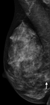
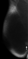
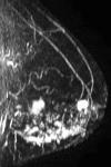
Bilateral contrast-enhanced dual-energy digital mammography: feasibility and comparison with conventional digital mammography and MR imaging in women with known breast carcinoma
- PMID: 23220903
- PMCID: PMC5673037
- DOI: 10.1148/radiol.12121084
Bilateral contrast-enhanced dual-energy digital mammography: feasibility and comparison with conventional digital mammography and MR imaging in women with known breast carcinoma
Abstract
Purpose: To determine feasibility of performing bilateral dual-energy (DE) contrast agent-enhanced (CE) digital mammography and to evaluate its performance compared with conventional digital mammography and breast magnetic resonance (MR) imaging in women with known breast cancer.
Materials and methods: This study was approved by the institutional review board and was HIPAA compliant. Written informed consent was obtained. Patient accrual began in March 2010 and ended in August 2011. Mean patient age was 49.6 years (range, 25-74 years). Feasibility was evaluated in 10 women with newly diagnosed breast cancer who were injected with 1.5 mL per kilogram of body weight of iohexol and imaged between 2.5 and 10 minutes after injection. Once feasibility was confirmed, 52 women with newly diagnosed cancer who had undergone breast MR imaging gave consent to undergo DE CE digital mammography. Positive findings were confirmed with pathologic findings.
Results: Feasibility was confirmed with no adverse events. Visualization of tumor enhancement was independent of timing after contrast agent injection for up to 10 minutes. MR imaging and DE CE digital mammography both depicted 50 (96%) of 52 index tumors; conventional mammography depicted 42 (81%). Lesions depicted by using DE CE digital mammography ranged from 4 to 67 mm in size (median, 17 mm). DE CE digital mammography depicted 14 (56%) of 25 additional ipsilateral cancers compared with 22 (88%) of 25 for MR imaging. There were two false-positive findings with DE CE digital mammography and 13 false-positive findings with MR imaging. There was one contralateral cancer, which was not evident with either modality.
Conclusion: Bilateral DE CE digital mammography was feasible and easily accomplished. It was used to detect known primary tumors at a rate comparable to that of MR imaging and higher than that of conventional digital mammography. DE CE digital mammography had a lower sensitivity for detecting additional ipsilateral cancers than did MR imaging, but the specificity was higher. © RSNA, 2012.
Figures


References
-
- Jemal A, Bray F, Center MM, Ferlay J, Ward E, Forman D. Global cancer statistics. CA Cancer J Clin 2011;61(2):69–90. - PubMed
-
- Kerlikowske K, Carney PA, Geller B, et al. Performance of screening mammography among women with and without a first-degree relative with breast cancer. Ann Intern Med 2000;133(11):855–863. - PubMed
-
- Kuhl CK, Schmutzler RK, Leutner CC, et al. Breast MR imaging screening in 192 women proved or suspected to be carriers of a breast cancer susceptibility gene: preliminary results. Radiology 2000;215(1):267–279. - PubMed
-
- Warner E, Plewes DB, Hill KA, et al. Surveillance of BRCA1 and BRCA2 mutation carriers with magnetic resonance imaging, ultrasound, mammography, and clinical breast examination. JAMA 2004;292(11):1317–1325. - PubMed
-
- Leach MO, Boggis CR, Dixon AK, et al. Screening with magnetic resonance imaging and mammography of a UK population at high familial risk of breast cancer: a prospective multicentre cohort study (MARIBS). Lancet 2005;365(9473):1769–1778. - PubMed
Publication types
MeSH terms
Substances
LinkOut - more resources
Full Text Sources
Other Literature Sources
Medical

