. 2013 Jan;123(1):335-9.
doi: 10.1172/JCI63193. Epub 2012 Dec 10.

Sustained MEK inhibition abrogates myeloproliferative disease in Nf1 mutant mice

Affiliations

Sustained MEK inhibition abrogates myeloproliferative disease in Nf1 mutant mice

Tiffany Chang et al. J Clin Invest. 2013 Jan.

Erratum in

Abstract

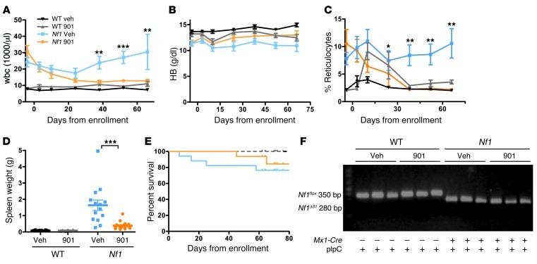
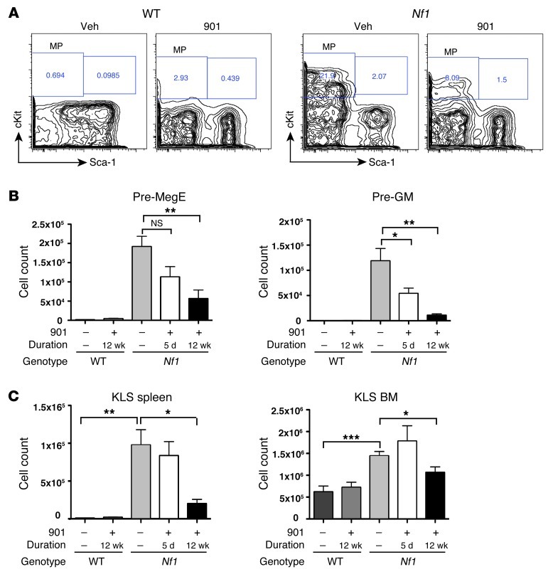
Children with neurofibromatosis type 1 (NF1) are predisposed to juvenile myelomonocytic leukemia (JMML), an aggressive myeloproliferative neoplasm (MPN) that is refractory to conventional chemotherapy. Conditional inactivation of the Nf1 tumor suppressor in hematopoietic cells of mice causes a progressive MPN that accurately models JMML and chronic myelomonocytic leukemia (CMML). We characterized the effects of Nf1 loss on immature hematopoietic populations and investigated treatment with the MEK inhibitor PD0325901 (hereafter called 901). Somatic Nf1 inactivation resulted in a marked expansion of immature and lineage-committed myelo-erythroid progenitors and ineffective erythropoiesis. Treatment with 901 induced a durable drop in leukocyte counts, enhanced erythropoietic function, and markedly reduced spleen sizes in mice with MPN. MEK inhibition also restored a normal pattern of erythroid differentiation and greatly reduced extramedullary hematopoiesis. Remarkably, genetic analysis revealed the persistence of Nf1-deficient hematopoietic cells, indicating that MEK inhibition modulates the proliferation and differentiation of Nf1 mutant cells in vivo rather than eliminating them. These data provide a rationale for performing clinical trials of MEK inhibitors in patients with JMML and CMML.


Figures

Figure 1
Figure 1. 901 reduces myeloproliferation and enhances erythropoiesis in Mx1-Cre;Nf1flox/flox (Nf1) mice.
Nf1 and WT mice were treated with vehicle (Veh) or 901 for 10 weeks. (A) wbc counts, (B) hemoglobin (HB) concentrations, (C) and percentage of reticulocytes in the blood of Nf1 mice given vehicle (blue) or 901 (orange) and of WT mice treated with vehicle (black) or 901 (gray). (D) Spleen weights at the end of the trial. Mean and SEM are shown (n = 11–13 per group). Asterisks indicate significant differences between Nf1 mice that received 901 and vehicle (*P < 0.05; **P < 0.001; ***P < 0.0001). (E) Kaplan-Meier analysis revealed a trend toward enhanced survival in 901-treated Nf1 mice (P = NS). (F) PCR amplification of BM DNA from individual WT and Nf1 mice at the end of the trial (n = 11–13 per group). Notably, both 901- and vehicle-treated Nf1 mutant mice demonstrated complete excision of exon 31.
Figure 2
Figure 2. 901 restores normal erythroid differentiation in vivo.
Hematopoietic tissues from 6-month-old Mx1-Cre;Nf1flox/flox (Nf1) and WT mice treated with 901 or vehicle were analyzed at the end of the trial. (A) BFU-E and (B) CFU-E colonies were grown from splenocytes in methylcellulose medium–containing erythropoietin (100 ng/ml). Mean and SEM are shown (n = 11–13 per group). (C) Erythroid differentiation was analyzed by flow cytometry using the cell-surface markers CD71 and Ter119. Contour plots of splenocytes from representative WT and Nf1 mutant mice that were treated for 12 weeks are shown. The relative number of cells in each region as a percentage of viable, nucleated erythroid cells is shown at the far right. The error bars show SEM (n = 5–6 per group), and asterisks indicate significant differences between these groups (**P < 0.001;***P < 0.0001).
Figure 3
Figure 3. 901 normalizes early myelo-erythroid populations in Mx1-Cre;Nf1flox/flox (Nf1) mice.
(A) Representative profiles of myeloid progenitors (MP) in the spleens of WT and Nf1 mice treated with vehicle or 901 for 12 weeks. (B) Total numbers of splenic myeloid progenitor cells in WT and Nf1 mice treated with 901 for 5 days or 12 weeks. (C) Total numbers of KLS cells in spleen and BM. In B and C, the error bars show SEM (n = 5–10 per group), and asterisks indicate significant differences between vehicle-and 901-treated Nf1 mice (*P < 0.05, **P < 0.01, ***P < 0.0001).

References

    1. Cichowski K, Jacks T. NF1 tumor suppressor gene function: narrowing the GAP. Cell. 2001;104(4):593–604. doi: 10.1016/S0092-8674(01)00245-8. - DOI - PubMed
    1. Loh ML. Recent advances in the pathogenesis and treatment of juvenile myelomonocytic leukaemia. Br J Haematol. 2011;152(6):677–687. doi: 10.1111/j.1365-2141.2010.08525.x. - DOI - PubMed
    1. Side L, et al. Homozygous inactivation of the NF1 gene in bone marrow cells from children with neurofibromatosis type 1 and malignant myeloid disorders. N Engl J Med. 1997;336(24):1713–1720. doi: 10.1056/NEJM199706123362404. - DOI - PubMed
    1. Bollag G, et al. Loss of NF1 results in activation of the Ras signaling pathway and leads to aberrant growth in murine and human hematopoietic cells. . Nat Genet. 1996;12(2):144–148. - PubMed
    1. Ward AF, Braun BS, Shannon KM. Targeting oncogenic Ras signaling in hematologic malignancies. Blood. 2012;120(17):3397–3406. doi: 10.1182/blood-2012-05-378596. - DOI - PMC - PubMed

Publication types

MeSH terms