. 2013 Feb;87(4):1997-2010.
doi: 10.1128/JVI.02853-12. Epub 2012 Dec 5.

Hypervariable domains of nsP3 proteins of New World and Old World alphaviruses mediate formation of distinct, virus-specific protein complexes

Affiliations

Hypervariable domains of nsP3 proteins of New World and Old World alphaviruses mediate formation of distinct, virus-specific protein complexes

Niall J Foy et al. J Virol. 2013 Feb.

Abstract

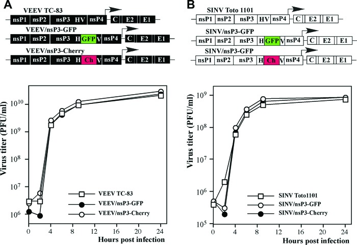
Alphaviruses are a group of single-stranded RNA viruses with genomes of positive polarity. They are divided into two geographically isolated groups: the Old World and the New World alphaviruses. Despite their similar genome organizations and virion structures, they differ in many aspects of pathogenesis and interaction with the host cell. Here we present new data highlighting previously unknown differences between these two groups. We found that nsP3 proteins of Sindbis virus (SINV) and Venezuelan equine encephalitis virus (VEEV) form cytoplasmic complexes with different morphologies and protein compositions. Unlike the amorphous aggregates formed by SINV nsP3 and other Old World alphavirus-specific nsP3s, VEEV nsP3 forms unique, large spherical structures with striking symmetry. Moreover, VEEV nsP3 does not interact with proteins previously identified as major components of SINV nsP3 complexes, such as G3BP1 and G3BP2. Importantly, the morphology of the complexes and the specificity of the interaction with cellular proteins are largely determined by the hypervariable domain (HVD) of nsP3. Replacement of the VEEV nsP3 HVD with the corresponding domain of SINV nsP3 rendered this protein capable of interaction with G3BPs. Conversely, replacement of the SINV nsP3 HVD with that of VEEV abolished SINV nsP3's interaction with G3BPs. The replacement of natural HVDs with those from heterologous viruses did not abrogate virus replication, despite these fragments demonstrating very low levels of sequence identity. Our data suggest that in spite of the differences in morphology and composition of the SINV- and VEEV-specific nsP3 complexes, it is likely that they have similar functions in virus replication and modification of the cellular environment.


Figures

Fig 1
Fig 1
In-frame insertions of different fluorescent proteins into alphavirus nsP3-specific HVDs do not interfere with virus replication. Schematic representations of VEEV TC-83 (A) and SINV (B) genomes containing GFP and Cherry fluorescent protein insertions in their nsP3-specific HVDs, indicated as HV, are shown, as well as the rates of their replication in BHK-21 cells. The GFP and Cherry coding sequences were inserted into nsP3 after aa 391 and 389 in VEEV and SINV, respectively. BHK-21 cells (5 × 10⁵) in 6-well Costar plates were infected with the indicated viruses at an MOI of 20 PFU/cell. At the indicated times postinfection, the medium was replaced, and virus titers were determined by plaque assay on BHK-21 cells.
Fig 2
Fig 2
VEEV- and SINV-specific nsP3 proteins demonstrate different distributions in infected cells. (A) BHK-21 cells in Ibidi 8-well μ-slides were infected with the indicated viruses at an MOI of 20 PFU/cell. At 4 h postinfection, cells were fixed with 4% paraformaldehyde, permeabilized, and stained with a dsRNA-specific MAb, and nuclei were stained with Hoechst dye. Images are presented as multiple-intensity projections (MIPs) of 3D stacks after deconvolution. Insets (6× zoom) in the first panels demonstrate the morphologies of the complexes formed by VEEV and SINV nsP3-GFP. The arrowheads point to the colocalization of SINV nsP3-GFP with dsRNA (red). (B) BHK-21 cells were infected with VEEV TC-83 encoding GFP under the control of the subgenomic promoter. At 6 h postinfection, cells were fixed, permeabilized, and stained with a VEEV nsP3-specific MAb and an Alexa Fluor 555-labeled secondary antibody. The lower panel presents stained mock-infected cells, which were processed and imaged identically to infected cells. Images are presented as MIPs of 6 optical sections. (C) Enlarged, representative single optical section images presenting one of the spheroids demonstrated in the cell in panel B. Note that antibodies did not penetrate efficiently into the nsP3-GFP-containing spheroid. (D) Fluorescence intensity profile of the spheroid image presented in panel C.
Fig 3
Fig 3
nsP3-containing spheroids are gradually formed at early times post-VEEV infection and contain no dsRNA. BHK-21 cells in Ibidi 8-well μ-slides were infected with VEEV/nsP3-GFP at an MOI of 20 PFU/cell, fixed at the indicated times postinfection, and, after permeabilization, stained with the dsRNA-specific MAb followed by the Alexa Fluor 555-labeled secondary antibody and Hoechst dye (nuclei). Images are presented as MIPs of 3D stacks after deconvolution. The overlap between nsP3-GFP and dsRNA structures appears as yellow staining at 1 h p.i. (see bottom panels). Very few overlapping spots could be detected at 2 and 3 h p.i.
Fig 4
Fig 4
SINV and VEEV nsP3-specific cytoplasmic complexes do not colocalize. BHK-21 cells in Ibidi 8-well μ-slides were coinfected at an MOI of 20 PFU/cell with pairs of recombinant VEEV and SINV encoding different fluorescent proteins within the nsP3 HVDs. At 6 h postinfection, cells were fixed and counterstained with Hoechst dye (nuclei). Images are presented as MIPs of 3D stacks after deconvolution. nsP3-GFP and nsP3-Cherry expressed by homologous viruses were completely colocalized, as shown in yellow in the overlay images and evident from the presented colocalization maps and Pearson's coefficient values (indicated below the columns). No nsP3-GFP and nsP3-Cherry colocalization was detected in the case of infection with the heterologous viruses SINV/nsP3-GFP and VEEV/nsP3-Cherry.
Fig 5
Fig 5
Domain structure and sequence alignment of nsP3 proteins of representative members of the New World and Old World alphaviruses. The schematic representation of the nsP3 protein shows the three predicted structural domains: the macro domain, the alpha domain, and the HVD. The sequence alignment of nsP3 proteins of different alphaviruses was performed with Clustal Omega (http://www.ebi.ac.uk/Tools/msa/clustalo/), and the figure was prepared with Jalview (http://www.jalview.org). The domain sequences are underlined with the same colors as those used in the schematic presentation. Sequences were derived from the following viruses: VEEV (GenBank accession no. P27282.2), SINV (GenBank accession no. P03317.1), SFV (GenBank accession no. NP_740667.1), CHIKV (GenBank accession no. NP_690588.1), and EEEV (GenBank accession no. Q4QXJ8.2).
Fig 6
Fig 6
Alphaviruses with heterologous HVDs are capable of efficient replication in a variety of cell lines. (A) Schematic representation of VEEV genomes encoding either wt nsP3 or chimeric nsP3 with the SINV-specific HVD, infectivities of the in vitro-synthesized RNAs in the infectious-center assay, and replication rates of the rescued viruses in the indicated cell lines. (B) Schematic representation of SINV genomes encoding either wt nsP3 or chimeric nsP3 with the VEEV-specific HVD, infectivities of the in vitro-synthesized RNAs in the infectious-center assay, and replication rates of the rescued viruses in the indicated cell lines. In the experiments presented in both panels, subconfluent cells in 6-well Costar plates were infected with the indicated viruses at an MOI of 20 PFU/cell. At the indicated time points, the medium was replaced, and virus titers were measured by plaque assay on BHK-21 cells.
Fig 7
Fig 7
The HVD determines the formation of virus-specific nsP3 complexes. The viruses used in this study encoded either homologous nsP3 HVDs with Cherry insertions (VEEV/nsP3-Cherry and SINV/nsP3-Cherry) or heterologous nsP3 HVDs with GFP insertions (VEEV/HVDsinv-GFP and SINV/HVDveev-GFP). BHK-21 cells in Ibidi 8-well μ-slides were coinfected with the indicated pairs of viruses at an MOI of 20 PFU/cell. Cells were fixed at 6 h postinfection and counterstained with Hoechst dye (nuclei). Images are presented as MIPs of 3D stacks after deconvolution. Pearson's coefficient values for each pair are indicated below the columns.
Fig 8
Fig 8
Colocalization of G3BP1 with nsP3 is determined by the HVD and is specific for SINV, not VEEV. BHK-21 cells in Ibidi 8-well μ-slides were coinfected at an MOI of 20 PFU/cell with the indicated viruses, encoding either homologous or heterologous nsP3 HVDs with GFP insertions. At 6 h postinfection, cells were fixed, permeabilized, and stained with a G3BP1-specific antibody, a secondary Alexa Fluor 555-labeled antibody, and Hoechst dye (nuclei). Colocalization of nsP3-GFP and G3BP1 is indicated in yellow in the overlay images.

Similar articles

Cited by

References

    1. Strauss JH, Strauss EG. 1994. The alphaviruses: gene expression, replication, and evolution. Microbiol. Rev. 58:491–562 - PMC - PubMed
    1. Rulli NE, Melton J, Wilmes A, Ewart G, Mahalingam S. 2007. The molecular and cellular aspects of arthritis due to alphavirus infections: lesson learned from Ross River virus. Ann. N. Y. Acad. Sci. 1102:96–108 - PubMed
    1. Staples JE, Breiman RF, Powers AM. 2009. Chikungunya fever: an epidemiological review of a re-emerging infectious disease. Clin. Infect. Dis. 49:942–948 - PubMed
    1. Toivanen A. 2008. Alphaviruses: an emerging cause of arthritis? Curr. Opin. Rheumatol. 20:486–490 - PubMed
    1. Weaver SC, Ferro C, Barrera R, Boshell J, Navarro JC. 2004. Venezuelan equine encephalitis. Annu. Rev. Entomol. 49:141–174 - PubMed

Publication types

Substances

LinkOut - more resources