. 2013 Feb;87(4):2206-14.
doi: 10.1128/JVI.02097-12. Epub 2012 Dec 5.

ATF6 signaling is required for efficient West Nile virus replication by promoting cell survival and inhibition of innate immune responses

Affiliations

ATF6 signaling is required for efficient West Nile virus replication by promoting cell survival and inhibition of innate immune responses

Rebecca L Ambrose et al. J Virol. 2013 Feb.

Abstract

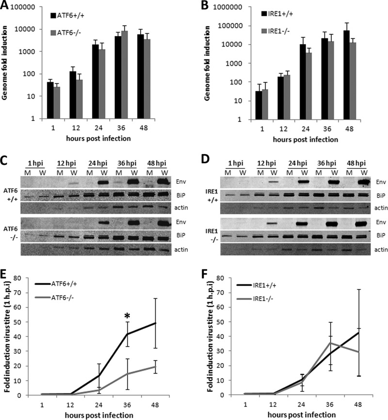
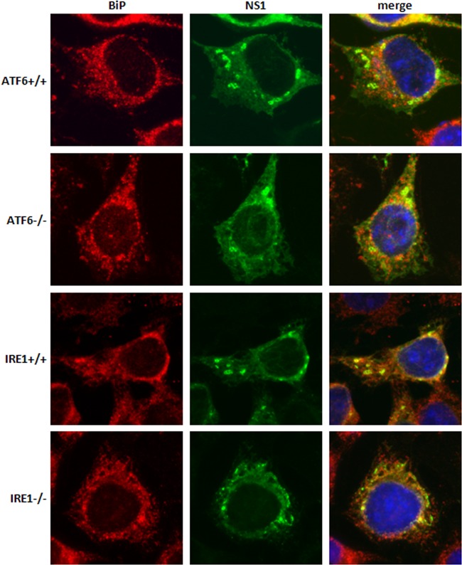
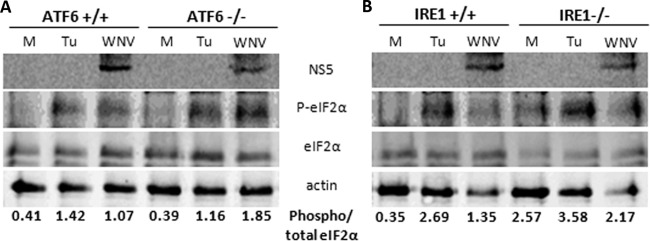
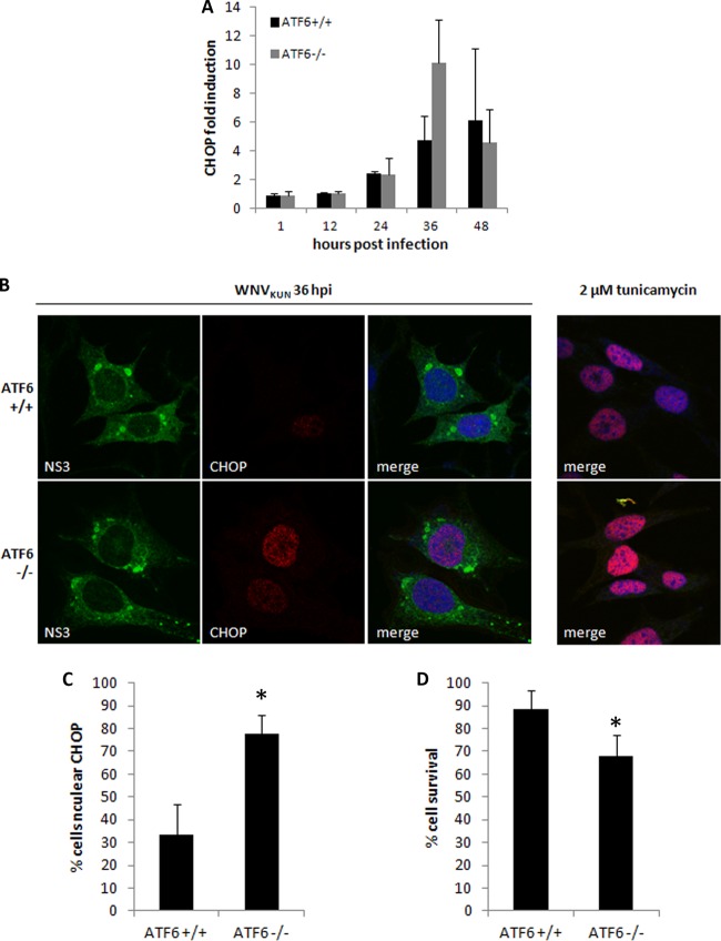
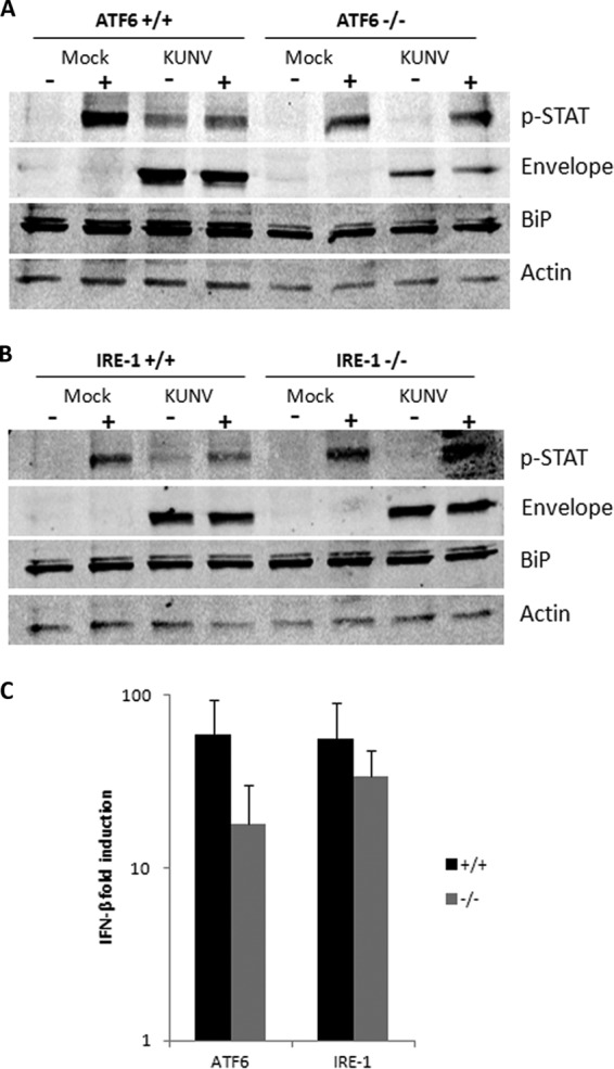
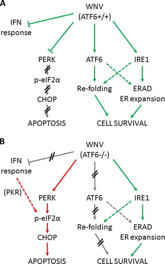
West Nile virus strain Kunjin (WNV(KUN)) is an enveloped, positive-sense RNA virus within the virus family Flaviviridae. Many flaviviruses have been shown to manipulate multiple signaling pathways, including autophagic, innate immune, and stress responses, in order to benefit replication. In particular, we have demonstrated that WNV(KUN) regulates the unfolded protein response (UPR), skewing the downstream effectors toward chaperone expression and Xbp-1 activation while preventing PERK-mediated translation attenuation and C/EBP homologous protein (CHOP) upregulation. WNV(KUN)-regulated UPR signaling can then be hijacked in order to affect type I interferon (IFN) responses, preventing IFN-mediated STAT1 phosphorylation and nuclear translocation. To extend our previous observations, we aimed to investigate the contribution of ATF6- and IRE1-mediated signaling during WNV(KUN) replication and how the two sensors contribute to the inhibition of IFN signaling. ATF6-deficient cells infected with WNV(KUN) showed decreased protein and virion production. These cells also demonstrated increased eIF2α phosphorylation and CHOP transcription, absent in infected matched control cells. Thus, we propose that in the absence of ATF6, WNV(KUN) is incapable of manipulating the PERK-mediated response to infection. In contrast, infection of IRE1(-/-) knockout cells showed no discernible differences compared to IRE1(+/+) cells. However, both ATF6 and IRE1 were required for WNV(KUN)-induced inhibition of STAT1 phosphorylation. We suggest that the combination of abhorrent UPR signaling, promotion of cell death, and increased innate immune responses contributes to the replication defects in ATF6-deficient cells, thus demonstrating the dual importance of ATF6 in maintaining cell viability and modulating immune responses during WNV(KUN) infection.


Figures

Fig 1
Fig 1
WNVKUN production is decreased in ATF6−/− cells. ATF6 and IRE1 knockout MEFs and their matched controls were infected with WNVKUN (MOI, 10) for 1, 12, 24, 36, and 48 h, and supernatants, protein, and RNA samples were collected at each time point. (A and B) Viral genomic RNA was quantified by qRT-PCR using gene-specific primers and compared to that for the internal control GAPDH. Error bars indicate 1 standard deviation of three independent experiments. (C and D) Protein lysates collected at each time point were analyzed for viral protein expression (Env) and chaperone upregulation (BiP) using Western blotting, with actin used as a loading control. (E and F) Infectious virion secretion (from supernatant samples) was analyzed by plaque assay on Vero cells. Error bars indicate 1 standard deviation of three independent experiments, and asterisks show statistically significant differences in samples, as determined by a Student t test (P < 0.05).
Fig 2
Fig 2
WNVKUN replication complex formation is not altered in knockout cell lines. ATF6−/−, IRE1−/−, and their matched control cells were infected with WNVKUN as indicated above, fixed at 36 h p.i., and then labeled with anti-NS1 (green) and anti-BiP (red) antibodies. Cells were viewed on an Olympus epifluorescence microscope.
Fig 3
Fig 3
eIF2α phosphorylation is elevated in ATF6−/− MEFs. ATF6−/−, IRE1−/−, and their matched control cell lines were infected with WNVKUN (MOI, 10) over the time course indicated or treated with 2 μM tunicamycin for 12 h. (A) At 36 h p.i., protein lysates were collected and probed for levels of phospho-eIF2α (Ser51), total eIF2α, NS5 (as an infection control), and actin by Western blotting. The ratio of phospho to total eIF2α levels was determined by densitometry analysis (Quantity One; Bio-Rad) using the Western blot analysis shown.
Fig 4
Fig 4
Increased CHOP activity in WNVKUN-infected ATF6−/− MEFs leads to cell death. (A) Total RNA extracted from infected and mock cells at each time point was analyzed for CHOP transcription using qRT-PCR, and fold induction was calculated by comparison to mock cells at the same time point. Error bars indicate 1 standard deviation of three independent experiments. (B) Knockout and wild-type MEFs were infected with WNVKUN for 36 h (MOI, 10) or treated with 2 μM tunicamycin for 12 h. Cells were fixed and subsequently labeled with anti-CHOP (red) and anti-NS3 (green) antibodies before analysis by immunofluorescence. Cells were also counterstained with the nuclear dye DRAQ5 and viewed on a Zeiss confocal microscope. The right-hand-side panels indicate the efficiency of CHOP nuclear translocation in the presence of tunicamycin alone. (C) CHOP nuclear translocation was quantified as a percentage of infected cells, and error bars indicate 1 standard deviation of two independent quantitations (of ∼150 cells each). Asterisks denote statistically significant differences between samples, as determined by a Student t test (P < 0.05). (D) Knockout and wild-type MEFs were infected or mock infected with WNVKUN in a 96-well microtiter plate for 48 h. Cells were lysed in 5% (vol/vol) Triton X-100, and then lactate dehydrogenase activity was assessed as a measure of cell viability using a cytotoxicity kit. Error bars indicate 1 standard deviation of 3 independent experiments with triplicates, and asterisks denote P values of <0.05.
Fig 5
Fig 5
Late-phase IFN signaling is partially restored in ATF6−/− and IRE1−/− MEFs. (A and B) Knockout ATF6 and IRE1 MEFs and their matched controls were infected with WNVKUN (MOI, 10) for 36 h and then stimulated with 100 U/ml of mouse IFN-α for 1 h. Protein lysates were collected and probed for phospho-STAT1 (Y701), envelope, BiP, and actin using Western blotting. Results are indicative of at least two independent experiments. (C) RNA collected from infected cells was analyzed for IFN-β transcription by qRT-PCR using gene-specific primers. Error bars indicate 1 standard deviation of three independent experiments.
Fig 6
Fig 6
ATF6 is required during WNV replication to maintain cell viability. (A) During WNVKUN replication in WT cells, PERK and IFN signaling is actively inhibited to prevent cell death (red arrows) while ATF6- and IRE1-mediated effectors are induced (green arrows) to assist with viral protein and virion production (via chaperone expression, expansion of ER membranes). (B) However, in ATF6-deficient cells, the modulation of UPR signaling is lost, resulting in uncontrolled signaling through PERK (red arrows) as well as resumption of IFN signaling. Although WNVKUN can still signal via IRE1 (green arrows), the cells must override this manipulation to aid in viral clearance. The proviral effects of ATF6-induced chaperone expression are also ablated, resulting in translational defects and premature cell death in WNVKUN-infected cells. We propose that ATF6 (via a downstream effector) is required to modulate PERK and IFN responses.

Similar articles

Cited by

References

    1. Berridge MJ. 2002. The endoplasmic reticulum: a multifunctional signaling organelle. Cell Calcium 32:235–249 - PubMed
    1. Bernales S, Papa FR, Walter P. 2006. Intracellular signaling by the unfolded protein response. Annu. Rev. Cell Dev. Biol. 22:487–508 - PubMed
    1. Kimata Y, Oikawa D, Shimizu Y, Ishiwata-Kimata Y, Kohno K. 2004. A role for BiP as an adjustor for the endoplasmic reticulum stress-sensing protein Ire1. J. Cell Biol. 167:445–456 - PMC - PubMed
    1. Cui WJ, Li JZ, Ron D, Sha BD. 2011. The structure of the PERK kinase domain suggests the mechanism for its activation. Acta Crystallogr. D Biol. Crystallogr. 67:423–428 - PMC - PubMed
    1. Liu CY, Schroder M, Kaufman RJ. 2000. Ligand-independent dimerization activates the stress response kinases IRE1 and PERK in the lumen of the endoplasmic reticulum. J. Biol. Chem. 275:24881–24885 - PubMed

Publication types