

Proteomics reveals plastid- and periplastid-targeted proteins in the chlorarachniophyte alga Bigelowiella natans
- PMID: 23221610
- PMCID: PMC3542566
- DOI: 10.1093/gbe/evs115
Proteomics reveals plastid- and periplastid-targeted proteins in the chlorarachniophyte alga Bigelowiella natans
Abstract
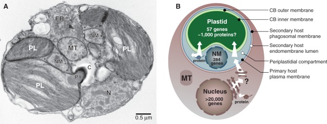
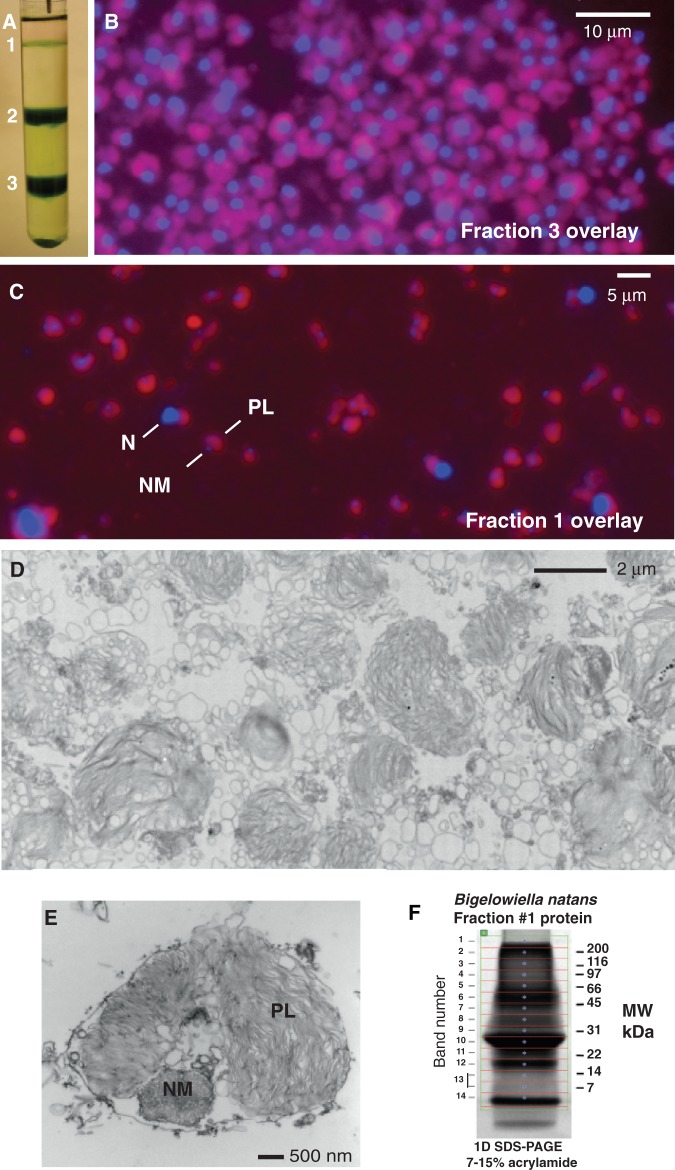
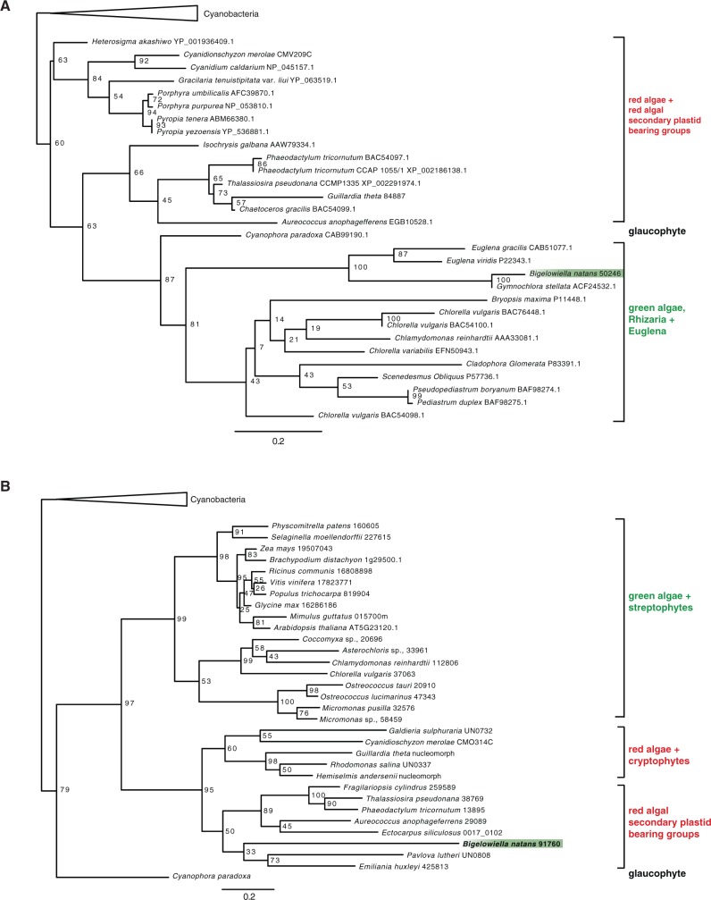
Chlorarachniophytes are unicellular marine algae with plastids (chloroplasts) of secondary endosymbiotic origin. Chlorarachniophyte cells retain the remnant nucleus (nucleomorph) and cytoplasm (periplastidial compartment, PPC) of the green algal endosymbiont from which their plastid was derived. To characterize the diversity of nucleus-encoded proteins targeted to the chlorarachniophyte plastid, nucleomorph, and PPC, we isolated plastid-nucleomorph complexes from the model chlorarachniophyte Bigelowiella natans and subjected them to high-pressure liquid chromatography-tandem mass spectrometry. Our proteomic analysis, the first of its kind for a nucleomorph-bearing alga, resulted in the identification of 324 proteins with 95% confidence. Approximately 50% of these proteins have predicted bipartite leader sequences at their amino termini. Nucleus-encoded proteins make up >90% of the proteins identified. With respect to biological function, plastid-localized light-harvesting proteins were well represented, as were proteins involved in chlorophyll biosynthesis. Phylogenetic analyses revealed that many, but by no means all, of the proteins identified in our proteomic screen are of apparent green algal ancestry, consistent with the inferred evolutionary origin of the plastid and nucleomorph in chlorarachniophytes.
Figures
Similar articles
-
Nucleus-encoded periplastid-targeted EFL in chlorarachniophytes.Mol Biol Evol. 2008 Sep;25(9):1967-77. doi: 10.1093/molbev/msn147. Epub 2008 Jul 3. Mol Biol Evol. 2008. PMID: 18599495
-
Nucleus- and nucleomorph-targeted histone proteins in a chlorarachniophyte alga.Mol Microbiol. 2011 Jun;80(6):1439-49. doi: 10.1111/j.1365-2958.2011.07643.x. Epub 2011 Apr 6. Mol Microbiol. 2011. PMID: 21470316
-
Nucleomorph and plastid genome sequences of the chlorarachniophyte Lotharella oceanica: convergent reductive evolution and frequent recombination in nucleomorph-bearing algae.BMC Genomics. 2014 May 15;15(1):374. doi: 10.1186/1471-2164-15-374. BMC Genomics. 2014. PMID: 24885563 Free PMC article.
-
Bilateral communication between plastid and the nucleus: plastid protein import and plastid-to-nucleus retrograde signaling.Biosci Biotechnol Biochem. 2010;74(3):471-6. doi: 10.1271/bbb.90842. Epub 2010 Mar 7. Biosci Biotechnol Biochem. 2010. PMID: 20208345 Review.
-
Nucleomorph genomes.Annu Rev Genet. 2009;43:251-64. doi: 10.1146/annurev-genet-102108-134809. Annu Rev Genet. 2009. PMID: 19686079 Review.
Cited by
-
Kingdom Chromista and its eight phyla: a new synthesis emphasising periplastid protein targeting, cytoskeletal and periplastid evolution, and ancient divergences.Protoplasma. 2018 Jan;255(1):297-357. doi: 10.1007/s00709-017-1147-3. Epub 2017 Sep 5. Protoplasma. 2018. PMID: 28875267 Free PMC article.
-
Genomic perspectives on the birth and spread of plastids.Proc Natl Acad Sci U S A. 2015 Aug 18;112(33):10147-53. doi: 10.1073/pnas.1421374112. Epub 2015 Apr 20. Proc Natl Acad Sci U S A. 2015. PMID: 25902528 Free PMC article.
-
Prospective function of FtsZ proteins in the secondary plastid of chlorarachniophyte algae.BMC Plant Biol. 2015 Nov 10;15:276. doi: 10.1186/s12870-015-0662-7. BMC Plant Biol. 2015. PMID: 26556725 Free PMC article.
-
Tracking N-terminal protein processing from the Golgi to the chromatophore of a rhizarian amoeba.Plant Physiol. 2022 Jun 27;189(3):1226-1231. doi: 10.1093/plphys/kiac173. Plant Physiol. 2022. PMID: 35485189 Free PMC article.
-
Subcellular Compartments Interplay for Carbon and Nitrogen Allocation in Chromera velia and Vitrella brassicaformis.Genome Biol Evol. 2019 Jul 1;11(7):1765-1779. doi: 10.1093/gbe/evz123. Genome Biol Evol. 2019. PMID: 31192348 Free PMC article.
References
-
- Altschul SF, Gish W, Miller W, Myers EW, Lipman DJ. Basic local alignment search tool. J Mol Biol. 1990;215:403–410. - PubMed
-
- Archibald JM. Nucleomorph genomes: structure, function, origin and evolution. Bioessays. 2007;29:392–402. - PubMed
-
- Archibald JM. The puzzle of plastid evolution. Curr Biol. 2009;19:R81–R88. - PubMed
-
- Archibald JM, Logsdon JM, Jr, Doolittle WF. Origin and evolution of eukaryotic chaperonins: phylogenetic evidence for ancient duplications in CCT genes. Mol Biol Evol. 2000;17:1456–1466. - PubMed
Publication types
MeSH terms
Substances
LinkOut - more resources
Full Text Sources
