Review
. 2013 Jan;8(1):e22598.
doi: 10.4161/psb.22598. Epub 2012 Dec 6.

Role of LysM receptors in chitin-triggered plant innate immunity

Affiliations
Review

Role of LysM receptors in chitin-triggered plant innate immunity

Kiwamu Tanaka et al. Plant Signal Behav. 2013 Jan.

Abstract

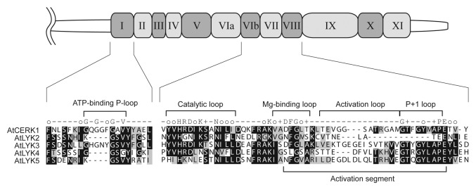
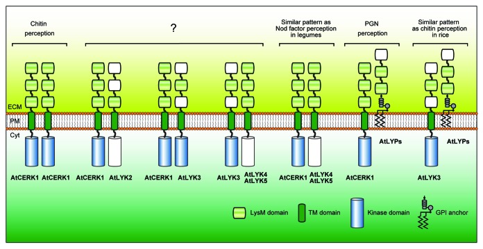
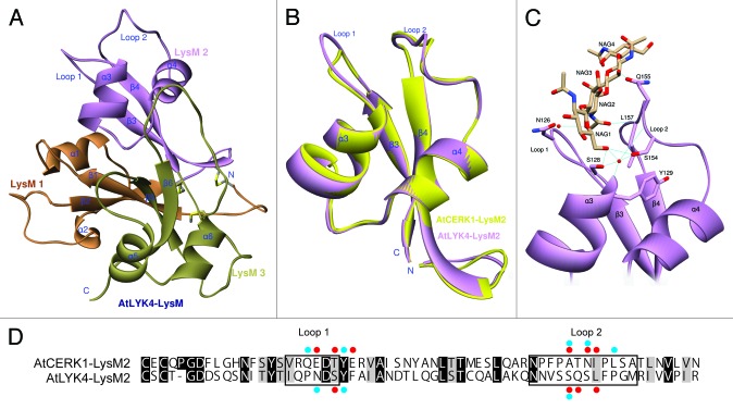
Recent research findings clearly indicate that lysin motif (LysM)-containing cell surface receptors are involved in the recognition of specific oligosaccharide elicitors (chitin and peptidoglycan), which trigger an innate immunity response in plants. These receptors are either LysM-containing receptor-like kinases (LYKs) or LysM-containing receptor proteins (LYPs). In Arabidopsis, five LYKs (AtCERK1/AtLYK1 and AtLYK2-5) and three LYPs (AtLYP1-3) are likely expressed on the plasma membrane. In this review, we summarize recent research results on the role of these receptors in plant innate immunity, including the recent structural characterization of AtCERK1 and composition of the various receptor complexes in Arabidopsis.

Keywords: Arabidopsis; chitin (N-acetylchitooligosaccharide); lysin motif; lysin motif-containing receptors; microbe-associated molecular patterns; plant innate immunity.


Figures

None
Figure 1. Sequence comparison of the kinase domain of Arabidopsis LYK proteins. Schematic representation shows the kinase domain of AtCERK1 (drawn to scale). Kinase subdomain I and VIb to VIII are selectively highlighted below the drawing to highlight the five AtLYK proteins. Identical and similar residues throughout the alignment are shown in black and gray, respectively. The consensus line among the eukaryotic protein kinase superfamily is given according to the following code: uppercase letters, invariant residues; o, conserved nonpolar residues; +, conserved small residues with near neutral polarity. Note that AtCERK1 and AtLYK3 have an intact intracellular kinase domain.
None
Figure 3. Possible combinations of Arabidopsis LysM receptor proteins involved in MAMP recognition. The picture represents theoretical combinations of LysM receptors with a prerequisite condition that two or more LysM domains are present in the extracellular region and that at least one of the receptors possess an active, intracellular kinase domain. Putative LysM domains with less sequence conservation and pseudo kinase domains are drawn colorless. Note that AtCERK1 is a unique LysM receptor in Arabidopsis since it alone can form a homodimer given the prerequisites imposed. Abbreviations: LysM, lysin motif; TM, transmembrane; GPI, glycosylphosphatidylinositol; ECM, extracellular matrix; PM, plasma membrane; Cyt, cytoplasm.
None
Figure 2. Three-dimensional model prediction of the structure of the AtLYK4 extracellular domain, including a ligand docking model. (A)The 3D model of AtLYK4 extracellular domain was built based on the crystal structure of AtCERK1 (PDB code: 4EBY). Each LysM domain is represented in a different color: first LysM (orange), second LysM (purple) and third LysM (green). (B)The second LysM domains of AtLYK4 (purple) and AtCERK1 (yellow) are superimposed to highlight the similarity in structure. Note that the overall folds are highly conserved between the two models, although the AtLYK4 has the longer extended Loop 1 which is a constitutive part of the cleft where the predicted chitin-binding site is found. (C) Docking model between the second LysM domain of AtLYK4 and chitotetraose. (D) Pairwise sequence comparison of the second LysM domains of AtLYK4 and AtCERK1. Identical and similar residues throughout the alignment are shown in black and gray, respectively. Enclosed boxes represent Loop 1 and Loop 2. Red and blue dots indicate the residues involved in direct interactions and water molecule-mediated interactions with chitotetraose, respectively. The residues involved in van der Waals interactions were neglected here.

References

    1. Chisholm ST, Coaker G, Day B, Staskawicz BJ. Host-microbe interactions: shaping the evolution of the plant immune response. Cell. 2006;124:803–14. doi: 10.1016/j.cell.2006.02.008. - DOI - PubMed
    1. Janeway CA, Jr., Medzhitov R. Innate immune recognition. Annu Rev Immunol. 2002;20:197–216. doi: 10.1146/annurev.immunol.20.083001.084359. - DOI - PubMed
    1. Jones JD, Dangl JL. The plant immune system. Nature. 2006;444:323–9. doi: 10.1038/nature05286. - DOI - PubMed
    1. Silipo A, Erbs G, Shinya T, Dow JM, Parrilli M, Lanzetta R, et al. Glyco-conjugates as elicitors or suppressors of plant innate immunity. Glycobiology. 2010;20:406–19. doi: 10.1093/glycob/cwp201. - DOI - PubMed
    1. Silipo A, Molinaro A, Sturiale L, Dow JM, Erbs G, Lanzetta R, et al. The elicitation of plant innate immunity by lipooligosaccharide of Xanthomonas campestris. J Biol Chem. 2005;280:33660–8. doi: 10.1074/jbc.M506254200. - DOI - PubMed

Publication types

MeSH terms