

Differential effects of deep sedation with propofol on the specific and nonspecific thalamocortical systems: a functional magnetic resonance imaging study
- PMID: 23221862
- PMCID: PMC4080838
- DOI: 10.1097/ALN.0b013e318277a801
Differential effects of deep sedation with propofol on the specific and nonspecific thalamocortical systems: a functional magnetic resonance imaging study
Abstract
Background: The current state of knowledge suggests that disruption of neuronal information integration may be a common mechanism of anesthetic-induced unconsciousness. A neural system critical for information integration is the thalamocortical system whose specific and nonspecific divisions may play the roles for representing and integrating information, respectively. How anesthetics affect the function of these systems individually is not completely understood. The authors studied the effect of propofol on thalamocortical functional connectivity in the specific and nonspecific systems, using functional magnetic resonance imaging.
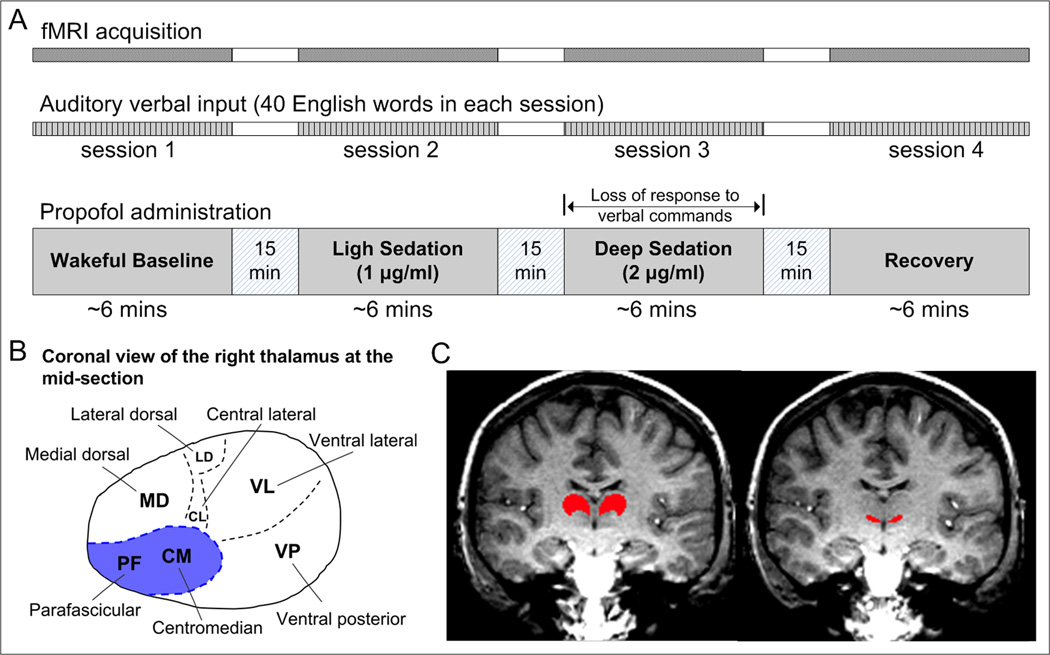
Methods: Eight healthy volunteers were instructed to listen to and encode 40 English words during wakeful baseline, light sedation, deep sedation, and recovery in the scanner. Functional connectivity was determined as the temporal correlation of blood oxygen level-dependent signals with seed regions defined within the specific and nonspecific thalamic nuclei.
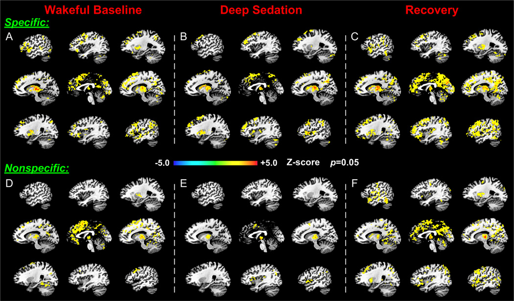
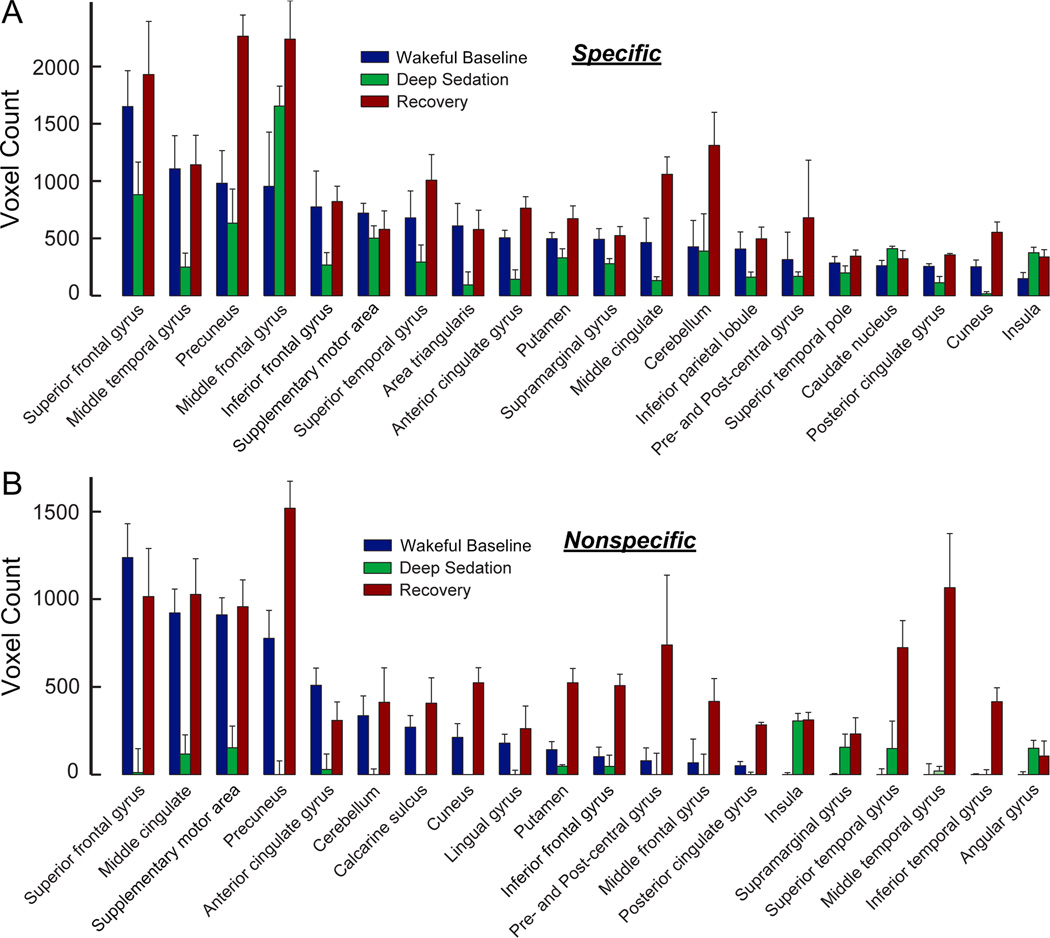
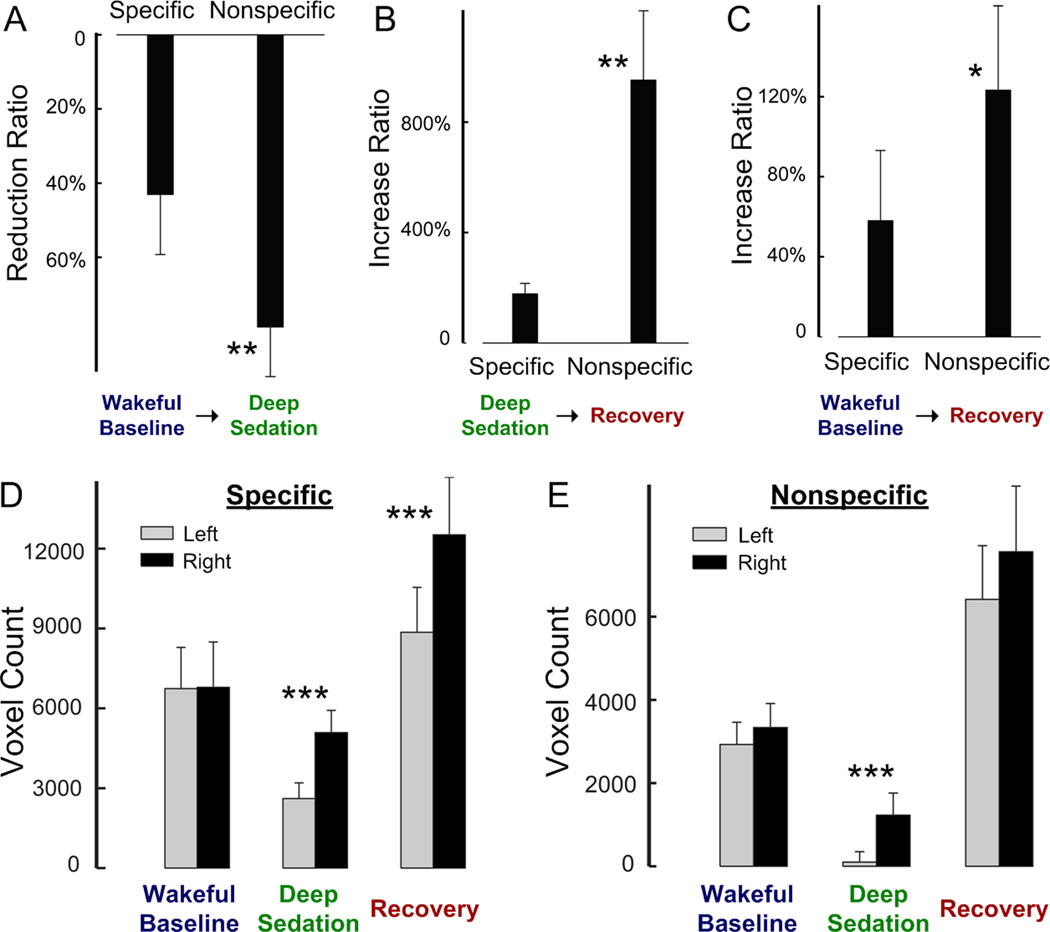
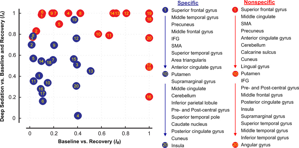
Results: Thalamocortical connectivity at baseline was dominantly medial and bilateral frontal and temporal for the specific system, and medial frontal and medial parietal for the nonspecific system. During deep sedation, propofol reduced functional connectivity by 43% (specific) and 79% (nonspecific), a significantly greater reduction in the nonspecific than in the specific system and in the left hemisphere than in the right. Upon regaining consciousness, functional connectivity increased by 58% (specific) and 123% (nonspecific) during recovery, exceeding their values at baseline.
Conclusions: Propofol conferred differential changes in functional connectivity of the specific and nonspecific thalamocortical systems, particularly in left hemisphere, consistent with the verbal nature of stimuli and task. The changes in nonspecific thalamocortical connectivity may correlate with the loss and return of consciousness.
Figures
Comment in
-
Consciousness, anesthesia, and the thalamocortical system.Anesthesiology. 2013 Jan;118(1):13-5. doi: 10.1097/ALN.0b013e318277a9c6. Anesthesiology. 2013. PMID: 23208518 No abstract available.
-
Laterality of motor control and consciousness shares the same hemisphere.Anesthesiology. 2013 Sep;119(3):727-8. doi: 10.1097/ALN.0b013e31829e4b54. Anesthesiology. 2013. PMID: 23962933 No abstract available.
-
In reply.Anesthesiology. 2013 Sep;119(3):728-9. doi: 10.1097/ALN.0b013e31829e6d29. Anesthesiology. 2013. PMID: 23962934 Free PMC article. No abstract available.
-
Postanesthesia evaluation of neuromuscular function.Anesthesiology. 2013 Sep;119(3):729. doi: 10.1097/ALN.0b013e31829ff1f3. Anesthesiology. 2013. PMID: 23962935 No abstract available.
-
In reply.Anesthesiology. 2013 Sep;119(3):729-30. doi: 10.1097/ALN.0b013e31829fff78. Anesthesiology. 2013. PMID: 23962936 No abstract available.
References
-
- Schrouff J, Perlbarg V, Boly M, Marrelec G, Boveroux P, Vanhaudenhuyse A, Bruno MA, Laureys S, Phillips C, Pelegrini-Issac M, Maquet P, Benali H. Brain functional integration decreases during propofol-induced loss of consciousness. Neuroimage. 2011;57:198–205. - PubMed
-
- Tononi G. Consciousness as integrated information: a provisional manifesto. Biol Bull. 2008;215:216–242. - PubMed
-
- Hudetz AG. Suppressing Consciousness: mechanisms of general anesthesia. Seminars in Anesthesia, Perioperative Medicine and Pain. 2006;25:196–204.
Publication types
MeSH terms
Substances
Grants and funding
LinkOut - more resources
Full Text Sources
Other Literature Sources
Medical
