. 2012 Dec 15;26(24):2780-801.
doi: 10.1101/gad.208926.112. Epub 2012 Dec 7.

FoxO6 regulates memory consolidation and synaptic function

Affiliations

FoxO6 regulates memory consolidation and synaptic function

Dervis A M Salih et al. Genes Dev. .

Abstract

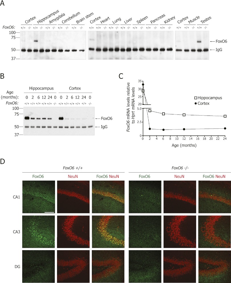
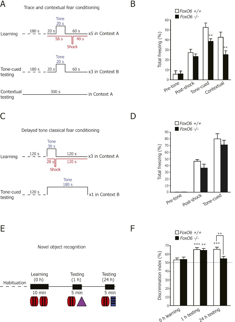
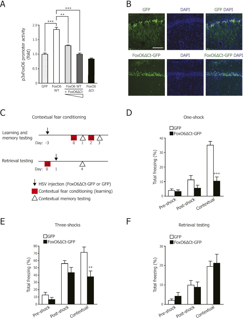
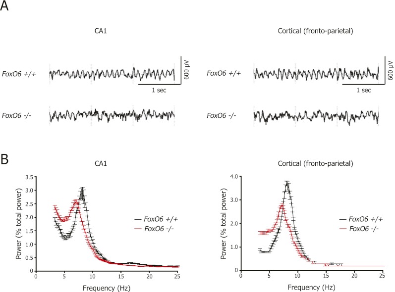
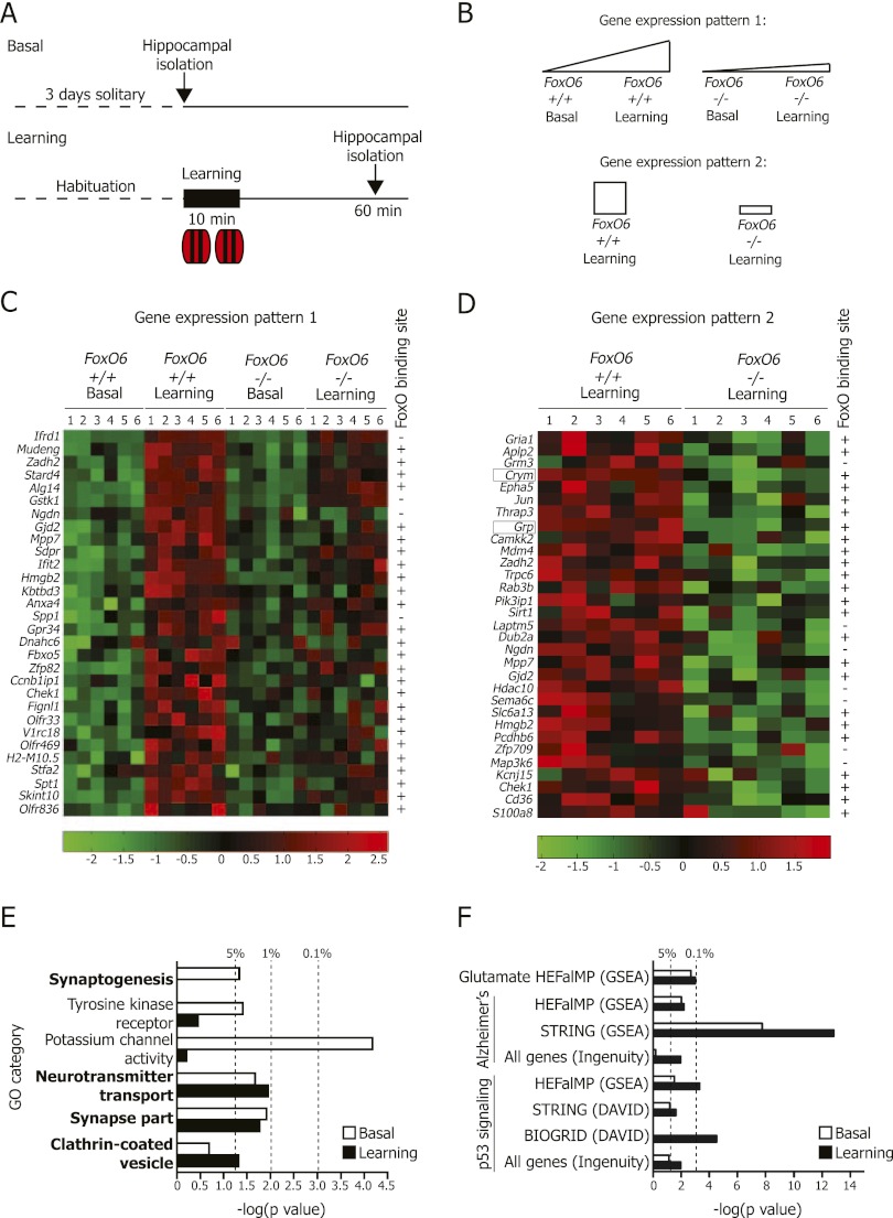
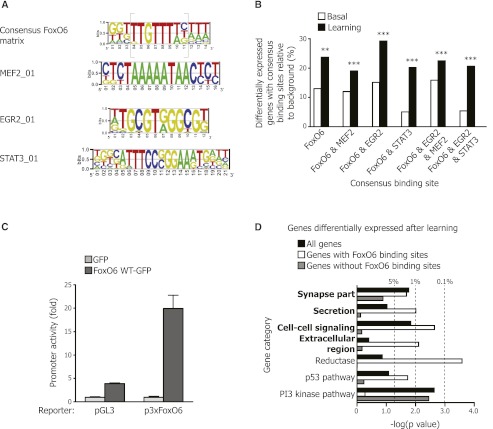
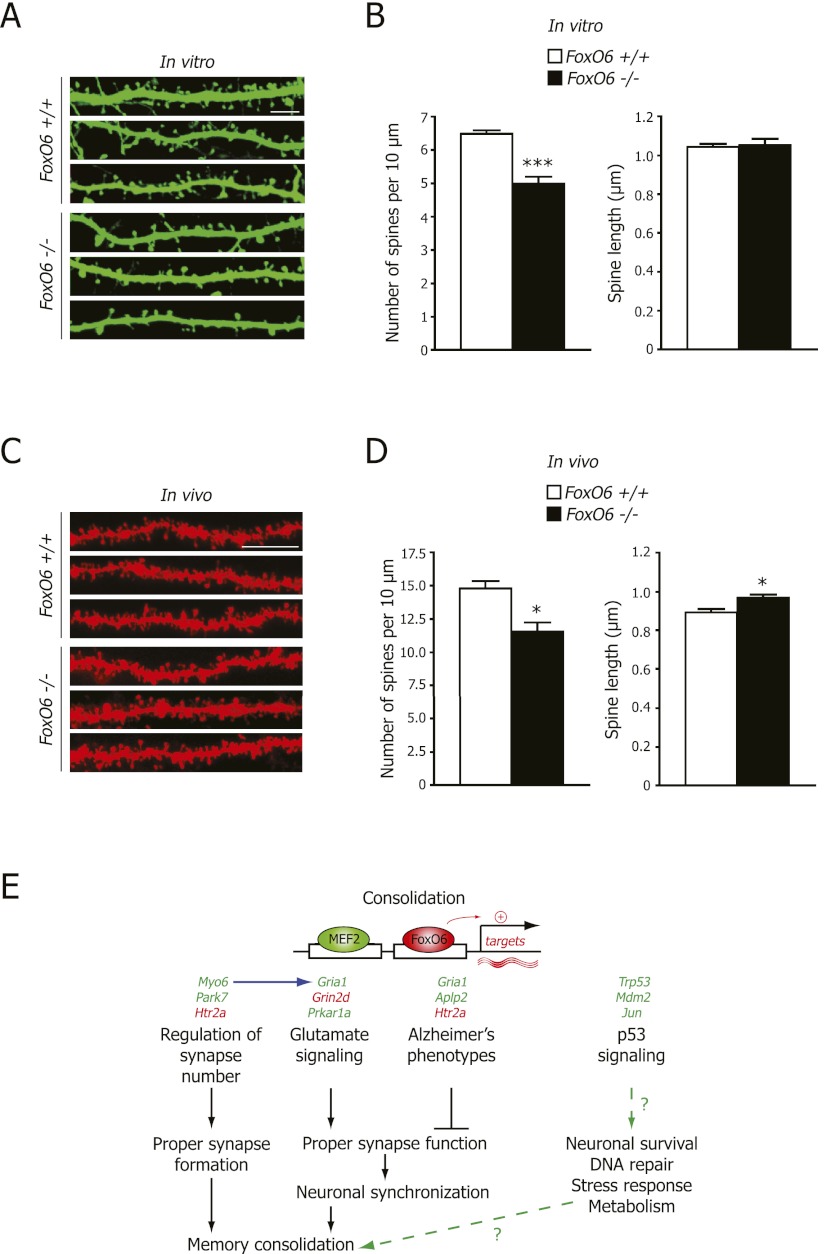
The FoxO family of transcription factors is known to slow aging downstream from the insulin/IGF (insulin-like growth factor) signaling pathway. The most recently discovered FoxO isoform in mammals, FoxO6, is highly enriched in the adult hippocampus. However, the importance of FoxO factors in cognition is largely unknown. Here we generated mice lacking FoxO6 and found that these mice display normal learning but impaired memory consolidation in contextual fear conditioning and novel object recognition. Using stereotactic injection of viruses into the hippocampus of adult wild-type mice, we found that FoxO6 activity in the adult hippocampus is required for memory consolidation. Genome-wide approaches revealed that FoxO6 regulates a program of genes involved in synaptic function upon learning in the hippocampus. Consistently, FoxO6 deficiency results in decreased dendritic spine density in hippocampal neurons in vitro and in vivo. Thus, FoxO6 may promote memory consolidation by regulating a program coordinating neuronal connectivity in the hippocampus, which could have important implications for physiological and pathological age-dependent decline in memory.


Figures

Figure 1.
Figure 1.
FoxO6 is expressed predominantly in the CA1 and CA3 regions of the hippocampus in adult mice. (A) FoxO6 protein is highly expressed in the hippocampus of the adult mouse brain. Tissues from 8-wk-old FoxO6 mutant (−/−) and wild-type (+/+) siblings were tested by immunoprecipitation with an antibody to full-length FoxO6 and Western blotting with an antibody to an N-terminal FoxO6 peptide. (B,C) FoxO6 protein (B) and mRNA (C) are expressed abundantly in the hippocampus and cortex at birth and decrease while becoming enriched in the hippocampus relative to the cortex in adult mice. (D) FoxO6 protein is expressed in the nuclei of neurons of the pyramidal cell layer of the CA1 and CA3 hippocampal fields of the adult mouse brain but not in the dentate gyrus (DG). Immunohistochemistry on coronal brain sections from 2.5-mo-old FoxO6 mutant and wild-type siblings with antibodies to full-length FoxO6 and the neuronal marker NeuN. Bar, 100 μm.
Figure 2.
Figure 2.
FoxO6 mutant mice have impaired expression of contextual and recognition memories. (A) Scheme to test contextual learning and memory using trace tone-cued and contextual fear conditioning. Learning (day 1), tone-cued memory testing (day 2), and contextual memory testing (day 3). (B) FoxO6 mutant mice are significantly impaired in the consolidation of both trace tone-cued and contextual fear memories. Results are expressed as the percentage of time for which freezing behavior was shown prior to receiving the shock (pretone), during the learning stage (post-shock), during tone-cued memory testing (tone-cued), and during contextual memory testing (contextual). Mean ± SEM. n = 16–19 mice (4- to 7-mo-old males) per genotype. P < 0.0001 for the stage of the fear-conditioning test, two-way repeated measures ANOVA with genotype and stage of the fear conditioning as the factors, F3,99 = 64.29; (**) P < 0.01 FoxO6 wild-type versus null, unpaired Student's t-test. (C) Scheme to test auditory (tone) fear learning and memory using delayed tone fear conditioning. Learning (day 1) and tone-cued memory testing (day 2). (D) FoxO6 mutant mice are not impaired in the consolidation of tone-cued fear memories. Results are expressed as the percentage of time for which freezing behavior was shown prior to receiving the shock (pretone), during the learning stage (post-shock), and during tone-cued memory testing (tone-cued). Mean ± SEM. n = 7–9 mice (2- to 3-mo-old males) per genotype. P < 0.0001 for the stage of the fear-conditioning test, two-way repeated measures ANOVA with genotype and stage of the fear conditioning as the factors, F2,28 = 163.6. (E) Scheme to test object learning and memory using the novel object recognition task. (F) FoxO6 mutant mice could learn the novel object recognition task (1-h testing), but FoxO6 mutant mice are significantly impaired in the consolidation or retrieval of object recognition memories at 24 h. The discrimination index (percent) is shown for the time spent exploring the novel object versus the total time spent exploring both objects. Mean ± SEM. n = 20–21 mice (2- to 5 mo-old males) per genotype. P < 0.0001 for the stage of the novel object recognition test, two-way repeated measures ANOVA with genotype and stage of the test as the factors, F2,78 = 13.21. FoxO6 mutant and wild-type mice both show a preference for the novel object at 1 h. (**) P < 0.01; (***) P < 0.001 for 0 h versus 1 h, paired Student's t-test. Wild-type mice, but not FoxO6 mutant mice, show a preference for the novel object at 24 h. (***) P < 0.001 for FoxO6 wild-type 0 h versus 24 h, paired Student's t-test; (**) P < 0.01 for FoxO6 mutant versus wild-type at 24 h, unpaired Student's t-test.
Figure 3.
Figure 3.
Disrupting FoxO6 function locally and acutely in the CA1 region of the dorsal hippocampus impairs the consolidation of contextual fear memories. (A) FoxO6 with a deletion of the C-terminal region acts as a dominant negative. Luciferase assays in U2OS cells using constructs expressing wild-type FoxO6-GFP (FoxO6 WT) alone or with increasing amounts of FoxO6ΔCt-GFP (FoxO6ΔCt) and a luciferase reporter driven by three consensus FoxO6-binding sites (p3xFoxO6). Results are normalized to renilla and to GFP construct alone. Mean ± SEM from three independent experiments performed in triplicate. (**) P < 0.01; (***) P < 0.001, one-way ANOVA with the Bonferroni post hoc test. (B) HSV vectors expressing FoxO6ΔCt-GFP or GFP were microinjected into the CA1 region of the hippocampus in adult wild-type mice. FoxO6ΔCt-GFP was predominantly in the nucleus of neurons from the CA1 pyramidal cell layer. Counterstained with the nuclear marker DAPI. Bar, 100 μm. (C) Scheme to test contextual learning and memory in adult wild-type mice injected with HSV expressing FoxO6ΔCt-GFP or GFP. Contextual fear conditioning was performed with one or three foot shocks as indicated in the squares. (D) Wild-type mice expressing FoxO6ΔCt-GFP are significantly impaired in contextual fear memory following one foot shock. Mice were microinjected with viral vectors expressing FoxO6ΔCt-GFP or GFP in the CA1 region of the hippocampus, and contextual fear conditioning was performed 3 d following surgery. Results are expressed as the percentage of time for which freezing behavior was shown prior to receiving the shock (preshock), during the learning stage (post-shock), and during contextual memory testing (contextual). Mean ± SEM. n = 7 mice (2- to 3-mo-old) per viral construct. P < 0.0001 for the stage of the fear-conditioning test, two-way repeated measures ANOVA with viral construct and stage of the fear conditioning as the factors, F2,24 = 38.26; (***) P < 0.001 FoxO6ΔCt-GFP versus GFP, unpaired Student's t-test. (E) Adult mice injected with FoxO6ΔCt-GFP are significantly impaired in stronger contextual fear memory (in response to three foot shocks). Results are expressed as in D. Mean ± SEM. n = 7 mice (2- to 3-mo-old) per viral construct. P < 0.0001 for the stage of the fear-conditioning test, two-way repeated measures ANOVA with viral construct and stage of the fear conditioning as the factors, F2,24 = 56.95; (**) P < 0.01 FoxO6ΔCt-GFP versus GFP, unpaired Student's t-test. (F) Adult mice injected with FoxO6ΔCt-GFP after learning and consolidation can retrieve contextual fear memory normally. The learning phase of the contextual fear conditioning was performed on wild-type mice; 1 d later, the mice were microinjected with viral vectors expressing FoxO6ΔCt-GFP or GFP in the CA1 region of the hippocampus, and contextual fear conditioning was performed 3 d following surgery. Results are expressed as in D. Mean ± SEM. n = 6–7 mice (2- to 3-mo-old) per viral construct. P < 0.0001 for the stage of the fear-conditioning test, two-way repeated measures ANOVA with viral construct and stage of the fear conditioning as the factors, F2,22 = 20.72.
Figure 4.
Figure 4.
FoxO6 is necessary for neural network synchronicity in the hippocampus. (A) Representative spectrograms of EEG recordings from the hippocampal CA1 area (left panels) and the frontoparietal cortex region (right panels) for wild-type (top) and FoxO6 mutant (bottom) siblings. (B) Theta frequency oscillations are irregular in the FoxO6 mutant mice within the hippocampus and the frontoparietal cortex region. Normalized and averaged power spectra of EEG recordings from the hippocampal CA1 area (left panel) and the frontoparietal cortex region (right panel) for FoxO6 wild-type and mutant siblings. The X-axis represents the EEG frequency scale for 0.2-Hz frequency bins from 2 to 25 Hz. Mean ± SEM. n = 7–8 mice (3- to 4-mo-old males) per genotype. Peak theta frequency for wild-type siblings at 7.9 ± 0.2 Hz and for FoxO6 mutant mice at 7.0 ± 0.3 Hz in the CA1 region (P < 0.05 for FoxO6 mutant vs. wild-type mice, unpaired Student's t-test). Peak theta frequency for wild-type at 8.1 ± 0.1 Hz and for FoxO6 mutant mice at 7.3 ± 0.2 Hz in the frontoparietal cortex region (P < 0.01 for FoxO6 mutant vs. wild-type mice; unpaired Student's t-test).
Figure 5.
Figure 5.
FoxO6 is necessary for the expression of a program of genes involved in synaptic function in response to novel object learning. (A) Scheme to test the hippocampal gene expression of the FoxO6 mutant and wild-type mice under basal conditions (basal) or after a novel object learning task. n = 6 mice (8- to 9-wk-old males) per genotype per condition. (B) The differentially expressed genes in the hippocampus of adult FoxO6 mutant versus wild-type mice before and after object learning were divided into two gene expression patterns: (1) genes significantly induced by learning in wild-type mice relative to FoxO6 mutant mice and (2) genes down-regulated in FoxO6 mutant mice after learning. (C,D) Heat map of selected genes significantly up-regulated by object learning in the hippocampus of wild-type mice relative to FoxO6 mutant mice (C) or significantly decreased in the hippocampus of FoxO6 mutant mice compared with wild-type siblings following object learning (D). P < 0.05 for interaction between genotype and learning, two-way ANOVA; P < 0.05 for wild-type siblings basal versus learning, one-way ANOVA; P < 0.05 for FoxO6 mutant versus wild-type, one-way ANOVA. The entire set of genes with this expression profile is presented in Supplemental Table 1. Colors represent the Z score of the expression level for each gene (red is high expression, and green is low expression). (+) Presence of a consensus FoxO-binding site in the gene promoters (5 kb upstream of or downstream from the transcriptional start site). (E) Genes regulated by FoxO6 after novel object learning are enriched for genes involved in synaptic function. Genes differentially expressed in the FoxO6 mutant mice under basal (white bars) or learning conditions (black bars) were compared with GO categories using DAVID version 6.7 (P < 0.05 for FoxO6 mutant vs. wild-type; one-way ANOVA). GO category information is presented in Supplemental Table 1. (Dashed lines) P = 0.05, P = 0.01, and P = 0.001, modified Fisher's exact test. (F) Genes regulated by FoxO6 are enriched for genes involved in glutamate signaling, Alzheimer's disease, and the p53 signaling pathway. Genes differentially expressed in the FoxO6 mutant mice under basal (white bars) or learning conditions (black bars) (P < 0.05 for FoxO6 mutant vs. wild-type, one-way ANOVA) were compared. (Dashed lines) P = 0.05 and P = 0.001.
Figure 6.
Figure 6.
The promoters of genes that are positively regulated by FoxO6 after novel object learning contain consensus binding sites for FoxO and for the activity-dependent transcription factor MEF2. (A) Consensus matrix for FoxO6-binding sites constructed by aligning the promoter sequences from the genes differentially expressed in the FoxO6 mutant and wild-type mice both in basal conditions (basal) and after object learning (learning). The known FoxO-binding site is shown in gray brackets. Consensus matrices for MEF2_01-, EGR2_01-, and STAT3_01-binding sites from the TRANSFAC database shown to co-occur with the consensus FoxO6 matrix are represented. (B) Proportion of genes down-regulated in FoxO6 mutant versus wild-type mice containing the consensus binding sites described in A in basal conditions (white bars) and after learning (black bars). (**) P < 0.01; (***) P < 0.001, log rank test. (C) The consensus FoxO6 matrix drives FoxO6-dependent transcription in cultured neurons. Luciferase assays in primary cultures of cerebellar granule neurons (CGNs) transfected with constructs expressing a control luciferase reporter (pGL3), a luciferase reporter driven by three consensus FoxO6-binding sites (p3xFoxO6), and a construct to ectopically express FoxO6-GFP. Results are normalized to renilla. Mean ± SD of a representative experiment performed in triplicate. (D) Genes regulated by FoxO6 after novel object learning and containing FoxO-binding sites are enriched for genes involved in the synapse compartment, secretion, and cell–cell signaling. All genes differentially expressed in the FoxO6 mutant mice after learning (black bars), the subset of genes containing the FoxO-binding site (white bars), or the subset of genes without a FoxO-binding site (gray bars) were compared with gene categories using DAVID version 6.7 (P < 0.05 for FoxO6 mutant vs. wild-type, one-way ANOVA). DAVID category information is presented in Supplemental Table 1. (Dashed lines) P = 0.05, P = 0.01, and P = 0.001, modified Fisher's exact test.
Figure 7.
Figure 7.
FoxO6 deficiency leads to decreased spine density in vitro and in vivo. (A) The dendrites and spines from cultured embryonic hippocampal neurons transfected with GFP were visualized after 18–20 d by staining with an antibody against GFP. Representative images of spines in FoxO6 mutant and wild-type neurons are shown. Bar, 5 μm. (B) Spine density is decreased in hippocampal neurons from FoxO6 mutant animals compared with wild-type siblings, but spine length is not affected. (Left panel) Quantification of spine number per length of dendrite of neurons prepared as in A. (Right panel) Quantification of dendritic spine length of neurons prepared as in A. Mean ± SEM. n = 7–8 mice per genotype. Spine density: 125 neurons analyzed in total; spine length: eight–10 neurons per mouse and 50 spines per neuron, resulting in 2950–3200 spines per genotype. (***) P < 0.001 for FoxO6 mutant versus wild-type mice, unpaired Student's t-test. (C) The dendrites and spines in the stratum radiatum of the CA1 region were visualized by sporadically labeling hippocampal neurons by diolistic delivery of the fluorescent carbocyanine dye DiI. Representative images are shown. Bar, 10 μm. (D) Spine density is decreased in vivo in hippocampal CA1 pyramidal neurons from adult FoxO6 mutants compared with wild-type siblings, and spine length is increased in the FoxO6 mutant hippocampal neurons. (Left panel) Quantification of spine number per length of dendrite of neurons visualized as in C. (Right panel) Quantification of dendritic spine length of neurons visualized as in C. Mean ± SEM. n = 4 mice (4- to 4.5-mo-old) per genotype. Spine density: Eight to 10 neurons per mouse were analyzed by counting the number of spines in 30- to 160-μm lengths of dendrite with two to five replicates per neuron; spine length: eight to 10 neurons per mouse and 50 spines per neuron, resulting in 1600–1650 spines per genotype. (*) P < 0.05 for FoxO6 mutant versus wild-type mice, unpaired Student's t-test. (E) Model of FoxO6 action. Upon learning, FoxO6 would induce the expression of genes that coordinate proper synaptic number (e.g., Myo6 and Park7), glutamate signaling (e.g., Gria1 and Prkar1a), and p53 signaling (e.g., Trp53 and Jun) in the hippocampus, which in turn would allow proper synapse formation/function and neuronal stress responses, which are required for the consolidation of contextual and object memories. Accordingly, genes dysregulated in Alzheimer's disease are also regulated by FoxO6 (e.g., Gria1 and Aplp2). (Green) Genes down-regulated in FoxO6 mutant mice; (red) genes up-regulated in FoxO6 mutant mice.

References

    1. Abel T, Lattal KM 2001. Molecular mechanisms of memory acquisition, consolidation and retrieval. Curr Opin Neurobiol 11: 180–187 - PubMed
    1. Agis-Balboa RC, Arcos-Diaz D, Wittnam J, Govindarajan N, Blom K, Burkhardt S, Haladyniak U, Agbemenyah HY, Zovoilis A, Salinas-Riester G, et al. 2011. A hippocampal insulin-growth factor 2 pathway regulates the extinction of fear memories. EMBO J 30: 4071–4083 - PMC - PubMed
    1. Alarcon JM, Malleret G, Touzani K, Vronskaya S, Ishii S, Kandel ER, Barco A 2004. Chromatin acetylation, memory, and LTP are impaired in CBP+/− mice: A model for the cognitive deficit in Rubinstein-Taybi syndrome and its amelioration. Neuron 42: 947–959 - PubMed
    1. Aleman A, Torres-Aleman I 2009. Circulating insulin-like growth factor I and cognitive function: Neuromodulation throughout the lifespan. Prog Neurobiol 89: 256–265 - PubMed
    1. Anagnostaras SG, Maren S, Sage JR, Goodrich S, Fanselow MS 1999. Scopolamine and Pavlovian fear conditioning in rats: Dose-effect analysis. Neuropsychopharmacology 21: 731–744 - PubMed

Publication types

Substances