. 2013 Jan;41(2):1071-80.
doi: 10.1093/nar/gks1209. Epub 2012 Dec 7.

The Type ISP Restriction-Modification enzymes LlaBIII and LlaGI use a translocation-collision mechanism to cleave non-specific DNA distant from their recognition sites

Affiliations

The Type ISP Restriction-Modification enzymes LlaBIII and LlaGI use a translocation-collision mechanism to cleave non-specific DNA distant from their recognition sites

Eva Šišáková et al. Nucleic Acids Res. 2013 Jan.

Abstract

The Type ISP Restriction-Modification (RM) enzyme LlaBIII is encoded on plasmid pJW566 and can protect Lactococcus lactis strains against bacteriophage infections in milk fermentations. It is a single polypeptide RM enzyme comprising Mrr endonuclease, DNA helicase, adenine methyltransferase and target-recognition domains. LlaBIII shares >95% amino acid sequence homology across its first three protein domains with the Type ISP enzyme LlaGI. Here, we determine the recognition sequence of LlaBIII (5'-TnAGCC-3', where the adenine complementary to the underlined base is methylated), and characterize its enzyme activities. LlaBIII shares key enzymatic features with LlaGI; namely, adenosine triphosphate-dependent DNA translocation (∼309 bp/s at 25°C) and a requirement for DNA cleavage of two recognition sites in an inverted head-to-head repeat. However, LlaBIII requires K(+) ions to prevent non-specific DNA cleavage, conditions which affect the translocation and cleavage properties of LlaGI. By identifying the locations of the non-specific dsDNA breaks introduced by LlaGI or LlaBIII under different buffer conditions, we validate that the Type ISP RM enzymes use a common translocation-collision mechanism to trigger endonuclease activity. In their favoured in vitro buffer, both LlaGI and LlaBIII produce a normal distribution of random cleavage loci centred midway between the sites. In contrast, LlaGI in K(+) ions produces a far more distributive cleavage profile.


Figures

Figure 1.
Figure 1.
LlaGI and LlaBIII are related RM enzymes with similar protein domain structures. (A) Pairwise amino acid sequence alignment of LlaGI and LlaBIII by Basic Local Alignment Search Tool was used to identify identical, similar and different amino acids and regions with inserts/deletions. These are shown as vertical lines within horizontal rows, which merge into solid areas at high density. Below is shown a cartoon representation of the putative domain structure for LlaGI/LlaBIII. Vertical dotted lines show domain boundaries within loop regions identified from secondary structure predictions using PSIPRED (6) (data not shown). Numbering is for LlaGI. Some principal conserved amino acid motifs are labelled within the helicase and methyltransferase domains. (B) Recognition sites for LlaBIII (identified here) and LlaGI (3). The adenine residue modified by the MTase is underlined in bold. The arrows indicate the direction of translocation. Also see Figure 6 for further discussion of the site format.
Figure 2.
Figure 2.
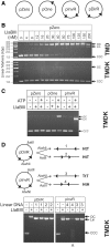
DNA substrate requirements for LlaBIII endonuclease activity. TMD/TMDK refers to the buffers used in the reactions (see main text). (A) Plasmid DNA substrates. The arrowheads represent the orientation of the LlaBIII site as in Figure 1B. (B) Non-specific DNA cleavage by LlaBIII in the absence of K+ ions. Five nanomolars of pZero was incubated with the concentrations of LlaBIII shown for 10 min in either TMD or TMDK with 4 mM ATP and the reactions separated by agarose gel electrophoresis. CCC (covalently closed circular), open circle (OC) and Lin (linear) bands are labelled. (C) dsDNA cleavage requires two sites, but a one-site DNA is nicked. pZero, pOne or pInvR (5 nM) were incubated with 200 nM LlaBIII for 10 min. The reactions separated by agarose gel electrophoresis. (D) dsDNA cleavage requires two sites in inverted HtH repeat. Plasmid DNA, or DNA pre-linearized with AlwNI or AatII as shown (DNA 1–4) (5 nM), was incubated with 200 nM LlaBIII for 10 min with 4 mM ATP. The reactions were separated by agarose gel electrophoresis. Linear DNA contained pairs of sites in head-to-head (HtH) (DNA 4), tail-to-tail (TtT) (DNAs 1 and 2) or tail-to-tail (TtT) (DNA 3) repeat. On linear DNA, only the HtH arrangement produces cleavage (highlighted by the grey arrowhead).
Figure 3.
Figure 3.
Determining the methylation sensitivity of LlaBIII. (A) Plasmid DNA substrates ‘top’ and ‘bottom’ used in the assays in which the sequence of one of the two HtH LlaBIII sites (in bold) overlapped with an EcoAI site (in grey). Adenines methylated by M.EcoAI are underlined. (B) Pre-incubation with EcoAI blocks LlaGI cleavage of the ‘bottom’ DNA but not the ‘top’ DNA. DNA was either used directly or pre-incubated with M.EcoAI or LlaGI in the absence of ATP to allow methylation (indicated by dots above the lanes). The DNA was then incubated with LlaBIII and 4 mM of ATP for 10 min, and the reactions were separated by agarose gel electrophoresis. See main text for full details.
Figure 4.
Figure 4.
Cleavage and translocation kinetics for LlaGI and LlaBIII in TMDK buffer at 25°C. (A) Quantification of cleavage reactions using 5 nM HtH DNA, 200 nM LlaBIII or LlaGI and 4 mM ATP. The reactions were separated by agarose gel electrophoresis, and the CCC (covalently closed circular), open circle (OC) and Lin (linear) bands were quantified. Mean values and standard deviations are shown for at least two repeat experiments. (B) Triplex displacement reactions on linear DNA. Linear triplex DNA was pre-incubated with LlaGI or LlaBIII, and the reaction initiated by mixing with ATP, to give final concentrations of 1 nM DNA, 100 nM LlaBIII or LlaGI and 4 mM ATP. Triplex displacement profiles (left-hand graphs) were fitted to Equation 1 (solid lines) to obtain the estimated lag time (Tlag). The linear relationship between Tlag and d, (the distance between the binding site and triplex—see cartoon) was used to estimate the translocation rate, kstep (right-hand graphs). Mean values and standard deviations at each spacing are from at least three repeat experiments.
Figure 5.
Figure 5.
Mapping the positions of linear DNA cleavage by LlaBIII and LlaGI. (A–C) Maps of the linear DNA are shown, with the yellow sun symbols indicating the 5′-ends labelled with 32-phopshorous. The smaller 255/261-bp fragments were side products of substrate production. Two nanomolars of DNA was incubated with 200 nM of enzyme in either TMD or TMDK buffer, as indicated, with 4 mM of ATP. Aliquots were removed and quenched at the times indicated and then separated by alkaline denaturing agarose gel electrophoresis alongside labelled linear DNA markers. (D) Gel images from A–C were quantified using ImageQuant TL. The DNA marker lanes were used to calibrate and correct the pixel positions to DNA lengths. The y-axis intensity values were calculated as a percentage relative to the uncut FLL bands. The ‘linear range’ of the gel, that is, the range in which fragment sizes can be confidently estimated, is 2.7—1.0 kb (see ‘Materials and Methods’ section).
Figure 6.
Figure 6.
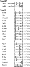
Similar recognition sequence formats in Type ISP and Type IIL RM enzymes. Sequences (3,17,18) are aligned according to the single methylated adenine residue (bold). Cleavage loci are indicated by the brackets (top/bottom strand positions indicated). Variable residues highlighted in grey. The Type IIL enzymes are split into two main phylogenetic branches based on amino acid sequence (18).

References

    1. Wilson GG, Murray NE. Restriction and modification systems. Annu. Rev. Genet. 1991;25:585–627. - PubMed
    1. Labrie SJ, Samson JE, Moineau S. Bacteriophage resistance mechanisms. Nat. Rev. Microbiol. 2010;8:317–327. - PubMed
    1. Smith RM, Diffin FM, Savery NJ, Josephsen J, Szczelkun MD. DNA cleavage and methylation specificity of the single polypeptide restriction-modification enzyme LlaGI. Nucleic Acids Res. 2009;37:7206–7218. - PMC - PubMed
    1. Smith RM, Josephsen J, Szczelkun MD. An Mrr-family nuclease motif in the single polypeptide restriction-modification enzyme LlaGI. Nucleic Acids Res. 2009;37:7231–7238. - PMC - PubMed
    1. Smith RM, Josephsen J, Szczelkun MD. The single polypeptide restriction-modification enzyme LlaGI is a self-contained molecular motor that translocates DNA loops. Nucleic Acids Res. 2009;37:7219–7230. - PMC - PubMed

Publication types

MeSH terms