. 2013 Jan;41(1):159-70.
doi: 10.1097/CCM.0b013e3182676322.

Cecal ligation and puncture-induced murine sepsis does not cause lung injury

Affiliations

Cecal ligation and puncture-induced murine sepsis does not cause lung injury

Kendra N Iskander et al. Crit Care Med. 2013 Jan.

Abstract

Objective: The cause of death in murine models of sepsis remains unclear. The primary purpose of this study was to determine if significant lung injury develops in mice predicted to die after cecal ligation and puncture-induced sepsis compared with those predicted to live.

Design: Prospective, laboratory controlled experiments.

Setting: University research laboratory.

Subjects: Adult, female, outbred Institute of Cancer Research mice.

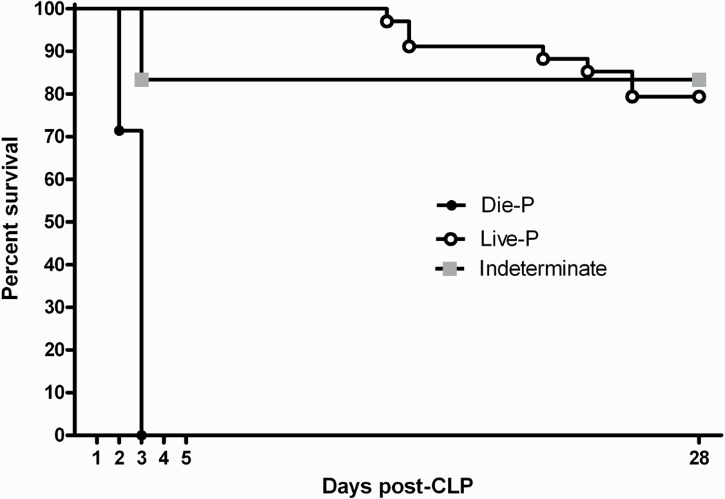
Interventions: Mice underwent cecal ligation and puncture to induce sepsis. Two groups of mice were euthanized at 24 and 48 hrs postcecal ligation and puncture and samples were collected. These mice were further stratified into groups predicted to die (Die-P) and predicted to live (Live-P) based on plasma interleukin-6 levels obtained 24 hrs postcecal ligation and puncture. Multiple measures of lung inflammation and lung injury were quantified in these two groups. Results from a group of mice receiving intratracheal normal saline without surgical intervention were also included as a negative control. As a positive control, bacterial pneumonia was induced with Pseudomonas aeruginosa to cause definitive lung injury. Separate mice were followed for survival until Day 28 postcecal ligation and puncture. These mice were used to verify the interleukin-6 cutoffs for survival prediction.

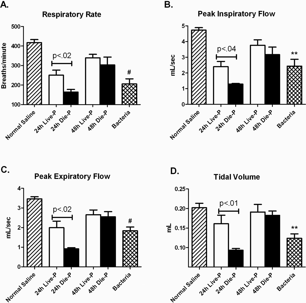
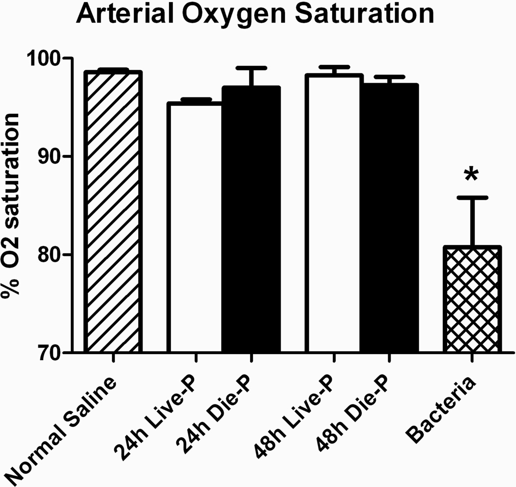
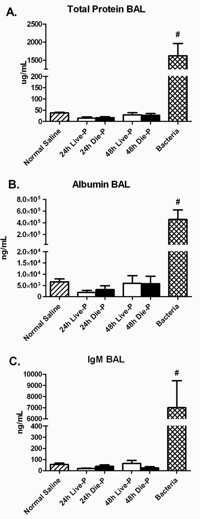
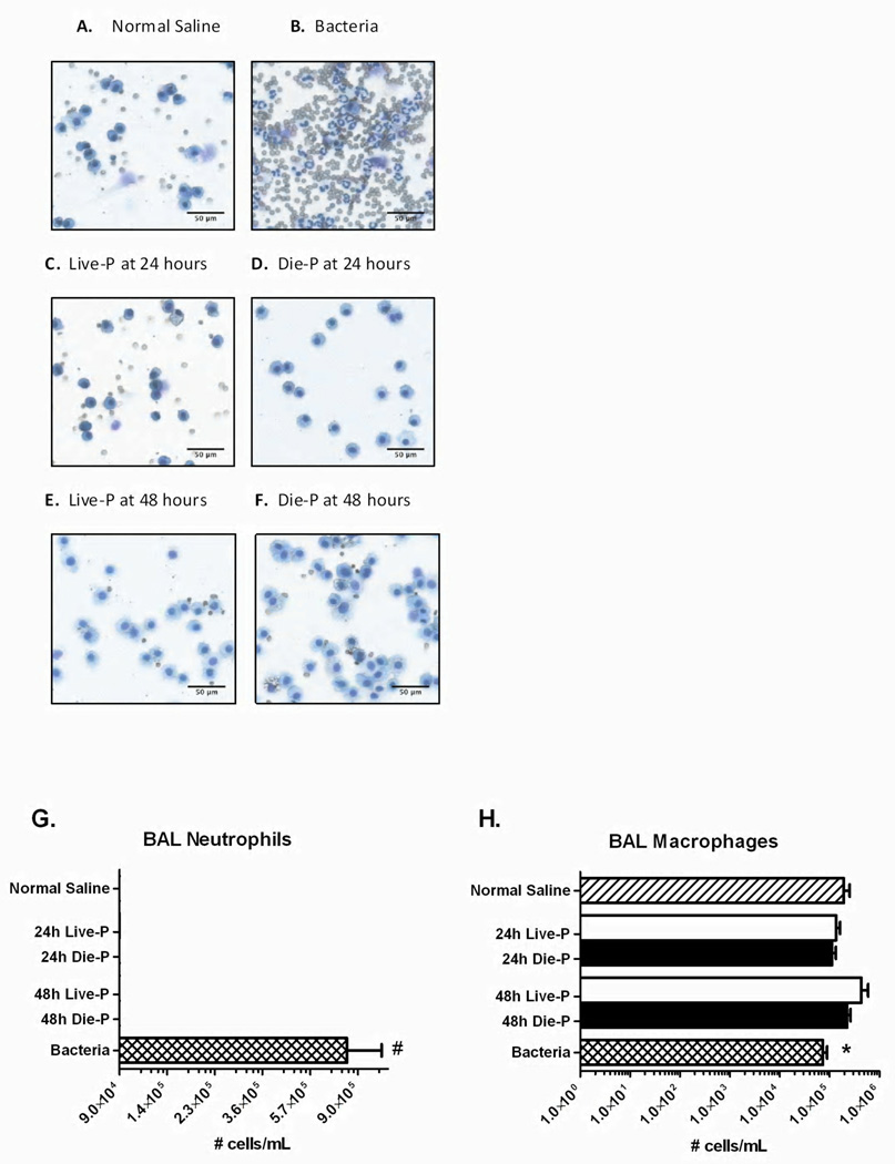
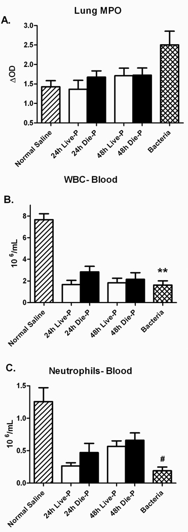
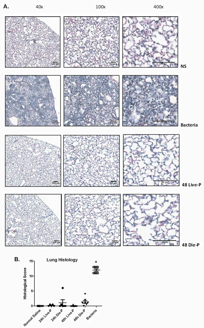
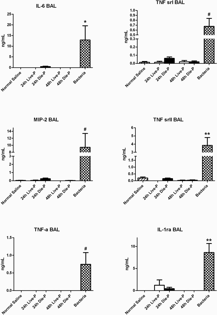
Measurements and main results: After sepsis, both the Die-P and Live-P mice had significantly suppressed measures of respiratory physiology but maintained normal levels of arterial oxygen saturation. Bronchoalveolar lavage levels of pro- and anti-inflammatory cytokines were not elevated in the Die-P mice compared with the Live-P. In addition, there was no increase in the recruitment of neutrophils to the lung, pulmonary vascular permeability, or histological evidence of damage. In contrast, all of these pulmonary injury and inflammatory parameters were increased in mice with Pseudomonas pneumonia.

Conclusions: These data demonstrate that mice predicted to die during sepsis have no significant lung injury. In murine intra-abdominal sepsis, pulmonary injury cannot be considered the etiology of death in the acute phase.


Conflict of interest statement

The authors have not disclosed any potential conflicts of interest

Figures

Figure 1
Figure 1
Confirmation of the accuracy of survival prediction based on IL-6 concentrations in plasma collected at 24 hours after cecal ligation and puncture. Blood was collected by facial bleeding and the plasma IL-6 levels were determined. Die-P mice (n=7) had plasma IL-6 levels > 12ng/ml, Live-P mice (n=34) had levels <1.5 ng/ml while the Indeterminate group (n=6) had IL-6 levels between those values. For predicting survival to day 5, the 24 hour IL-6 levels had 100% specificity and 100% sensitivity using the two distinct discrimination points. All of the Die-P mice succumbed within 72 hours of CLP.
Figure 2
Figure 2
Pulmonary respiratory parameters measured by whole body plethysmography 24 and 48 hours after CLP. Septic mice were separated in predicted to live until 5 days post cecal ligation and puncture, 24h Live-P (n=5) and 48h Live-P (n=7), or die by day 5, 24h Die-P (n=10) and 48h Die-P (n=4), based on the plasma IL-6 cut-offs. Two control groups of mice that were not subjected to cecal ligation and puncture, normal saline (n=8) and bacteria (n=8) were included in the analysis. The respiratory rate (A), peak inspiratory flow (B), and peak expiratory flow (C) were decreased in the septic mice predicted to die compared to the predicted to live group at 24 hours. By 48 hours, respiratory parameters for Live-P and Die-P mice were improving. Despite these changes, the tidal volume was equivalent for CLP mice compared to normal saline (D), except for 24h Die-P whose death was usually imminent. Values are mean ± SEM. There were significant differences between all the groups for all panels, p<.001. The differences between paired groups is indicated by the line. ** = p<0.01, #p<0.001 for bacteria versus normal saline.
Figure 3
Figure 3
Arterial oxygen saturation in early sepsis. Carotid artery puncture was performed to measure oxygen saturation of mice immediately prior to sacrifice 24 or 48 hours in post CLP mice and at 18 hours for control mice receiving P. aeruginosa (Bacteria) or normal saline. Group size was 24h Live-P (n=5), 24h Die-P (n=3); 48h Live-P (n=4); 48h Die=P (n=4), normal saline (n=5), bacteria (n=4). Values are mean ± SEM. There were significant differences between all the groups, p<0.01, but there was no difference between Live-P and Die-P mice at either 24 or 48 hours. * = p<0.05 bacteria versus normal saline.
Figure 4
Figure 4
Pulmonary alveolar protein content 24 and 48 hours after cecal ligation and puncture compared to normal saline and bacteria. Bronchoalveolar lavage was performed and the total protein (Panel A), albumin (Panel B) and IgM (Panel C) levels in the BA fluid were measured for 24h Live-P (n=5), 24h Die-P (n=6); 48h Live-P (n=7); 48h Die=P (n=7), normal saline (n=8), bacteria (n=8). Values are mean ± SEM. There were significant differences between all the groups, p<0.001, but there was no difference between Live-P and Die-P mice at either 24 or 48 hours. #p<0.001 for bacteria versus normal saline.
Figure 5
Figure 5
BAL cells 24 and 48 hours after cecal ligation and puncture. Panels A (normal saline), B (bacteria), C (24h Live-P), D (24h Die-P), E (48h Live-P), F (48h Die-P) show representative cytospin slides from each group of mice (Diff-Quick stain; 1000X magnification). Neutrophil extravasation in the airways did not occur in either mice predicted to die or live septic mice. Panel G shows the total neutrophil count, and Panel H the macrophages recovered from the airways by bronchoalveolar lavage 24h Live-P (n=5), 24h Die-P (n=5); 48h Live-P (n=7); 48h Die=P (n=9), normal saline (n=8), bacteria (n=8). Values are mean ± SEM. There were significant differences between all the groups, p<0.001, but there was no difference between Live-P and Die-P mice at either 24 or 48 hours. * = p<.05, #p<0.001 for bacteria versus normal saline.
Figure 6
Figure 6
Pulmonary neutrophil sequestration and peripheral blood counts 24 and 48 hours after cecal ligation and puncture. Myeloperoxidase (MPO) levels in the lung homogenate were comparable to normal saline for Die-P and Live-P mice in Panel A (24h Live-P (n=5), 24h Die-P (n=5); 48h Live-P (n=6); 48h Die=P (n=7), normal saline (n=8), bacteria (n=8). The peripheral white blood cell and neutrophil counts were lower for the groups of septic mice compared to normal saline in Panel B (24h Live-P (n=5), 24h Die-P (n=5); 48h Live-P (n=6); 48h Die=P (n=7), normal saline (n=8), bacteria (n=8)). Septic mice had a decreased number of neutrophils in the blood 24 and 48 hours after cecal ligation and puncture but this did not translate in an increased recruitment to the lung. Values are mean ± SEM. There was no significant difference between the groups for MPO by Kruskal Wallis. For the other panels there was a significant difference between the groups, p<0.001, but there was no difference between Live-P and Die-P mice at either 24 or 48 hours. ** = p<0.01, #p<0.001 for bacteria versus normal saline.
Figure 7
Figure 7
Histopathological evaluation of the lung 24 and 48 hours after cecal ligation and puncture. In Figure 7 A, images of hematoxylin and eosin stained slides at increasing magnifications are shown for 48 hour Die-P and Live-P mice, as well as a mouse given normal saline or bacteria. No appreciable signs of edema, protein leakage or alveolar neutrophil infiltration could be detected at any magnification in either Live-P or Die-P mice. Histological scoring is shown for each group in Figure 7 B (24h Live-P (n=5), 24h Die-P (n=5); 48h Live-P (n=6); 48h Die-P (n=7), normal saline (n=8), bacteria (n=8). The sections were evaluated in a blinded manner by a board certified pathologist. Values are mean ± SEM. There were significant differences between all the groups, p<0.001, but there was no difference between Live-P and Die-P mice at either 24 or 48 hours. * = p<.05 for bacteria versus normal saline.
Figure 8
Figure 8
Local levels of cytokines at 24 and 48 hours after cecal ligation and puncture. There was no difference in BAL levels of IL-6, MIP-2, TNF-α, TNF sr I, TNF sr II, IL-1rα. BAL group size was 24h Live-P (n=5), 24h Die-P (n=8); 48h Live-P (n=3); 48h Die-P (n=6), normal saline (n=8), bacteria (n=8). Values are mean ± SEM. There were significant differences between all the groups, p<0.001, but there was no difference between Live-P and Die-P mice at either 24 or 48 hours. * = p<0.05, ** = p<.01, #p<0.001 for bacteria versus normal saline.

Comment in

References

    1. Angus DC, Wax RS. Epidemiology of sepsis: an update. Crit Care Med. 2001;29(7 Suppl):S109–S116. - PubMed
    1. Parrillo JE, Parker MM, Natanson C, et al. Septic shock in humans. Advances in the understanding of pathogenesis, cardiovascular dysfunction, and therapy. Ann Intern Med. 1990;113(3):227–242. - PubMed
    1. Levy MM, Fink MP, Marshall JC, et al. 2001 SCCM/ESICM/ACCP/ATS/SIS International Sepsis Definitions Conference. Crit Care Med. 2003;31(4):1250–1256. - PubMed
    1. Martin GS, Mannino DM, Eaton S, et al. The epidemiology of sepsis in the United States from 1979 through 2000. N Engl J Med. 2003;348(16):1546–1554. - PubMed
    1. Vincent JL, Sakr Y, Sprung CL, et al. Sepsis in European intensive care units: results of the SOAP study. Crit Care Med. 2006;34(2):344–353. - PubMed

Publication types