

Direct evidence for encoding of motion streaks in human visual cortex
- PMID: 23222445
- PMCID: PMC3574303
- DOI: 10.1098/rspb.2012.2339
Direct evidence for encoding of motion streaks in human visual cortex
Abstract
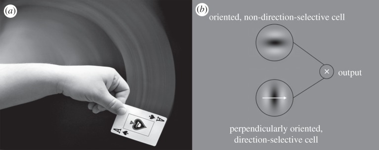
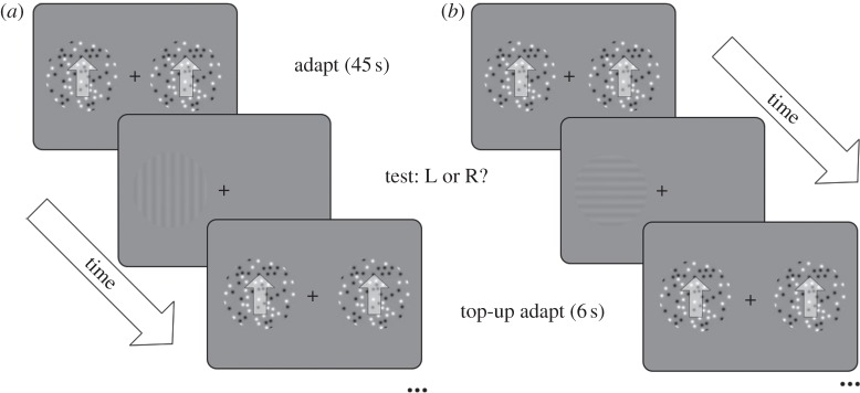
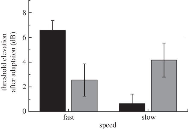
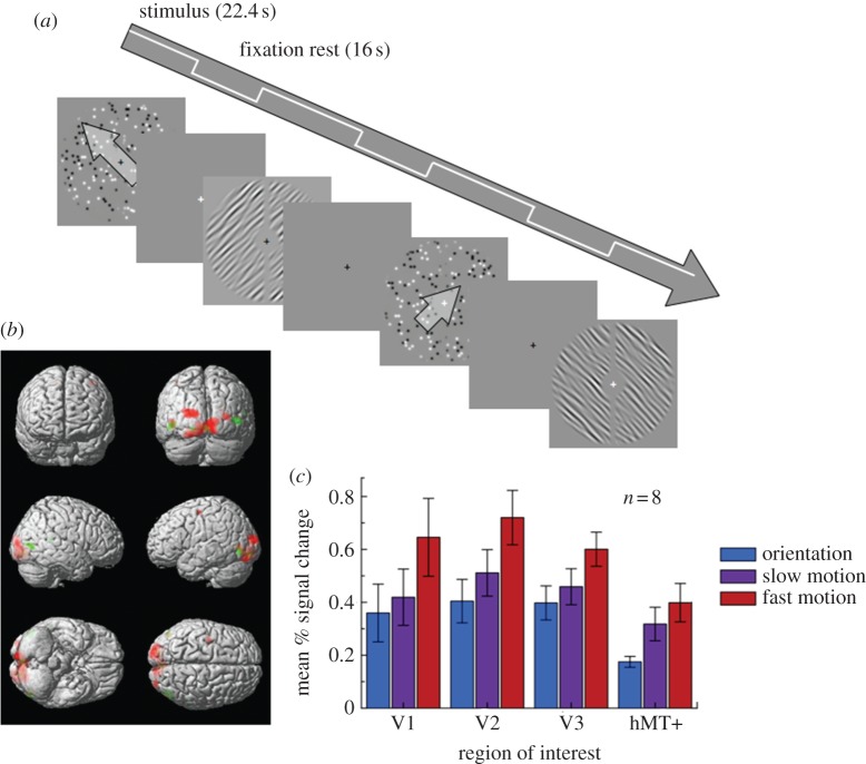
Temporal integration in the visual system causes fast-moving objects to generate static, oriented traces ('motion streaks'), which could be used to help judge direction of motion. While human psychophysics and single-unit studies in non-human primates are consistent with this hypothesis, direct neural evidence from the human cortex is still lacking. First, we provide psychophysical evidence that faster and slower motions are processed by distinct neural mechanisms: faster motion raised human perceptual thresholds for static orientations parallel to the direction of motion, whereas slower motion raised thresholds for orthogonal orientations. We then used functional magnetic resonance imaging to measure brain activity while human observers viewed either fast ('streaky') or slow random dot stimuli moving in different directions, or corresponding static-oriented stimuli. We found that local spatial patterns of brain activity in early retinotopic visual cortex reliably distinguished between static orientations. Critically, a multivariate pattern classifier trained on brain activity evoked by these static stimuli could then successfully distinguish the direction of fast ('streaky') but not slow motion. Thus, signals encoding static-oriented streak information are present in human early visual cortex when viewing fast motion. These experiments show that motion streaks are present in the human visual system for faster motion.
Figures
Similar articles
-
Motion streaks in fast motion rivalry cause orientation-selective suppression.J Vis. 2009 May 14;9(5):10.1-14. doi: 10.1167/9.5.10. J Vis. 2009. PMID: 19757888
-
Temporal integration of movement: the time-course of motion streaks revealed by masking.PLoS One. 2011;6(12):e28675. doi: 10.1371/journal.pone.0028675. Epub 2011 Dec 20. PLoS One. 2011. PMID: 22205961 Free PMC article.
-
Tilt aftereffects and tilt illusions induced by fast translational motion: evidence for motion streaks.J Vis. 2009 Jan 21;9(1):27.1-11. doi: 10.1167/9.1.27. J Vis. 2009. PMID: 19271897
-
Why do parallel cortical systems exist for the perception of static form and moving form?Percept Psychophys. 1991 Feb;49(2):117-41. doi: 10.3758/bf03205033. Percept Psychophys. 1991. PMID: 2017350 Review.
-
The Meaning of Motion Lines?: A Review of Theoretical and Empirical Research on Static Depiction of Motion.Cogn Sci. 2023 Nov;47(11):e13377. doi: 10.1111/cogs.13377. Cogn Sci. 2023. PMID: 37966099 Review.
Cited by
-
Interactions between motion and form processing in the human visual system.Front Comput Neurosci. 2013 May 20;7:65. doi: 10.3389/fncom.2013.00065. eCollection 2013. Front Comput Neurosci. 2013. PMID: 23730286 Free PMC article.
-
Motion direction biases and decoding in human visual cortex.J Neurosci. 2014 Sep 10;34(37):12601-15. doi: 10.1523/JNEUROSCI.1034-14.2014. J Neurosci. 2014. PMID: 25209297 Free PMC article.
-
Integration of motion responses underlying directional motion anisotropy in human early visual cortical areas.PLoS One. 2013 Jun 28;8(6):e67468. doi: 10.1371/journal.pone.0067468. Print 2013. PLoS One. 2013. PMID: 23840711 Free PMC article.
-
Spatial and Temporal Selectivity of Translational Glass Patterns Assessed With the Tilt After-Effect.Iperception. 2021 May 21;12(3):20416695211017924. doi: 10.1177/20416695211017924. eCollection 2021 May-Jun. Iperception. 2021. PMID: 34104382 Free PMC article.
-
Intrasaccadic motion streaks jump-start gaze correction.Sci Adv. 2021 Jul 23;7(30):eabf2218. doi: 10.1126/sciadv.abf2218. Print 2021 Jul. Sci Adv. 2021. PMID: 34301596 Free PMC article.
References
-
- Geisler WS. 1999. Motion streaks provide a spatial code for motion direction. Nature 400, 65–6910.1038/21886 (doi:10.1038/21886) - DOI - DOI - PubMed
-
- Burr D. 1980. Motion smear. Nature 284, 164–16510.1038/284164a0 (doi:10.1038/284164a0) - DOI - DOI - PubMed
-
- Geisler WS, Albrecht DG, Crane AM, Stern L. 2001. Motion direction signals in the primary visual cortex of cat and monkey. Vis. Neurosci. 18, 501–51610.1017/S0952523801184014 (doi:10.1017/S0952523801184014) - DOI - DOI - PubMed
-
- Skottun BC, Zhang J, Grosof DH. 1994. On the directional selectivity of cells in the visual cortex to drifting dot patterns. Vis. Neurosci. 11, 885–89710.1017/S0952523800003849 (doi:10.1017/S0952523800003849) - DOI - DOI - PubMed
-
- Mante V, Carandini M. 2005. Mapping of stimulus energy in primary visual cortex. J. Neurophys. 94, 788–79810.1152/jn.01094.2004 (doi:10.1152/jn.01094.2004) - DOI - DOI - PubMed
Publication types
MeSH terms
Grants and funding
LinkOut - more resources
Full Text Sources
Other Literature Sources
