. 2013 Jan;20(1):111-8.
doi: 10.1038/nsmb.2462. Epub 2012 Dec 9.

FMNL3 FH2-actin structure gives insight into formin-mediated actin nucleation and elongation

Affiliations

FMNL3 FH2-actin structure gives insight into formin-mediated actin nucleation and elongation

Morgan E Thompson et al. Nat Struct Mol Biol. 2013 Jan.

Abstract

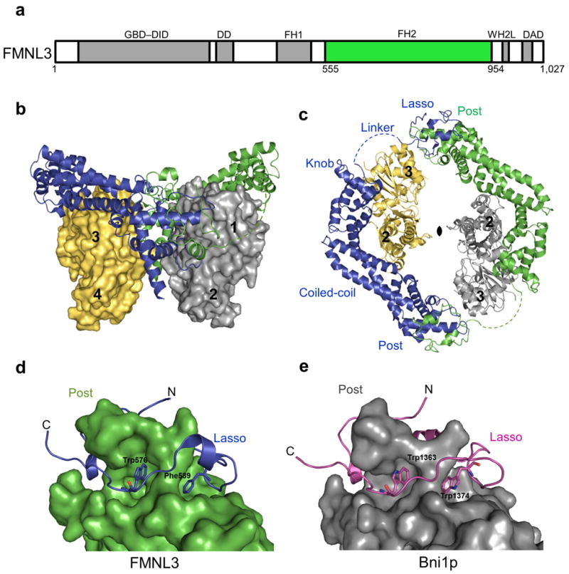
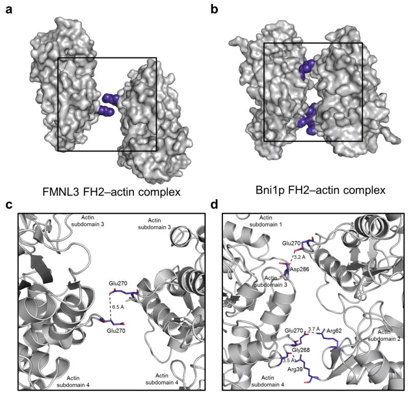
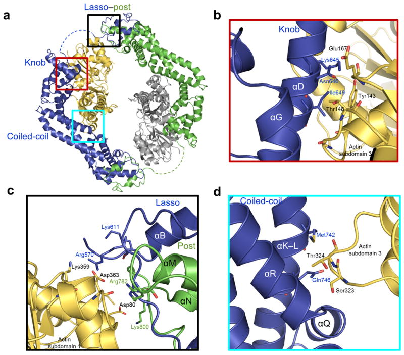
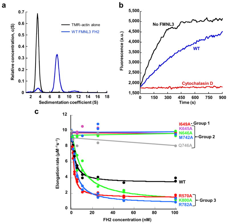
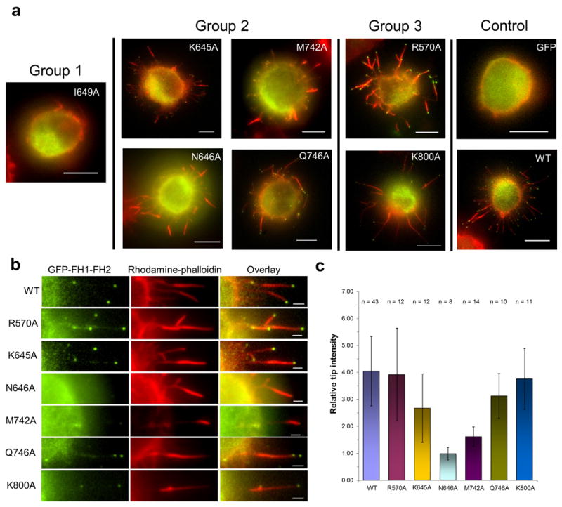
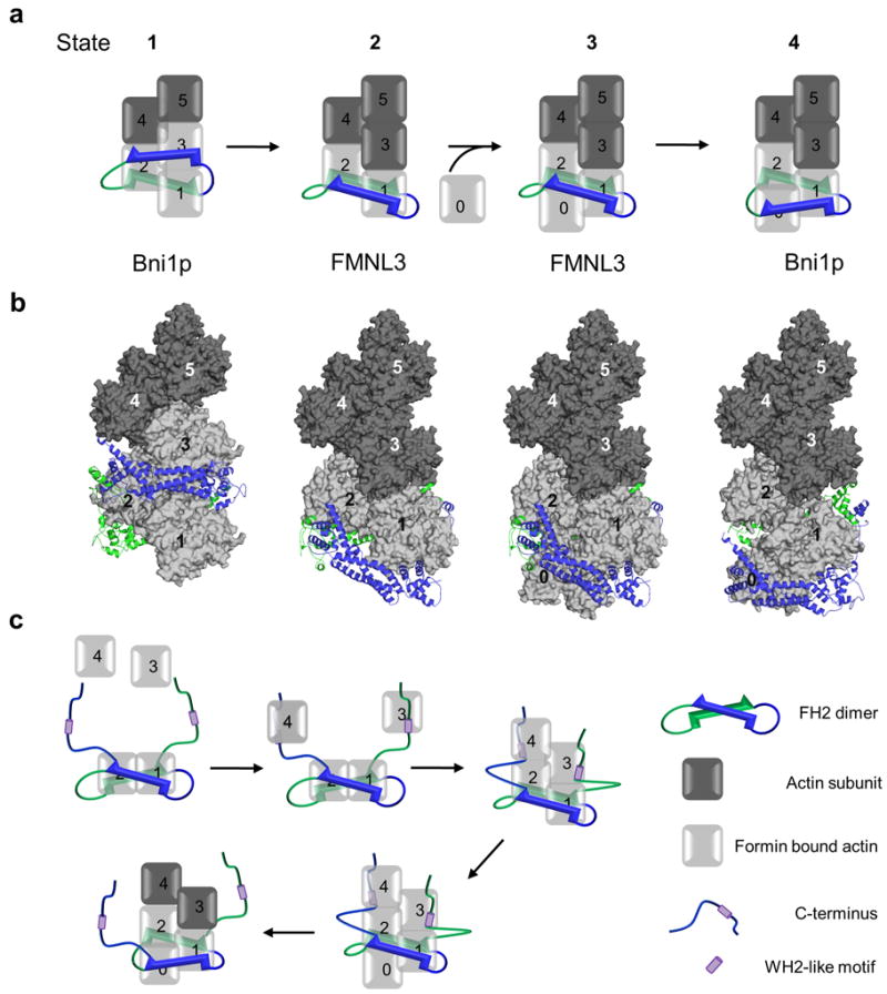
Formins are actin-assembly factors that act in a variety of actin-based processes. The conserved formin homology 2 (FH2) domain promotes filament nucleation and influences elongation through interaction with the barbed end. FMNL3 is a formin that induces assembly of filopodia but whose FH2 domain is a poor nucleator. The 3.4-Å structure of a mouse FMNL3 FH2 dimer in complex with tetramethylrhodamine-actin uncovers details of formin-regulated actin elongation. We observe distinct FH2 actin-binding regions; interactions in the knob and coiled-coil subdomains are necessary for actin binding, whereas those in the lasso-post interface are important for the stepping mechanism. Biochemical and cellular experiments test the importance of individual residues for function. This structure provides details for FH2-mediated filament elongation by processive capping and supports a model in which C-terminal non-FH2 residues of FMNL3 are required to stabilize the filament nucleus.


Conflict of interest statement

Competing Financial Interests

The authors declare no competing financial interests.

Figures

Figure 1
Figure 1. Structure of FMNL3 FH2 domain bound to TMR-actin
(a) Domain organization of FMNL3 (1027 amino acids) with the GTPase binding domain (GBD), diaphanous inhibitory domain (DID), dimerization domain (DD), formin homology 1 (FH1), WASP-homology 2-like (WH2L) motif, and diaphanous autoinhibitory domain (DAD) colored in grey and the formin homology 2 (FH2) domain (555-954) in green. (b-c) Overall structure of the FMNL3 FH2-actin biological unit with an FH2 dimer (blue and green) bound to two actin subunits (yellow and grey). Actin subdomains are numbered. (c) Top view of the complex with the 2-fold non-crystallographic symmetry axis indicated. The FH2 subdomains are labeled with the disordered linker represented as a dashed line. (d-e) FH2 domain dimerization through the lasso and post subdomains of FMNL3 and Bni1p (1Y64) respectively. Conserved hydrobphobic residues in the lasso of FMNL3 (W576, F589) and Bni1p (W1363, W1374) aid in dimerization.
Figure 2
Figure 2. Distinct actin-actin interactions in the FMNL3 FH2-actin complex compared to the Bni1p-actin complex
(a-b) Orientation of two actin subunits (gray surface) from the FMNL3 complex (panel a) and the Bni1p complex (panel b). Actin-actin closest contacts are highlighted in blue spheres and boxed with enlargement in panels c and d. The distances between the closest sidechain atoms are indicated by dotted lines to illustrate the difference in actin organization within the FMNL3 and Bni1p FH2-actin complexes.
Figure 3
Figure 3. FH2-actin binding interfaces
(a) Location of the post, knob, and coiled-coil actin binding regions are boxed in black, red, and cyan respectively. (b-d) Regions boxed in a highlight interacting residues in the FH2 domain binding interface within 3.5 Å of actin for the knob (panel b), lasso/post (panel c), and coiled-coil (panel d) subdomains of FMNL3. Coloring is the same as in Figure 1.
Figure 4
Figure 4. Effects of FMNL3 mutations on actin filament elongation
(a) Velocity analytical ultracentrifugation sedimentation profiles for 4.7 μM TMR-actin alone (black curve) and in the presence of 9.7 μM wild-type FMNL3 FH2 (WT) (blue curve) monitored at 557 nm, the peak absorbance wavelength for TMR-actin. TMR-actin alone sediments with a sedimentation coefficient of 3.4 S while a peak shift to 7.4 S is observed in the presence of the WT FH2 domain that form a stable complex with TMR-actin in solution. (b) Pyrene-actin elongation assay performed with 0.5 μM actin monomers (25% pyrene) from filament seeds stabilized with phalloidin. Actin elongation (black curve) is partially inhibited by 100 nM WT FMNL3 FH2 (blue curve) and completely inhibited b 40 nM cytochalasin D (red curve). (c) Concentration dependence of filament elongation inhibition by FMNL3 mutants. FH2 mutant actin binding (data found in Table 2) and elongation data were used to separate FH2 mutants into Group 1 (no TMR-actin binding, no elongation inhibition), Group 2 (binds TMR-actin, no elongation inhibition), or Group 3 (binds TMR-actin, enhanced elongation inhibition). Elongation rates are calculated from the slope of the first 10% of the time course and plotted versus concentration of FH2 domain.
Figure 5
Figure 5. Effects of FMNL3 mutants on formation of filopodia in Jurket cells
Expression of GFP alone or FH1-FH2 GFP-fusion constructs of FMNL3 (WT or Group 1, 2, or 3 mutants) in Jurkat cells for 6 hours followed by formaldehyde fixation and actin filament staining with rhodamine phalloidin. (a) Surface formation of filopodia was observed for all FMNL3 mutant constructs except for I649A (Scale bar is 5 μm). (b) Enlarged image of filopodia for WT and each of the mutant constructs. N646A and M742A do not display the characteristic GFP localization at the tips of the filopodia as seen for WT. Scale bar is 1 μm. (c) Quantification GFP intensity at tips of individual filopodia shows that intensity is reduced significantly for N646A and M742A. Error bars represent standard deviation (n > 10 from two experiments).
Figure 6
Figure 6. Model for FMNL3 mediated actin assembly
(a,b) Model of a 4-step elongation mechanism in which the FH2 dimer moves processively with the growing actin filament. An FH2 dimer (blue and green) forms a complex with either two (FMNL3 complex) or three (Bni1p complex) actin subunits at the barbed end (light grey surface). Numbered actin subunits aligned in a filament based on the Holmes model are shown in dark gray (surface). The translocation one subunit of the FH2 dimer (blue) is modeled while the second subunit (green) remains stationary. Panel a shows a schematic representation of the structure based filament model in b with the barbed ends of the actin filament oriented down. See Online Methods for a detailed description of the model design. (c) Model describing FMNL3 FH2-C mediated actin filament nucleation activity. The C-terminus alone is capable of binding actin monomers through a WH2-like motif allowing for the formation of an actin filament nucleus where the FH2 domain binds two actin subunits (as seen in the crystal structure) and the WH2-like motifs position additional monomers in close proximity to the FH2 bound actin subunits in a manner that favors nucleation. The WH2-like motifs then quickly release from their bound monomers in order for the FH2-mediated processive elongation to occur.

References

    1. Pollard TD, Cooper Ja. Actin, a central player in cell shape and movement. Science. 2009;326:1208–12. - PMC - PubMed
    1. Chang F, Drubin D, Nurse P. Cdc12P, a Protein Required for Cytokinesis in Fission Yeast, Is a Component of the Cell Division Ring and Interacts With Profilin. The Journal of cell biology. 1997;137:169–82. - PMC - PubMed
    1. Goode BL, Eck MJ. Mechanism and function of formins in the control of actin assembly. Annual review of biochemistry. 2007;76:593–627. - PubMed
    1. Xu Y, et al. Crystal structures of a Formin Homology-2 domain reveal a tethered dimer architecture. Cell. 2004;116:711–23. - PubMed
    1. Otomo T, et al. Structural basis of actin filament nucleation and processive capping by a formin homology 2 domain. Nature. 2005;433:488–94. - PubMed

MeSH terms

Associated data