. 2012 Dec 15;94(11):1103-10.
doi: 10.1097/TP.0b013e318270f3c0.

Cytotoxic effector function of CD4-independent, CD8(+) T cells is mediated by TNF-α/TNFR

Affiliations

Cytotoxic effector function of CD4-independent, CD8(+) T cells is mediated by TNF-α/TNFR

Jason M Zimmerer et al. Transplantation. .

Abstract

Background: Liver parenchymal cell allografts initiate both CD4-dependent and CD4-independent, CD8(+) T cell-mediated acute rejection pathways. The magnitude of allospecific CD8(+) T cell in vivo cytotoxic effector function is maximal when primed in the presence of CD4(+) T cells. The current studies were conducted to determine if and how CD4(+) T cells might influence cytotoxic effector mechanisms.

Methods: Mice were transplanted with allogeneic hepatocytes. In vivo cytotoxicity assays and various gene-deficient recipient mice and target cells were used to determine the development of Fas-, TNF-α-, and perforin-dependent cytotoxic effector mechanisms after transplantation.

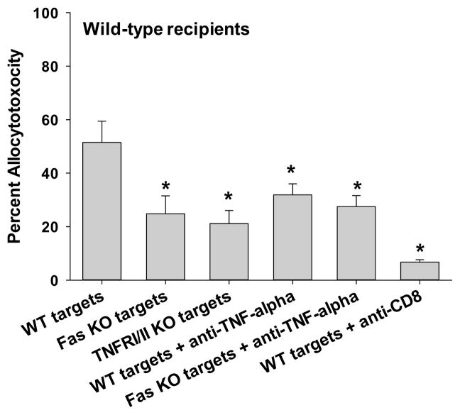
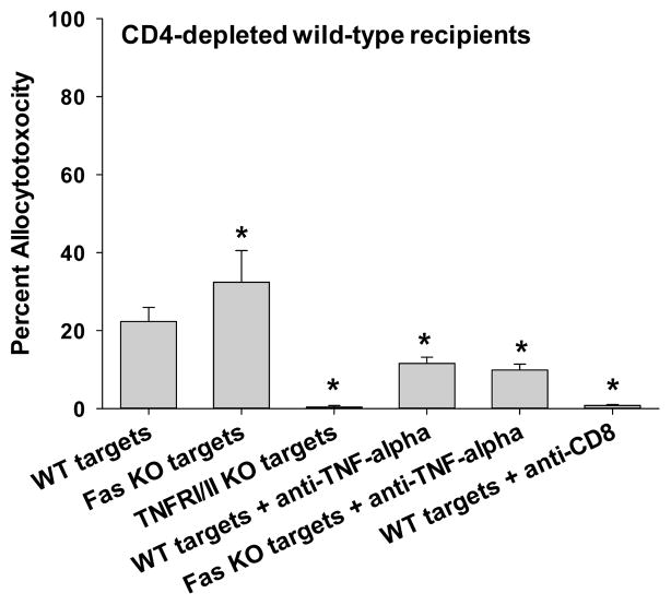
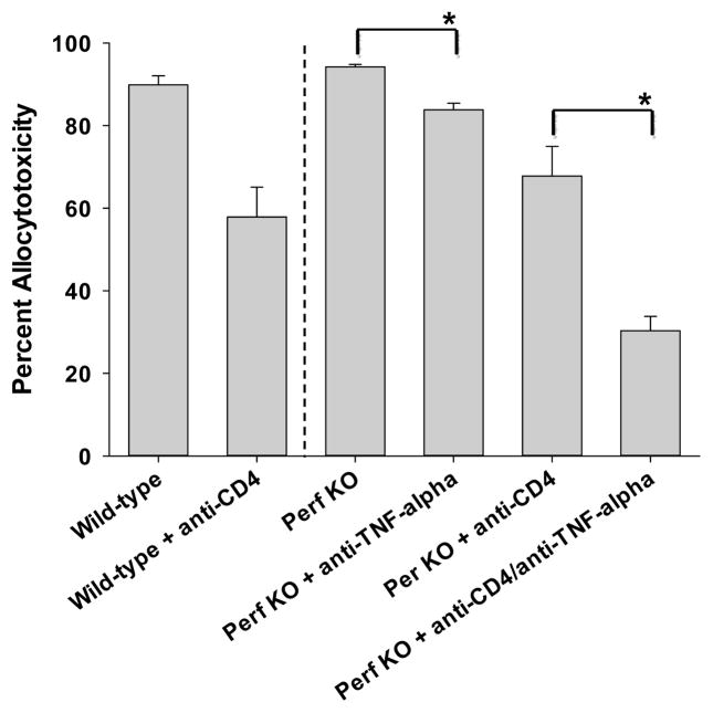
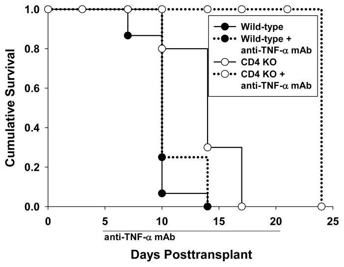
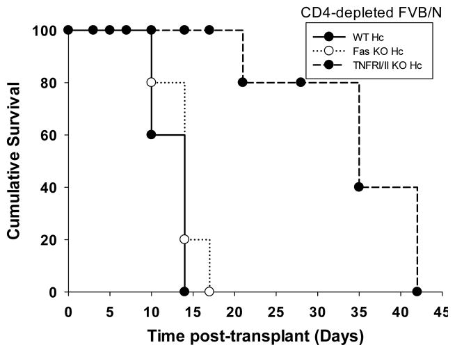
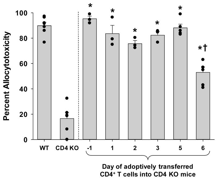
Results: CD8(+) T cells maturing in CD4-sufficient hepatocyte recipients develop multiple (Fas-, TNF-α-, and perforin-mediated) cytotoxic mechanisms. However, CD8(+) T cells, maturing in the absence of CD4(+) T cells, mediate cytotoxicity and transplant rejection that is exclusively TNF-α/TNFR-dependent. To determine the kinetics of CD4-mediated help, CD4(+) T cells were adoptively transferred into CD4-deficient mice at various times posttransplant. The maximal influence of CD4(+) T cells on the magnitude of CD8-mediated in vivo allocytotoxicityf occurs within 48 hours.

Conclusion: The implication of these studies is that interference of CD4(+) T cell function by disease or immunotherapy will have downstream consequences on both the magnitude of allocytotoxicity as well as the cytotoxic effector mechanisms used by allospecific CD8(+) cytolytic T cells.


Conflict of interest statement

Conflict of Interest

The authors of this manuscript have no conflicts of interest to disclose.

Figures

Figure 1
Figure 1. In vivo CD8-dependent cytotoxicity is mediated by FasL and TNF-α in wild-type recipients but only TNF-α in CD4-deficient recipients
C57BL/6 hepatocytes (H-2b) were transplanted into FVB/N (H-2q) recipients that were untreated (CD4-dependent CD8-mediated allocytotoxicity) or CD4-depleted (CD4-independent CD8-mediated allocytotoxicity) prior to transplantation. In vivo cytotoxicity was performed with different allogeneic targets: wild-type (susceptible to all mechanisms of cytotoxicity, wt); Fas mutant (resistant to FasL-mediated killing); or TNFR I/II KO (resistant to TNF-α-mediated killing) (all H-2b). A) In wild-type hosts, cytotoxic activity was significantly decreased against both Fas mutant (25±7%, n=5; p=0.003) and TNFR I/II KO targets (21±5%, n=5; p=0.01) relative to wild-type (52±8%, n=7) targets. B) In contrast, CD4-deficient hosts demonstrated similar killing of Fas mutant (32±8%, n=3; p>0.05) as compared to wild-type targets (22±4%, n=4), but significantly reduced killing of TNFR I/II KO targets (0±0%, n=5; p=0.0007). Similar results were observed in wild-type and CD4-deficient recipients when utilizing anti-TNF-α mAb treatment (400μg, i.p., day 5, 6) to block this effector mechanism (n≥4). Further downregulation of cytotoxicity was not observed in wild-type recipients when both FasL and TNF-α mechanisms were blocked (28±4%, n=4; Fas mutant targets and recipients treated with anti-TNF-α mAb) in comparison to control conditions of Fas mutant targets alone or TNF-α inhibition alone. Cytotoxicity was reduced or eliminated when CD8+ T cells were depleted (100 μg, day 5, 6) prior to the in vivo cytotoxicity assay in both untreated and CD4-depleted recipients, respectively (p<0.0009 for both). Significance is depicted by a “*” (p<0.05).
Figure 2
Figure 2. In vivo CD8+ T cell cytotoxic effector function does not require perforin
Wild-type (C57BL/6, H-2b) and perforin KO (H-2b) mice were transplanted with FVB/N (H-2q) hepatocytes. Cohorts of the recipients were depleted of CD4+ T cells prior to transplant (days -4 and -2). In vivo cytotoxicity was performed with wild-type (FVB/N) allogeneic targets. Perforin KO recipients (94±1%, n=3) exhibit similar cytotoxicity to wild-type mice (90±2%, n=10). Anti-TNF-α mAb reduces cytotoxicity in perforin KO recipients (84±2%, n=3; p=0.006). CD4-depleted wild-type (58±7%; n=4) and CD4-depleted perforin KO recipients (68±6%, n=3) exhibit similar cytotoxicity. However, CD4-depleted perforin KO recipients treated with anti-TNF-α mAb have significantly reduced cytotoxicity (30±4%, n=3) as compared to CD4-depleted perforin KO recipients (68±6%; p=0.01, as denoted by “*”).
Figure 3
Figure 3. Hepatocellular allograft survival is enhanced following TNF-α inhibition in CD4-deficient recipient mice
A) FVB/N hepatocyte allografts were transplanted into wild-type and CD4 KO (H-2b) hosts. On day 5 posttransplant, a cohort of recipients were treated with anti-TNF-α mAb (n=4). Some recipients were left untreated for comparison. Wild-type recipients rejected hepatocytes with a mean survival time of 10 days posttransplantation in both untreated (n=15) or anti-TNF-α mAb treated (n=4) recipients. In CD4 KO recipients, treatment with anti-TNF-α mAb (discontinued on day 20) resulted in significant enhancement of hepatocyte survival (MST=24 days; n=4) as compared to untreated mice (MST=14; p=0.002; n=10). B) To further determine the role of TNF-α/TNFR interactions, additional studies were performed with wild-type, Fas mutant, or TNFRI/II KO hepatocytes. Hepatocyte survival in untreated or CD4-depleted FVB/N recipients were monitored over time. CD4-sufficient recipients rejected wild-type, Fas mutant and TNFRI/II KO hepatocytes with median survival time of 10, 14, and 10 days, respectively (not shown). CD4-depleted recipients rejected wild-type and Fas mutant hepatocytes with a median survival time of 14 days posttransplant (n=3 for both). In contrast CD4-depleted recipients demonstrate prolonged survival of TNFRI/II KO donor hepatocytes (MST=35 days, n=3; p=0.003).
Figure 4
Figure 4. CD4+ T cells are not required at the time of transplant to develop maximal magnitude of CD8-mediated cytotoxic effector function
Allogeneic hepatocytes from FVBN mice were transplanted into either wild-type or CD4 KO mice on day 0. 10×106 CD4+ T cells were adoptively transferred on day −1, +1, +2, +3, +5, or +6 (n≥3 for all time points) into CD4 KO mice. The in vivo cytotoxicity assay was performed on day 7. The allocytotoxicity detected in all the CD4 KO recipient mice receiving adoptive transfer of CD4+ T cells was significantly higher than that of CD4 KO mice without adoptive transfer (collectively, p<0.0002, as denoted by “*”; “dots” represent individual samples). However, recipient CD4 KO mice receiving adoptive transfer of CD4+ T cells on day +6 posttransplant exhibited significantly less cytotoxicity than control wild-type recipients (p<0.0001, as denoted by “†”).

Similar articles

Cited by

References

    1. Barber DL, Wherry EJ, Ahmed R. Cutting edge: rapid in vivo killing by memory CD8 T cells. J Immunol. 2003;171 (1):27. - PubMed
    1. Strehlau J, Pavlakis M, Lipman M, et al. Quantitative detection of immune activation transcripts as a diagnostic tool in kidney transplantation. Proc Natl Acad Sci U S A. 1997;94 (2):695. - PMC - PubMed
    1. Halloran PF, Urmson J, Ramassar V, et al. Lesions of T-cell-mediated kidney allograft rejection in mice do not require perforin or granzymes A and B. Am J Transplant. 2004;4 (5):705. - PubMed
    1. O’Connell PJ, Pacheco-Silva A, Nickerson PW, et al. Unmodified pancreatic islet allograft rejection results in the preferential expression of certain T cell activation transcripts. J Immunol. 1993;150 (3):1093. - PubMed
    1. Han D, Xu X, Baidal D, et al. Assessment of cytotoxic lymphocyte gene expression in the peripheral blood of human islet allograft recipients: elevation precedes clinical evidence of rejection. Diabetes. 2004;53 (9):2281. - PubMed

Publication types

MeSH terms