Case Reports
. 2013 Jan;45(1):83-7.
doi: 10.1038/ng.2497. Epub 2012 Dec 9.

Recessive mutations in EPG5 cause Vici syndrome, a multisystem disorder with defective autophagy

Affiliations
Case Reports

Recessive mutations in EPG5 cause Vici syndrome, a multisystem disorder with defective autophagy

Thomas Cullup et al. Nat Genet. 2013 Jan.

Abstract

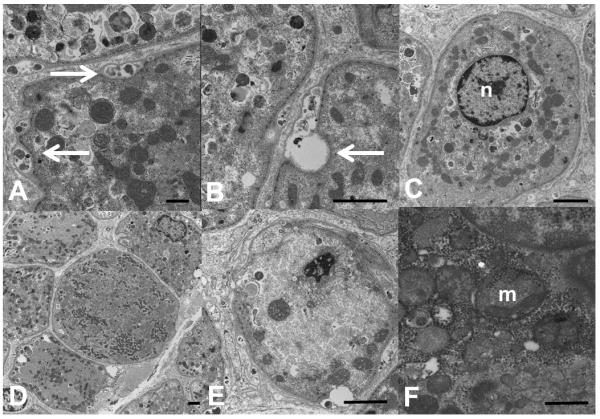
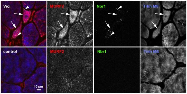
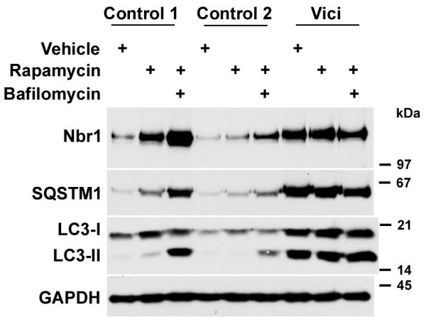
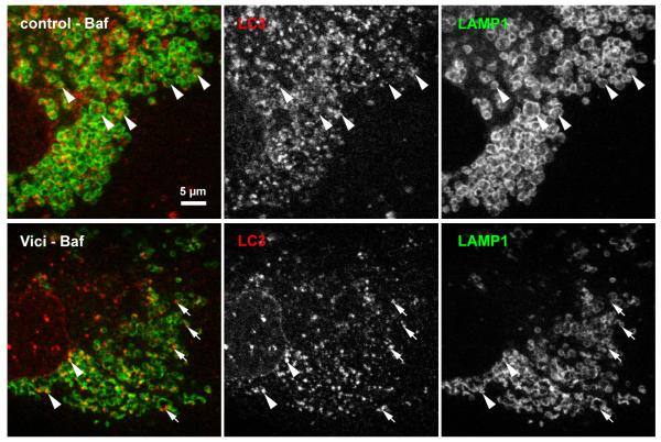
Vici syndrome is a recessively inherited multisystem disorder characterized by callosal agenesis, cataracts, cardiomyopathy, combined immunodeficiency and hypopigmentation. To investigate the molecular basis of Vici syndrome, we carried out exome and Sanger sequence analysis in a cohort of 18 affected individuals. We identified recessive mutations in EPG5 (previously KIAA1632), indicating a causative role in Vici syndrome. EPG5 is the human homolog of the metazoan-specific autophagy gene epg-5, encoding a key autophagy regulator (ectopic P-granules autophagy protein 5) implicated in the formation of autolysosomes. Further studies showed a severe block in autophagosomal clearance in muscle and fibroblasts from individuals with mutant EPG5, resulting in the accumulation of autophagic cargo in autophagosomes. These findings position Vici syndrome as a paradigm of human multisystem disorders associated with defective autophagy and suggest a fundamental role of the autophagy pathway in the immune system and the anatomical and functional formation of organs such as the brain and heart.


Figures

Figure 1
Figure 1. Ultrastructural abnormalities in Vici syndrome
Muscle biopsy from Patient 4.1, electron microscopy, transverse sections. In many fibres, there is material between layers of basal lamina (A, arrows, bar = 500 nm), or overt exocytic vacuoles (B, arrow, bar = 2 microns). Some fibres show a single centralized nucleus (C, bar = 2 microns). Mitochondria are of variable size and distribution. In some fibres, they form a loop around the nucleus in the periphery of the fibre resembling a necklace (C), in others they form clusters (D, bar = 2 microns). Appearance of cristae is often abnormal (E, bar = 2 microns; F, bar = 500 nm). n = nucleus, m = mitochondrion.
Figure 2
Figure 2. Accumulation of Nbr1-positive puncta in skeletal muscle of Vici patient
Transverse sections from muscle biopsies from a normal control (bottom panel) and Patient 3.1 (top panel) were stained with monoclonal antibody against Nbr1 (green channel), polyclonal MURF2 antibody (red channel), and counterstained with the anti-titin M-band antibody T-M8ra (blue channel). Accumulation of Nbr1 in puncta (autophagosomes, arrowheads in top panel), and of MURF2, as well as fibre inhomogeneity with marked fibre atrophy characterise Vici muscle. Numerous fibres of very small cross-sectional area (arrows in top panel) with high content of MURF2 and Nbr1 puncta are frequently seen. Scale bar: 10 μm.
Figure 3
Figure 3. Autophagy is blocked at a late stage in Vici syndrome
Accumulation of autophagy adaptors Nbr1, p62/SQSTM1 and the phagophore membrane component LC3- is induced in control and Vici-patient fibroblasts (Patient 4.1) by 12 h treatment with rapamycin, or dual treatment with rapamycin and bafilomycin. In control cells, rapamycin induces slight accumulation of unprocessed LC3-I and processed LC3-II, Nbr1 and p62/SQSTM1; dual induction of autophagy by rapamycin and block of autophagosomal degradation by bafilomycin leads to marked accumulation of all autophagosome components. In Vici patient fibroblasts, strong baseline accumulation of all components is visible that is largely unresponsive to both drugs.
Figure 4
Figure 4. Fusion of LC3-positive puncta with lysosomes in Vici syndrome
In control fibroblasts subjected to 6h bafilomycin treatment, lysosomal structures were detected by staining with monoclonal anti-LAMP1. Numerous LC3-positive autophagosomes are found engulfed by the LAMP1-positive vesicular structures (arrowheads). In contrast, Vici patients fibroblasts consistently show smaller LC3-positive puncta that only sporadically colocalise with LAMP1, with many isolated LC3-positive puncta (arrows). Note that LC3 signal in Vici cells occurs mostly at the rim of LAMP1-positive structures, not centrally. Scale bar: 5 μm

References

    1. Vici CD, et al. Agenesis of the corpus callosum, combined immunodeficiency, bilateral cataract, and hypopigmentation in two brothers. Am. J. Med. Genet. 1988;29:1–8. - PubMed
    1. del Campo M, et al. Albinism and agenesis of the corpus callosum with profound developmental delay: Vici syndrome, evidence for autosomal recessive inheritance. Am. J. Med. Genet. 1999;85:479–85. - PubMed
    1. Chiyonobu T, et al. Sister and brother with Vici syndrome: agenesis of the corpus callosum, albinism, and recurrent infections. Am. J. Med. Genet. 2002;109:61–6. - PubMed
    1. Miyata R, et al. Sibling cases of Vici syndrome: sleep abnormalities and complications of renal tubular acidosis. Am. J. Med. Genet. A. 2007;143:189–94. - PubMed
    1. McClelland V, et al. Vici syndrome associated with sensorineural hearing loss and evidence of neuromuscular involvement on muscle biopsy. Am. J. Med. Genet. A. 2010;152A:741–7. - PubMed

Publication types

Supplementary concepts