

Copy number variation analysis implicates the cell polarity gene glypican 5 as a human spina bifida candidate gene
- PMID: 23223018
- PMCID: PMC3578410
- DOI: 10.1093/hmg/dds515
Copy number variation analysis implicates the cell polarity gene glypican 5 as a human spina bifida candidate gene
Abstract
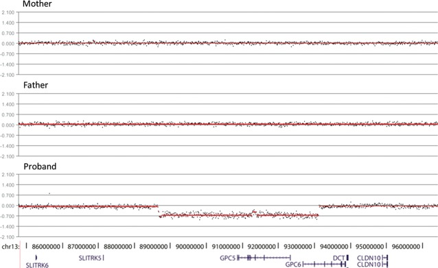
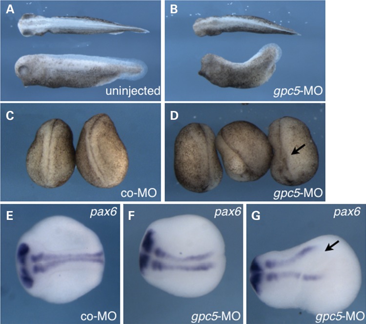
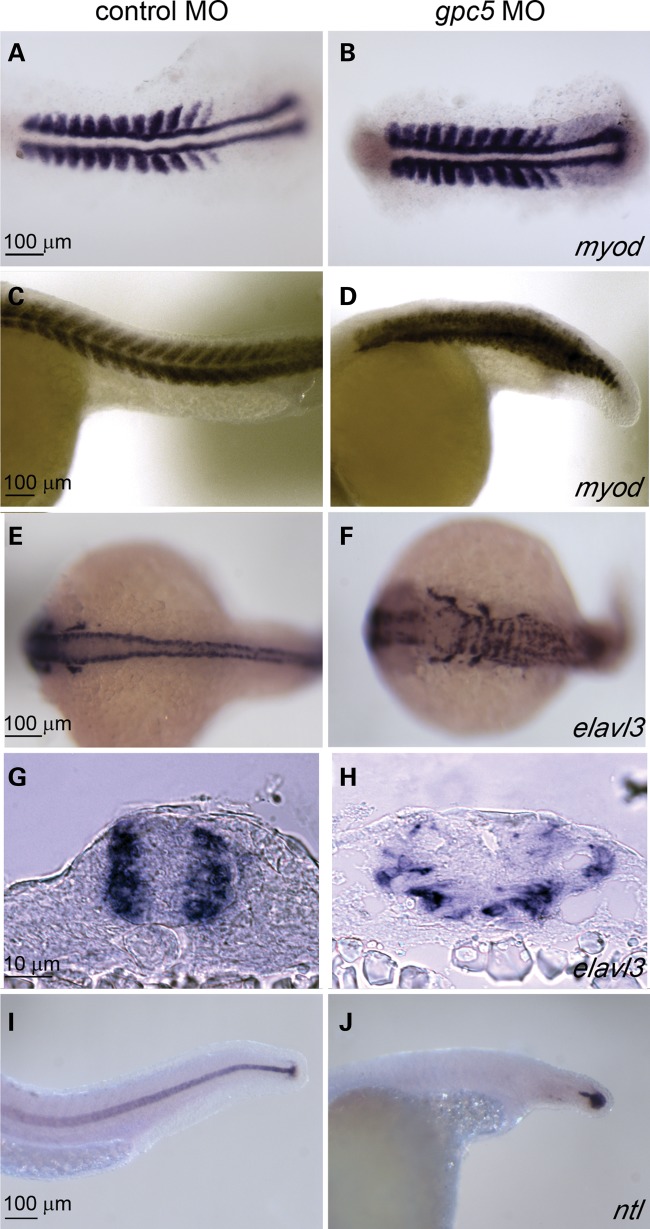
Neural tube defects (NTDs) are common birth defects of complex etiology. Family and population-based studies have confirmed a genetic component to NTDs. However, despite more than three decades of research, the genes involved in human NTDs remain largely unknown. We tested the hypothesis that rare copy number variants (CNVs), especially de novo germline CNVs, are a significant risk factor for NTDs. We used array-based comparative genomic hybridization (aCGH) to identify rare CNVs in 128 Caucasian and 61 Hispanic patients with non-syndromic lumbar-sacral myelomeningocele. We also performed aCGH analysis on the parents of affected individuals with rare CNVs where parental DNA was available (42 sets). Among the eight de novo CNVs that we identified, three generated copy number changes of entire genes. One large heterozygous deletion removed 27 genes, including PAX3, a known spina bifida-associated gene. A second CNV altered genes (PGPD8, ZC3H6) for which little is known regarding function or expression. A third heterozygous deletion removed GPC5 and part of GPC6, genes encoding glypicans. Glypicans are proteoglycans that modulate the activity of morphogens such as Sonic Hedgehog (SHH) and bone morphogenetic proteins (BMPs), both of which have been implicated in NTDs. Additionally, glypicans function in the planar cell polarity (PCP) pathway, and several PCP genes have been associated with NTDs. Here, we show that GPC5 orthologs are expressed in the neural tube, and that inhibiting their expression in frog and fish embryos results in NTDs. These results implicate GPC5 as a gene required for normal neural tube development.
Figures
References
-
- Copp A.J., Greene N.D., Murdoch J.N. The genetic basis of mammalian neurulation. Nat. Rev. Genet. 2003;4:784–793. doi:10.1038/nrg1181. - DOI - PubMed
-
- Wald N.J., Bower C. Folic acid, pernicious anaemia, and prevention of neural tube defects. Lancet. 1994;343:307. doi:10.1016/S0140-6736(94)91156-8. - DOI - PubMed
-
- Wald N.J., Law M.R., Jordan R. Folic acid food fortification to prevent neural tube defects. Lancet. 1998;351:834–835. doi:10.1016/S0140-6736(05)78966-2. - DOI - PubMed
-
- Wald N.J., Law M.R., Morris J.K., Wald D.S. Quantifying the effect of folic acid. Lancet. 2001;358:2069–2073. doi:10.1016/S0140-6736(01)07104-5. - DOI - PubMed
-
- Harris M.J., Juriloff D.M. An update to the list of mouse mutants with neural tube closure defects and advances toward a complete genetic perspective of neural tube closure. Birth Defects Res. A. 2010;88:653–669. doi:10.1002/bdra.20676. - DOI - PubMed
Publication types
MeSH terms
Substances
Grants and funding
LinkOut - more resources
Full Text Sources
Medical
