. 2013 Feb;12(2):244-53.
doi: 10.1128/EC.00214-12. Epub 2012 Dec 7.

The zinc cluster protein Sut1 contributes to filamentation in Saccharomyces cerevisiae

Affiliations

The zinc cluster protein Sut1 contributes to filamentation in Saccharomyces cerevisiae

Helen A Foster et al. Eukaryot Cell. 2013 Feb.

Abstract

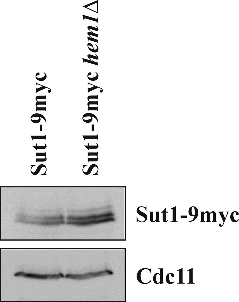
Sut1 is a transcriptional regulator of the Zn(II)(2)Cys(6) family in the budding yeast Saccharomyces cerevisiae. The only function that has been attributed to Sut1 is sterol uptake under anaerobic conditions. Here, we show that Sut1 is also expressed in the presence of oxygen, and we identify a novel function for Sut1. SUT1 overexpression blocks filamentous growth, a response to nutrient limitation, in both haploid and diploid cells. This inhibition by Sut1 is independent of its function in sterol uptake. Sut1 downregulates the expression of GAT2, HAP4, MGA1, MSN4, NCE102, PRR2, RHO3, and RHO5. Several of these Sut1 targets (GAT2, HAP4, MGA1, RHO3, and RHO5) are essential for filamentation in haploids and/or diploids. Furthermore, the expression of the Sut1 target genes, with the exception of MGA1, is induced during filamentous growth. We also show that SUT1 expression is autoregulated and inhibited by Ste12, a key transcriptional regulator of filamentation. We propose that Sut1 partially represses the expression of GAT2, HAP4, MGA1, MSN4, NCE102, PRR2, RHO3, and RHO5 when nutrients are plentiful. Filamentation-inducing conditions relieve this repression by Sut1, and the increased expression of Sut1 targets triggers filamentous growth.


Figures

Fig 1
Fig 1
Sut1 is expressed under aerobic conditions. Cells of the indicated strains were grown in YPD medium supplemented with ergosterol and Tween 80 and lysed, and equal amounts of protein were analyzed by immunoblotting using antibodies against the myc epitope and Cdc11 (loading control). Sut1 protein detected by immunoblotting usually exhibits multiple bands. Following dephosphorylation of precipitated Sut1, these bands disappeared (data not shown), indicating that Sut1 is a phosphoprotein.
Fig 2
Fig 2
SUT1 overexpression inhibits filamentous growth. (A) Cells overexpressing SUT1 have a defect in haploid invasive growth. Cells of the indicated strains carrying either a SUT1 overexpression construct (pNF1) or an empty plasmid (pNEV-N) were spotted onto a selective medium plate and were grown for 5 days at 30°C. Pictures were taken before (total growth) and after (invasive growth) rinsing with water. (B) SUT1 overexpression results in reduced FLO11 levels in haploid cells. Wild-type cells harboring a plasmid on which lacZ was fused to the promoter region of FLO11 (pSH13) and either a SUT1 overexpression construct (pNF1) or an empty plasmid (pNEV-N) were grown in selective medium. Shown is the mean β-galactosidase activity with the standard deviation (n ≥ 6). (C) Cells overexpressing SUT1 have a defect in diploid pseudohyphal growth. Wild-type cells carrying either an empty plasmid (pNEV-N) or a SUT1 overexpression plasmid (pNF1) were grown on low-nitrogen SLAD medium for 5 days at 30°C. (D) SUT1 overexpression in diploid cells results in decreased FLO11 expression levels. β-Galactosidase activity was determined for wild-type cells and cells overexpressing SUT1 from the GAL1 promoter, both carrying a FLO11-lacZ plasmid (B3782), grown in selective medium supplemented with galactose and raffinose (n ≥ 6). (E) Deletion of BAS1 leads to increased haploid invasive growth. Cells were grown on YPD medium for 3 days. Images were taken before (total growth) and after (invasive growth) rinsing with water.
Fig 3
Fig 3
SUT1 overexpression specifically reduces expression of its target genes. Cells harbored either a SUT1 overexpression construct (pNF1) or an empty plasmid (pNEV-N) in combination with the lacZ reporter fused to the indicated promoter regions. Shown is the mean with the standard deviation (n ≥ 6).
Fig 4
Fig 4
Characterization of downregulation of RHO5 expression by Sut1. (A) SUT1 overexpression reduces RHO5 levels in diploid cells. β-Galactosidase activity was determined for wild-type cells and cells overexpressing SUT1 from the GAL1 promoter, both carrying a RHO5-lacZ plasmid (pMC3) (n ≥ 6). (B) RHO5 expression is reduced in cells lacking HEM1. The wild-type strain and hem1Δ cells mimicking anaerobic conditions were grown in selective medium supplemented with ergosterol and Tween 80. Both strains carried a RHO5-lacZ plasmid (pHU35). Shown is the mean β-galactosidase activity with the standard deviation (n ≥ 6). (C) Mutation of the Sut1-binding site in the RHO5 promoter has no effect on the downregulation of RHO5 expression by SUT1 overexpression. Cells harbored plasmids on which lacZ was fused either to the wild-type promoter region of RHO5 (pHU35) or to the RHO5 promoter in which the Sut1-binding site 5′-CCGGCCCCCC-3′ located between positions −733 and −724 was mutated to 5′-GAGCTCATGC-3′ (pTH393). These cells also carried either a plasmid for SUT1 overexpression (pNF1) or an empty plasmid (pNEV-N). β-Galactosidase activity was determined for cells grown in selective medium (n ≥ 6).
Fig 5
Fig 5
Role of Sut1 target genes in filamentation. (A) Haploid invasive growth of Sut1 target gene deletion strains. Cells of the indicated strains were spotted onto YPD plates and were grown for 3 days at 30°C. Pictures were taken before (total growth) and after (invasive growth) rinsing with water. The ste20Δ strain was used as a control. (B) Diploid pseudohyphal phenotype of strains lacking Sut1 target genes. The indicated strains were grown on low-nitrogen SLAD medium for 5 days at 30°C. The ste20Δ/ste20Δ deletion strain served as a control.
Fig 6
Fig 6
Expression of Sut1 target genes during filamentation Relative expression levels of the indicated genes in cells grown on nutrient-poor (no glucose for haploid cells and low nitrogen for diploid cells) plates for 14 h were determined by quantitative real-time PCR. Cells grown in liquid nutrient-rich medium served as a reference. Three biological replicates were performed. Levels were normalized to the ACT1 gene expression level. Relative gene expression was calculated using the comparative ΔΔCT method. Shown is the mean relative quantity (RQ) value, which represents the relative expression level (fold change) compared to the reference sample. The RQ minimum and RQ maximum levels are displayed on the error bars and represent the standard error of the mean relative expression level at a 95% confidence level.
Fig 7
Fig 7
Regulation of SUT1 expression. (A) STE12 specifically downregulates SUT1 expression. SUT1-lacZ expression was determined for the wild-type strain and cells overexpressing the indicated transcriptional regulators from the GAL1 promoter. Bars indicate the mean with the standard deviation (n ≥ 6). (B) Overexpression of STE12, FLO8, MGA1, PHD1, and TEC1, respectively, leads to increased haploid invasive growth. The wild-type strain and cells overexpressing the indicated transcription factor were spotted onto a yeast extract-peptone plate supplemented with galactose and raffinose and were grown for 5 days at 30°C. Pictures were taken before (total growth) and after (invasive growth) rinsing with water. (C) Ste12 binds to the SUT1 promoter. Cells overexpressing 3HA-tagged STE12 from the GAL1 promoter and wild-type cells without an HA tag were subjected to ChIP. The immunoprecipitates (IP) were tested for the presence of the SUT1 promoter region. As a positive control for the PCR, cell lysates were tested without any anti-HA precipitation. (D) SUT1 expression is autoregulated. The SUT1-lacZ expression level was determined for the indicated strains. Shown is the mean with the standard deviation (n ≥ 6). (E) Sut1 associates with its own promoter. The wild-type strain and cells expressing 9myc-tagged Sut1 were subjected to ChIP and tested for the presence of the SUT1 promoter region. Lysates without any anti-myc precipitation served as the control for the PCR. (F) Sut1 protein levels are reduced during filamentation. Cells expressing Sut1-9myc in the wild-type or ste12Δ background were grown either in liquid SC medium with 2% glucose, promoting yeast growth (YG), or for 14 h on SC plates that lack glucose, inducing filamentous growth (FG). Cells were lysed, and equal amounts of protein were analyzed by immunoblotting using antibodies against the myc epitope and Cdc11 (loading control).
Fig 8
Fig 8
Model for the role of Sut1 in filamentous growth. (A) Under optimal growth conditions, Sut1 binds to its own promoter, allowing relatively high expression levels of SUT1. Binding of Sut1 to the promoters of various target genes, such as HAP4, MSN4, NCE102, PRR2, RHO3, and RHO5, partially represses their expression. (B) When cells are grown on semisolid medium and nutrients are scarce, the transcription factor Ste12 becomes activated and reduces SUT1 transcription. Reduced Sut1 protein levels further reduce SUT1 expression and increase transcription of Sut1 target genes. Expression of these genes is also under the control of the transcription factors Flo8, Mga1, Phd1, Sok2, Ste12, and Tec1, which are not shown here. The increased expression levels of all these factors together might trigger filamentous growth.

Similar articles

Cited by

References

    1. Ness F, Bourot S, Régnacq M, Spagnoli R, Bergès T, Karst F. 2001. SUT1 is a putative Zn[II]2Cys6-transcription factor whose upregulation enhances both sterol uptake and synthesis in aerobically growing Saccharomyces cerevisiae cells. Eur. J. Biochem. 268:1585–1595 - PubMed
    1. Schjerling P, Holmberg S. 1996. Comparative amino acid sequence analysis of the C6 zinc cluster family of transcriptional regulators. Nucleic Acids Res. 24:4599–4607 - PMC - PubMed
    1. Bourot S, Karst F. 1995. Isolation and characterization of the Saccharomyces cerevisiae SUT1 gene involved in sterol uptake. Gene 165:97–102 - PubMed
    1. Lewis TA, Taylor FR, Parks LW. 1985. Involvement of heme biosynthesis in control of sterol uptake by Saccharomyces cerevisiae. J. Bacteriol. 163:199–207 - PMC - PubMed
    1. Alimardani P, Régnacq M, Moreau-Vauzelle C, Ferreira T, Rossignol T, Blondin B, Bergès T. 2004. SUT1-promoted sterol uptake involves the ABC transporter Aus1 and the mannoprotein Dan1 whose synergistic action is sufficient for this process. Biochem. J. 381:195–202 - PMC - PubMed

Publication types

MeSH terms