. 2013 Apr;133(4):973-9.
doi: 10.1038/jid.2012.456. Epub 2012 Dec 6.

CD8(+) T cells in the lesional skin of atopic dermatitis and psoriasis patients are an important source of IFN-γ, IL-13, IL-17, and IL-22

Affiliations

CD8(+) T cells in the lesional skin of atopic dermatitis and psoriasis patients are an important source of IFN-γ, IL-13, IL-17, and IL-22

Dirkjan Hijnen et al. J Invest Dermatol. 2013 Apr.

Abstract

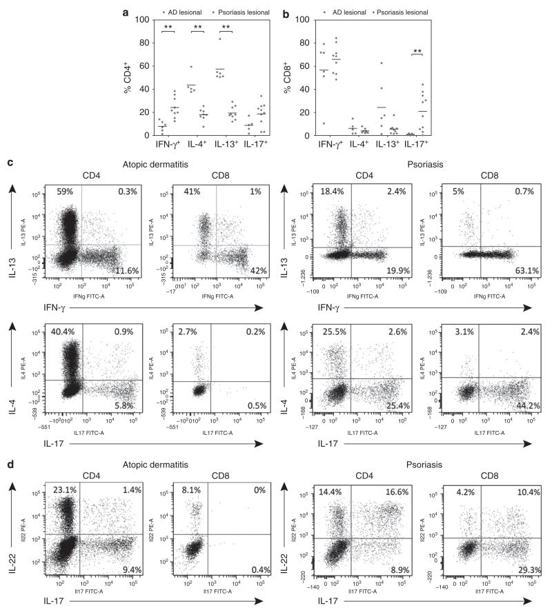
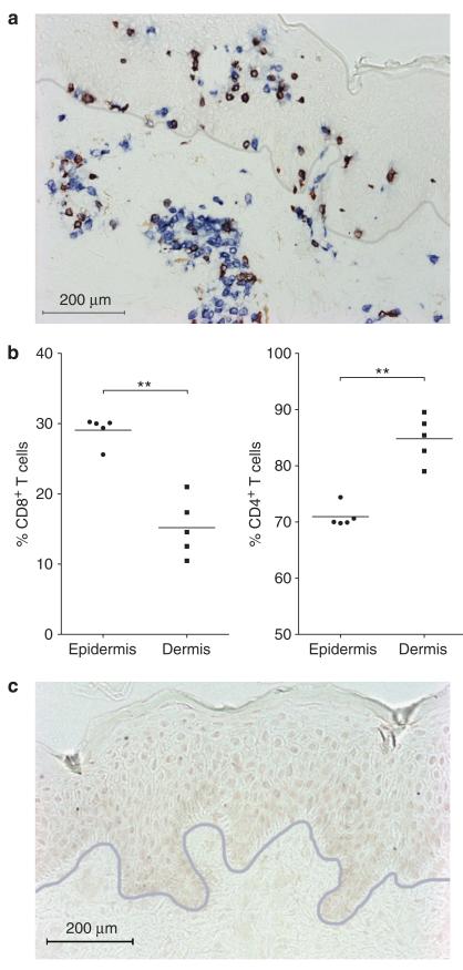
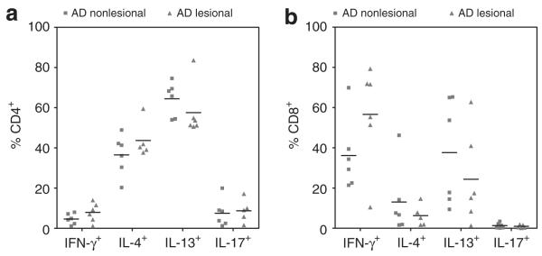
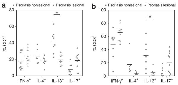
Although CD4(+) T cells are known to contribute to the pathology of atopic dermatitis (AD) and psoriasis, the role of CD8(+) T cells in these diseases remains poorly characterized. The aim of this study was to characterize the cytokine production of T cells from AD and psoriasis skin. We found that CD4(+) T cells isolated from AD skin were largely Th2 (T helper type 2) biased, in agreement with prior reports. However, we also observed large numbers of CD8(+) T cells producing IL-13, IFN-γ, and IL-22. We observed increased numbers of CD8(+) T cells isolated from AD skin, and immunohistochemistry studies confirmed the presence of CD8(+) T cells in the dermis and epidermis of AD skin lesions. Surprisingly, T-cell cytokine production was similar in the lesional and nonlesional skin of patients with AD. T cells from psoriatic lesional skin predominantly produced IFN-γ, IL-17, and IL-22, in agreement with prior studies. However, in addition to Th17 cells, we observed high percentages of CD8(+) T cells that produced both IL-22 and IL-17 in psoriatic skin lesions. Our findings demonstrate that CD8(+) T cells are a significant and previously unappreciated source of inflammatory cytokine production in both AD and psoriasis.


Figures

FIGURE 1
FIGURE 1
T cells from AD and psoriatic skin have distinct cytokine production profiles. (a) CD4+ T cells isolated from lesional skin of AD patients showed significantly increased expression of IL-4 and IL-13 (Th2), whereas CD4+ T cells isolated from lesional psoriasis skin showed increased expression of interferon-gamma (Th1). (b) CD8+ T cells isolated from lesional psoriasis skin demonstrated increased expression of IL-17 compared to CD8+ cells isolated from lesional AD skin, whereas CD8+ T cells from AD skin showed a trend towards increased percentages of IL-13 producing cells (P=0.088). Representative FACS plots: (c) expression of IL-13 and IFN-γ is mutually exclusive (upper plots), as is expression of IL-17 and IL-4 (lower plots); (d) expression of IL-22 and IL-17 in both CD4+ and CD8+ subsets. Whereas expression of IL-17 and IL-22 was largely mutually exclusive in AD patients, psoriasis patients showed IL-17+IL-22+ cells in both CD4+ and CD8+ T cell populations. Sample numbers may vary between groups (as a result of small numbers of cells isolated from specific samples). Horizontal bars represent median percentages for each group. ** P<0.01
FIGURE 2
FIGURE 2
CD8+ T cells are abundant in AD skin. (a) Representative immunohistochemical staining showing abundant CD8+ T cells in the dermis and epidermis of lesional AD skin (CD3 = blue, CD8 = red/brown). The gray line represents the dermo-epidermal junction. (b) The percentage of CD8+ T cells was significantly increased in the epidermis compared to the dermis of lesional AD skin. (c) Isotype control staining. Scale bar = 200 μm. Horizontal bars represents mean percentages. ** P<0.01
FIGURE 3
FIGURE 3
CD4+ T cells from both lesional and non-lesional AD skin had robust production of Th2 cytokines. (a) High numbers of CD4+ T cells isolated from lesional and non-lesional AD skin were found to produce IL-4 and IL-13. Strikingly, no significant differences were found between cells isolated from lesional and non-lesional AD skin. (b) High percentages CD8+ T cells from both lesional and non-lesional AD skin were found to express IFN-gamma or IL-13. Expression of IL-13 and interferon-gamma was mutually exclusive (Figure 1c). Sample numbers may vary between groups. Horizontal bars represent median percentages for each group.
FIGURE 4
FIGURE 4
IL-13 production is markedly reduced in lesional as compared to non-lesional skin in psoriasis. (a) CD4+ and (b) CD8+ T cells isolated from lesional psoriasis skin expressed significantly less IL-13 compared to non-lesional skin. Lesional CD4+ T cells from psoriasis patients showed a trend towards higher expression of IL-17 compared to non-lesional skin (P=0.15). Sample numbers may vary between groups. Horizontal bars represent median percentages for each group. * P<0.05

References

    1. Akdis CA, Akdis M, Simon D, et al. T cells and T cell-derived cytokines as pathogenic factors in the nonallergic form of atopic dermatitis. J Invest Dermatol. 1999;113:628–634. - PubMed
    1. Boniface K, Bernard FX, Garcia M, et al. IL-22 inhibits epidermal differentiation and induces proinflammatory gene expression and migration of human keratinocytes. J Immunol. 2005;174:3695–3702. - PubMed
    1. Clark RA, Chong BF, Mirchandani N, et al. A novel method for the isolation of skin resident T cells from normal and diseased human skin. J Invest Dermatol. 2006;126:1059–1070. - PubMed
    1. Conrad C, Boyman O, Tonel G, et al. Alpha1beta1 integrin is crucial for accumulation of epidermal T cells and the development of psoriasis. Nat Med. 2007;13:836–842. - PubMed
    1. De Benedetto A, Rafaels NM, McGirt LY, et al. Tight junction defects in patients with atopic dermatitis. J Allergy Clin Immunol. 2011;127:773–786. - PMC - PubMed

Publication types

MeSH terms