

IL-22 promotes fibroblast-mediated wound repair in the skin
- PMID: 23223145
- PMCID: PMC3610794
- DOI: 10.1038/jid.2012.463
IL-22 promotes fibroblast-mediated wound repair in the skin
Abstract
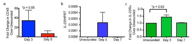
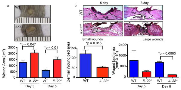
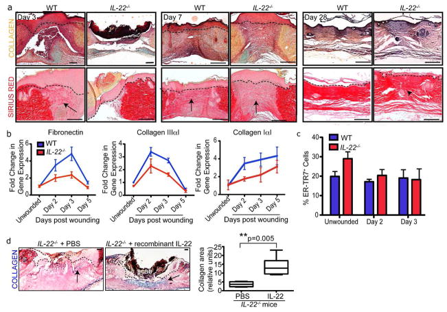
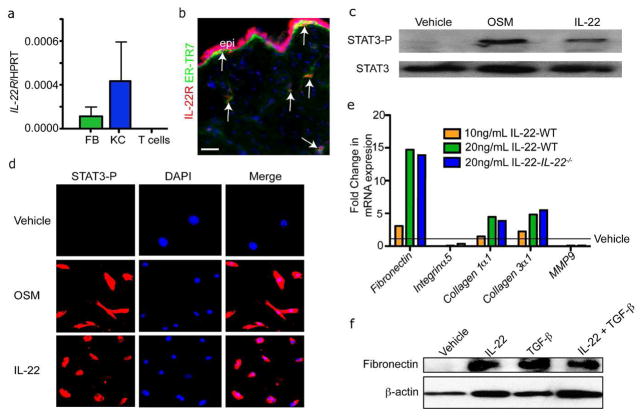
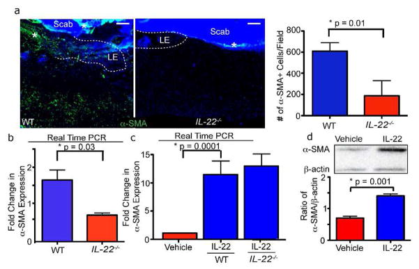
Skin wound repair requires complex and highly coordinated interactions between keratinocytes, fibroblasts, and immune cells to restore the epidermal barrier and tissue architecture after acute injury. The cytokine IL-22 mediates unidirectional signaling from immune cells to epithelial cells during injury of peripheral tissues such as the liver and colon, where IL-22 causes epithelial cells to produce antibacterial proteins, express mucins, and enhance epithelial regeneration. In this study, we used IL-22(-/-) mice to investigate the in vivo role for IL-22 in acute skin wounding. We found that IL-22(-/-) mice displayed major defects in the skin's dermal compartment after full-thickness wounding. We also found that IL-22 signaling is active in fibroblasts, using in vitro assays with primary fibroblasts, and that IL-22 directs extracellular matrix (ECM) gene expression and myofibroblast differentiation both in vitro and in vivo. These data define roles of IL-22 beyond epithelial cross talk, and suggest that IL-22 has a previously unidentified role in skin repair by mediating interactions between immune cells and fibroblasts.
Conflict of interest statement
The authors state no conflict of interest.
Figures
Similar articles
-
Interleukin (IL)-19 promoted skin wound healing by increasing fibroblast keratinocyte growth factor expression.Cytokine. 2013 Jun;62(3):360-8. doi: 10.1016/j.cyto.2013.03.017. Epub 2013 Apr 10. Cytokine. 2013. PMID: 23582717
-
Stimulation of skin repair is dependent on fibroblast source and presence of extracellular matrix.Tissue Eng. 2004 Jul-Aug;10(7-8):1054-64. doi: 10.1089/ten.2004.10.1054. Tissue Eng. 2004. PMID: 15363163
-
Inhibiting fibroblast aggregation in skin wounds unlocks developmental pathway to regeneration.Dev Biol. 2019 Nov 1;455(1):60-72. doi: 10.1016/j.ydbio.2019.07.001. Epub 2019 Jul 4. Dev Biol. 2019. PMID: 31279726
-
The complex dialogue between (myo)fibroblasts and the extracellular matrix during skin repair processes and ageing.Pathol Biol (Paris). 2012 Feb;60(1):20-7. doi: 10.1016/j.patbio.2011.10.002. Epub 2011 Nov 17. Pathol Biol (Paris). 2012. PMID: 22099331 Review.
-
Regeneration of Dermis: Scarring and Cells Involved.Cells. 2019 Jun 18;8(6):607. doi: 10.3390/cells8060607. Cells. 2019. PMID: 31216669 Free PMC article. Review.
Cited by
-
Umbilical Cord Mesenchymal Stromal/Stem Cells and Their Interplay with Th-17 Cell Response Pathway.Cells. 2024 Jan 16;13(2):169. doi: 10.3390/cells13020169. Cells. 2024. PMID: 38247860 Free PMC article.
-
Cytokines, Hormones and Cellular Regulatory Mechanisms Favoring Successful Reproduction.Front Immunol. 2021 Jul 28;12:717808. doi: 10.3389/fimmu.2021.717808. eCollection 2021. Front Immunol. 2021. PMID: 34394125 Free PMC article. Review.
-
Alteration of skin properties with autologous dermal fibroblasts.Int J Mol Sci. 2014 May 13;15(5):8407-27. doi: 10.3390/ijms15058407. Int J Mol Sci. 2014. PMID: 24828202 Free PMC article. Review.
-
Fibroblasts: Origins, definitions, and functions in health and disease.Cell. 2021 Jul 22;184(15):3852-3872. doi: 10.1016/j.cell.2021.06.024. Cell. 2021. PMID: 34297930 Free PMC article. Review.
-
IL-27 Facilitates Skin Wound Healing through Induction of Epidermal Proliferation and Host Defense.J Invest Dermatol. 2017 May;137(5):1166-1175. doi: 10.1016/j.jid.2017.01.010. Epub 2017 Jan 26. J Invest Dermatol. 2017. PMID: 28132857 Free PMC article.
References
-
- Barisic-Dujmovic T, Boban I, Clark SH. Fibroblasts/myofibroblasts that participate in cutaneous wound healing are not derived from circulating progenitor cells. J Cell Physiol. 2010;222:703–12. - PubMed
-
- Barrientos S, Stojadinovic O, Golinko MS, Brem H, Tomic-Canic M. Growth factors and cytokines in wound healing. Wound Repair Regen. 2008;16:585–601. - PubMed
-
- Boniface K, Bernard FX, Garcia M, Gurney AL, Lecron JC, Morel F. IL-22 inhibits epidermal differentiation and induces proinflammatory gene expression and migration of human keratinocytes. J Immunol. 2005;174:3695–702. - PubMed
-
- Cupedo T, Crellin NK, Papazian N, Rombouts EJ, Weijer K, Grogan JL, et al. Human fetal lymphoid tissue-inducer cells are interleukin 17-producing precursors to RORC+ CD127+ natural killer-like cells. Nat Immunol. 2009;10:66–74. - PubMed
Publication types
MeSH terms
Substances
Grants and funding
LinkOut - more resources
Full Text Sources
Other Literature Sources
Molecular Biology Databases
