. 2013 Jan 25;288(4):2118-31.
doi: 10.1074/jbc.M112.424291. Epub 2012 Dec 5.

Deletion of tetraspanin CD9 diminishes lymphangiogenesis in vivo and in vitro

Affiliations

Deletion of tetraspanin CD9 diminishes lymphangiogenesis in vivo and in vitro

Takeo Iwasaki et al. J Biol Chem. .

Abstract

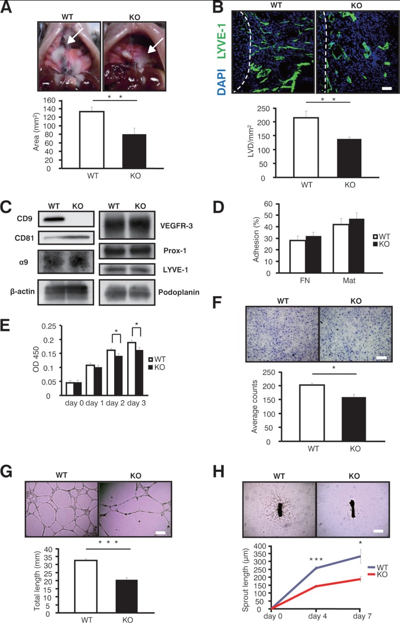
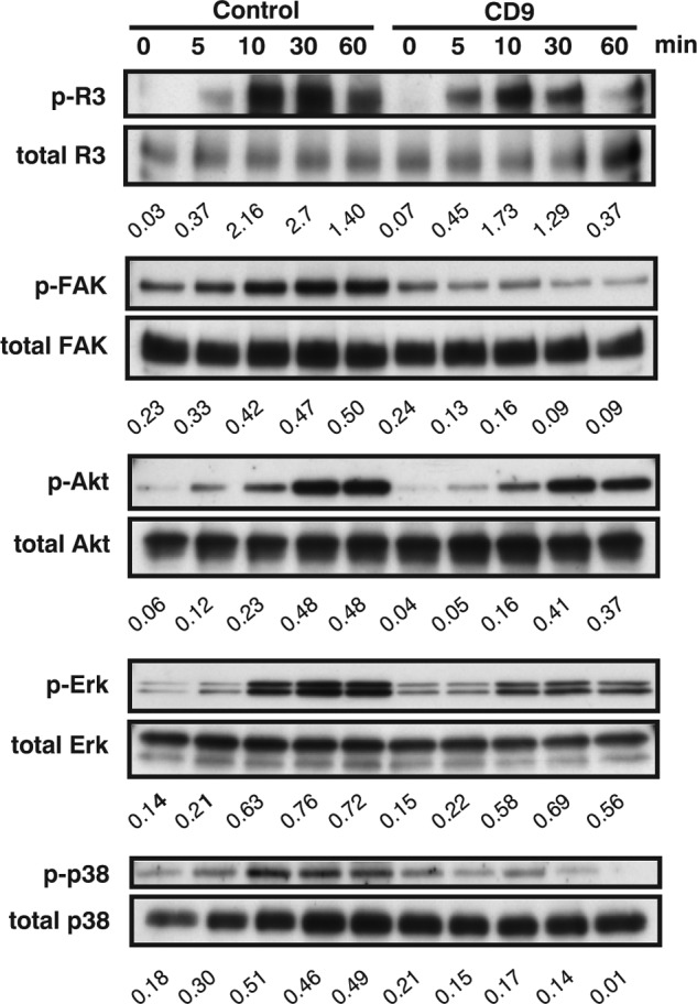
Tetraspanins have emerged as key players in malignancy and inflammatory diseases, yet little is known about their roles in angiogenesis, and nothing is known about their involvement in lymphangiogenesis. We found here that tetraspanins are abundantly expressed in human lymphatic endothelial cells (LEC). After intrathoracic tumor implantation, metastasis to lymph nodes was diminished and accompanied by decreased angiogenesis and lymphangiogenesis in tetraspanin CD9-KO mice. Moreover, lymphangiomas induced in CD9-KO mice were less pronounced with decreased lymphangiogenesis compared with those in wild-type mice. Although mouse LEC isolated from CD9-KO mice showed normal adhesion, lymphangiogenesis was markedly impaired in several assays (migration, proliferation, and cable formation) in vitro and in the lymphatic ring assay ex vivo. Consistent with these findings in mouse LEC, knocking down CD9 in human LEC also produced decreased migration, proliferation, and cable formation. Immunoprecipitation analysis demonstrated that deletion of CD9 in LEC diminished formation of functional complexes between VEGF receptor-3 and integrins (α5 and α9). Therefore, knocking down CD9 in LEC attenuated VEGF receptor-3 signaling, as well as downstream signaling such as Erk and p38 upon VEGF-C stimulation. Finally, double deletion of CD9/CD81 in mice caused abnormal development of lymphatic vasculature in the trachea and diaphragm, suggesting that CD9 and a closely related tetraspanin CD81 coordinately play an essential role in physiological lymphangiogenesis. In conclusion, tetraspanin CD9 modulates molecular organization of integrins in LEC, thereby supporting several functions required for lymphangiogenesis.


Figures

FIGURE 1.
FIGURE 1.
Expression of tetraspanins and integrins in HUVEC (A) and HDLEC (B). Expression of the indicated antigens (blue histograms) on the surface of HUVEC (A) and HDLEC (B) was determined by flow cytometry. Ten thousand cells were stained with CD9 (MM2/57), CD81 (JS64), CD151 (5C11), CD63 (MEM259), β1 (TS2/16), integrins α2 (BHA2.1), α3 (P1B5), α5 (P1D6), and α9 (2Q954) or podoplanin (NZ-1) at a concentration of 2 μg/ml and then labeled with FITC-conjugated goat anti-mouse, IgG (Vector Laboratories, Burlingame, CA). Normal mouse or rat IgG was used as a control. Stained cells were analyzed on a FACSCalibur (Becton Dickinson). Mean fluorescence intensity is shown in each histogram. Negative control peaks (green lines) were obtained with secondary antibody alone. Expression of podoplanin specific to LEC was observed in HDLEC, but not in HUVEC. Both HUVEC and HDLEC were analyzed between the third to fifth passages. The data are representative of three independent studies, which produced similar results.
FIGURE 2.
FIGURE 2.
Decreased lymph node metastasis and lymphangiogenesis in CD9-KO mice. A, growth of lymph node metastases and primary tumors in the lungs was assessed (n = 5). Dotted lines show the margins of the metastatic lymph nodes. Arrowheads show primary tumors in the lung. B, CD31-positive microvascular density (MVD) of the primary tumors was decreased in the CD9-KO mice. C, peritumoral LVD stained with LYVE-1 (green). The peritumor area was defined as a 1-mm distance from the tumor periphery. Dotted lines show the margins of the implanted tumors in the lungs. The cell nuclei are counterstained with Hoechst (blue). D, metastatic lymph nodes were diminished in CD9-KO mice, although the sizes of isolated lymph nodes were indistinguishable before tumor implantation (data not shown). The data are representative of three independent studies with similar results. The bars represent the means ± S.E. *, p < 0.05 versus WT; ***, p < 0.005 versus WT. Scale bar, 1 cm for D and 100 μm for B and C.
FIGURE 3.
FIGURE 3.
Decreased tumor growth and angiogenesis in CD9-KO mice. A, for primary tumor growth, mice were injected subcutaneously with Lewis lung carcinoma cells. Tumors were measured with Vernier calipers, and the volume was calculated (length × width2 × 0.52). After 20 days, tumors were collected and weighed (n = 10). Arrows show primary tumor growth in both sites at 20 days. B and C, cryosections of primary tumors were stained with anti-CD31 Ab (red) as a blood endothelial marker and with anti-LYVE-1 Ab (green) as a lymphatic endothelial marker. The data are representative of three independent studies with similar results. The bars represent the means ± S.E. *, p < 0.05 versus WT; **, p < 0.01 versus WT; ***, p < 0.005 versus WT. The scale bars represent 100 μm for B and C. MVD, microvascular density.
FIGURE 4.
FIGURE 4.
CD9 promotes angiogenesis in blood vascular endothelial cells from mice and humans. A, after HUVEC were transfected with siRNAs against CD9 (CD9) or random RNAs (Control), knockdown of CD9 protein was confirmed by immunoblotting in parallel with integrin β1 (TS2/16), CD9 (MM2/57), CD81 (JS64), CD151 (11G5a), and β-actin (C4). B, to measure random migration, HUVEC were plated on coverslips coated with Mat. Cell movements recorded by time lapse video microscopy were quantified for 1 h with MetaMorph (n = 10). C, for chemotaxis, HUVEC were plated in upper Transwell chambers coated with FN. Cells migrating through the filter were counted in four randomly selected fields after 6 h. D and E, mouse BEC were isolated from collagenase-digested lung tissue (with anti-mouse CD31-coated beads) and enriched (with anti-mouse ICAM-2) to more than 90% purity (positively stained for von Willebrand factor). Both HUVEC and mBEC were seeded on Matrigel, and the total length of cables was quantitated after 24 h. F, shown are phase contrast images of branching vessel-like structures in WT and CD9-KO explants (day 4). The data are representative of three independent studies with similar results. The bars represent the means ± S.E. *, p < 0.05 versus WT; **, p < 0.01 versus WT; ***, p < 0.005 versus WT. Scale bar, 200 μm for C–E and 500 μm for F.
FIGURE 5.
FIGURE 5.
CD9 in mice promotes lymphangiogenesis in vitro, ex vivo, and in vivo. A, shown are representative images of lymphangiomas in WT and CD9-KO mice (arrows). B, LVD defined with LYVE-1 Ab (green) was also decreased in the CD9-KO mice. The cell nuclei were counterstained with Hoechst (blue). Dotted lines show the margins of the lymphangiomas and the livers. C, isolated mLEC were confirmed by immunoblotting with the indicated antibodies: CD9 (KMC8), CD81 (Eat-2), integrin α9 (R&D Systems), VEGFR-3 (R&D Systems), Prox-1 (AngioBio), LYVE-1 (abcam), and podoplanin (8.1.1; eBioscience). D, static cell adhesion was performed on FN and Matrigel. E, isolated mLEC were stimulated with VEGF-C for the proliferation assay (300 ng/ml) (n = 3). F, for chemotaxis, mLEC were plated on upper Transwell chambers coated with FN. The bottom chambers contained VEGF-C (300 ng/ml). G, for cable formation, mLEC were seeded on Matrigel, and the total length was quantitated after 24 h. The cables were photographed in at least three random fields and quantified with MetaMorph imaging software. H, sprouting from thoracic rings was quantitated (n = 6). The data are representative of four independent studies with similar results. The bars represent the means ± S.E. *, p < 0.05 versus WT; **, p < 0.01 versus WT; ***, p < 0.005 versus WT. Scale bar, 200 μm for B and F and 500 μm for G and H.
FIGURE 6.
FIGURE 6.
CD9 promotes lymphangiogenesis in lymphatic endothelial cells from humans. A, after HDLEC were transfected with siRNAs against CD9 (CD9) or random RNAs (Control), knockdown of CD9 protein was confirmed by immunoblotting in parallel with integrin (β1, α5), CD9 (MM2/57), CD81 (JS64), CD151 (11G5a), VEGFR-3 (C20), LYVE-1 (RELIATech), podoplanin (NZ-1), and β-actin (C4). B, adhesion of HDLEC was quantified (n = 3). C, proliferation was stimulated with VEGF-C (300 ng/ml) (n = 3). D, for chemotactic migration, HDLEC were plated in upper Transwell chambers coated with FN. E, for the two-dimensional assay, HDLEC were seeded on Matrigel, and total cable length was quantitated after 24 h. The cables were photographed in at least three random fields and quantified with MetaMorph imaging software. The data are representative of three independent studies with similar results. The bars represent the means ± S.E. *, p < 0.05 versus WT; ***, p < 0.005 versus WT. Scale bar, 200 μm for D and E.
FIGURE 7.
FIGURE 7.
Deletion of CD9 impairs functional complexes between integrins and VEGFR-3 in LEC. A, to confirm the association between integrin β1, α5, CD9, and VEGFR-3, HDLEC lysed in 1% Brij-97 buffer were immunoprecipitated with the indicated antibody. B, after CD9 knockdown, immunoprecipitation with integrin β1 was compared between control siRNA (Control) and CD9-KD HDLEC (CD9). Note that co-precipitates with integrin β1 were down-regulated after CD9-KD. The antibodies used here were as follows: β1 (TS2/16), α5 (P1D6), α6(GOH3), α9 (2Q954), CD9 (MM2/57), CD151 (5C11), and VEGFR-3 (9D9F9). Whole cell lysates (WCL) were blotted with β-actin as a loading control. The data are representative of three independent studies with similar results. IP, immunoprecipitation; IB, immunoblotting.
FIGURE 8.
FIGURE 8.
Deletion of CD9 impairs VEGF-C-induced signaling in LEC. Serum-starved HDLEC were stimulated with human VEGF-C (200 μg/ml). Equal amounts of lysate were probed with Abs to activated VEGFR-3 (p-VEGFR-3), activated FAK (p-FAK), activated Akt (p-Akt), activated Erk (p-Erk), activated p38 (p-p38), and β-actin (to control for loading). The numbers under blots represent ratios of phosphorylated to total protein from densitometry analysis. The data are representative of three independent studies from different cell preparations.
FIGURE 9.
FIGURE 9.
CD9 and CD81 coordinately promote lymphangiogenesis in HDLEC. A, after HDLEC were transfected with siRNAs against CD9, CD81, and CD151, knockdown of these proteins was confirmed by immunoblotting in parallel with integrin β1 and β-actin. None of siRNAs modulated the expression of β1 integrin. β-Actin was blotted to control for loading. The numbers under the blots indicate ratios from densitometry analyses. The ratios were calculated by dividing densitometry values for actin by those for β1, CD9, CD81, and CD151. B, for the cable formation assay, HDLEC were seeded on Mat, and the total length of cellular cables was quantitated after 24 h. The scale bars represent 200 μm. The bars represent the means ± S.E. ***, p < 0.005 versus WT. The data are representative of three independent studies with similar results.
FIGURE 10.
FIGURE 10.
Impaired physiological lymphangiogenesis as well as angiogenesis in CD9/CD81-DKO mice. A, lymphatic vessels and blood vessels in trachea isolated from tetraspanin-KO mice. CD9, CD9-KO mice; CD81, CD81-KO mice; KO, CD9/CD81-DKO mice. Confocal microscopic images of immunoreactivities of tracheal whole mounts stained for CD31 (red, blood vessels) and LYVE-1 (green, lymphatic vessels). B, diaphragms were stained with LYVE-1. C, shown are pictures of LEC in tracheas from WT and DKO mice by electron microscopy. Arrowheads, protrusions from mLEC; arrows, anchoring filaments. D, shown are pictures of BEC from WT and DKO mice by electron microscopy. Arrows, basement membranes; arrowheads, pinocytes. Magnification: upper panel, ×20,000; lower panel, ×20,000. The bars represent the means ± S.E. ***, p < 0.005 versus WT. Scale bar, 200 μm for A and B.

References

    1. Adams R. H., Alitalo K. (2007) Molecular regulation of angiogenesis and lymphangiogenesis. Nat. Rev. Mol. Cell Biol. 8, 464–478 - PubMed
    1. Grothey A., Galanis E. (2009) Targeting angiogenesis: progress with anti-VEGF treatment with large molecules. Nat. Rev. Clin. Oncol. 6, 507–518 - PubMed
    1. Tammela T., Alitalo K. (2010) Lymphangiogenesis. Molecular mechanisms and future promise. Cell 140, 460–476 - PubMed
    1. Baluk P., Fuxe J., Hashizume H., Romano T., Lashnits E., Butz S., Vestweber D., Corada M., Molendini C., Dejana E., McDonald D. M. (2007) Functionally specialized junctions between endothelial cells of lymphatic vessels. J. Exp. Med. 204, 2349–2362 - PMC - PubMed
    1. Alitalo K., Tammela T., Petrova T. V. (2005) Lymphangiogenesis in development and human disease. Nature 438, 946–953 - PubMed

Publication types

MeSH terms

LinkOut - more resources