. 2012 Dec 5;32(49):17528-39.
doi: 10.1523/JNEUROSCI.2791-12.2012.

The relation between perception and brain activity in gaze-evoked tinnitus

Affiliations

The relation between perception and brain activity in gaze-evoked tinnitus

Margriet J van Gendt et al. J Neurosci. .

Abstract

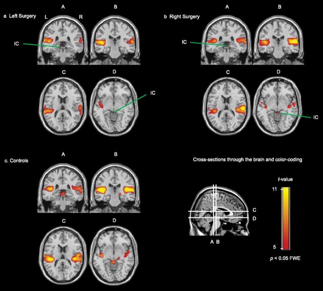
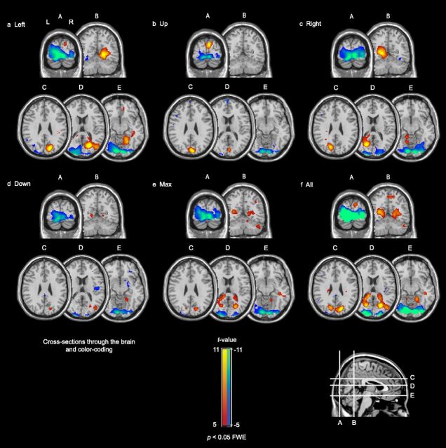
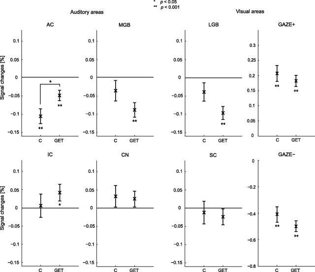
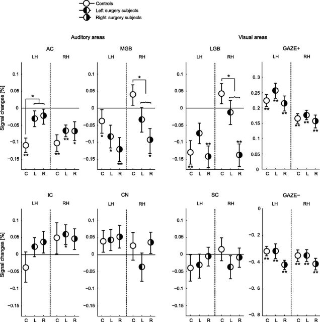
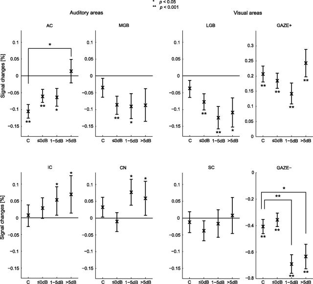
Tinnitus is a phantom sound percept that can be severely disabling. Its pathophysiology is poorly understood, partly due to the inability to objectively measure neural correlates of tinnitus. Gaze-evoked tinnitus (GET) is a rare form of tinnitus that may arise after vestibular schwannoma removal. Subjects typically describe tinnitus in the deaf ear on the side of the surgery that can be modulated by peripheral eye gaze. This phenomenon offers a unique opportunity to study the relation between tinnitus and brain activity. We used functional magnetic resonance imaging in humans to show that in normal-hearing control subjects, peripheral gaze results in inhibition of the auditory cortex, but no detectable response in the medial geniculate body (MGB) and inferior colliculus (IC). In patients with GET, peripheral gaze (1) reduced the cortical inhibition, (2) inhibited the MGB, and (3) activated the IC. Furthermore, increased tinnitus loudness is represented by increased activity in the cochlear nucleus (CN) and IC and reduced inhibition in the auditory cortex (AC). The increase of CN and IC activity with peripheral gaze is consistent with models of plastic reorganization in the brainstem following vestibular schwannoma removal. The activity decrease in the MGB and the reduced inhibition of the AC support a model that attributes tinnitus to a dysrhythmia of the thalamocortical loop, leading to hypometabolic theta activity in the MGB. Our data offer the first support of this loop hypothesis of tinnitus, independent of the initial experiments that led to its formulation.


Figures

Figure 1.
Figure 1.
Mean audiograms for the controls (n = 9), and the tinnitus subjects who underwent surgery on the left (n = 8) or right side (n = 10). For tinnitus subjects, only the audiograms of the ear contralateral to the surgery are shown. On the side of surgery, the patients were completely deaf, except two, for whom thresholds were between 90 and 120 dB HL (Hearing Level). Error bars indicate SDs.
Figure 2.
Figure 2.
The changes to tinnitus were investigated for five gaze directions (a): Left, Right, Up, Down (solid arrows), and the direction evoking the maximum subjective change of tinnitus (one of the solid or dashed arrows). Histograms showing the characteristics of the perceived tinnitus changes are shown in b–f. b, Shows the gaze direction with largest effect on the tinnitus. Gaze lateralization relative to surgery side with largest effect on the tinnitus is shown in c. Changes in, respectively, bandwidth, loudness, and pitch for the maximum tinnitus percept are shown in d–f. n.a., Not applicable.
Figure 3.
Figure 3.
Coronal and transversal cross sections of the brain through the AC (A–D), MGB (B), IC (A, D), and CN (A) showing significant responses to bilateral 90 dB SPL dynamic rippled sound in the subjects who underwent surgery on the left side (n = 8; a), the subjects who underwent surgery on the right side (n = 10; b), and the control subjects (n = 9; c). The red-yellow color-coded areas indicate areas with a significant response.
Figure 4.
Figure 4.
Brain responses to the five peripheral gaze directions, contrasted with gazing straight ahead. The responses of tinnitus and control subjects were combined (n = 27). Deactivation was found in primary visual cortex (A–E); activation was found in precuneus (a, c–e; cross sections B–E), and dorsally in the occipital lobe (b; cross sections A, C). f, Shows the average activation and deactivation across all subjects and all gaze directions. The red-yellow color-code indicates areas with a significantly increased activity to gazing; the blue-green color-coded areas indicate areas with a significantly decreased activity.
Figure 5.
Figure 5.
ROI responses to peripheral gaze for the controls (C; n = 9) and the tinnitus subjects (GET; n = 18). Left surgery and right surgery subjects were combined. Also, the left and the right hemisphere, as well as the gaze directions, were combined. ROI analyses were performed on bilateral AC, MGB, IC, CN, LGB, and SC, as well as the GAZE+ and GAZE−. Error bars indicate the group SEs around the mean. Statistical significances against baseline and differences between the GET group and the control group are represented by asterisks.
Figure 6.
Figure 6.
ROI responses to peripheral gaze for the controls (C; n = 9) and the left surgery (L; n = 8) and right surgery (R; n = 10) tinnitus groups. In contrast to Figure 5, the left and right surgery subjects and the left and the right hemispheres are shown separately. ROI analyses were performed on unilateral AC, MGB, IC, CN, LGB, and SC, as well as the GAZE+ and GAZE−. Error bars indicate the group SEs around the mean. Statistical significances against baseline and differences between the GET group and the control group are represented by asterisks. LH, left hemisphere; RH, right hemisphere.
Figure 7.
Figure 7.
ROI responses to peripheral gaze related to change in tinnitus loudness. Controls and tinnitus subjects are shown separately. Control subjects are copied from Figure 5. Responses are stratified with respect to the perceived amount of intensity increase of the modulated tinnitus (≤0 dB, 1–5 dB, and >5 dB). In total, 49, 19, and 22 responses were included in the sets ≤0 dB, 1–5 dB, and >5 dB, respectively. The number of right/left surgery subjects for each mentioned set equaled 7/7, 5/2, and 5/3 subjects, respectively. Error bars indicate the SEs around the mean. Statistical significances against baseline and differences between the sets and the control group are represented by asterisks.

References

    1. Andersson F, Joliot M, Perchey G, Petit L. Eye position-dependent activity in the primary visual area as revealed by fMRI. Hum Brain Mapp. 2007;28:673–680. - PMC - PubMed
    1. Baguley DM, Phillips J, Humphriss RL, Jones S, Axon PR, Moffat DA. The prevalence and onset of gaze modulation of tinnitus and increased sensitivity to noise after translabyrinthine vestibular schwannoma excision. Otol Neurotol. 2006;27:220–224. - PubMed
    1. Bateman N, Nikolopoulos TP, Robinson K, O'Donoghue GM. Impairments, disabilities, and handicaps after acoustic neuroma surgery. Clin Otolaryngol Allied Sci. 2000;25:62–65. - PubMed
    1. Baumgart F, Kaulisch T, Tempelmann C, Gaschler-Markefski B, Tegeler C, Schindler F, Stiller D, Scheich H. Electrodynamic headphones and woofers for application in magnetic resonance imaging scanners. Med Phys. 1998;25:2068–2070. - PubMed
    1. Biggs ND, Ramsden RT. Gaze-evoked tinnitus following acoustic neuroma resection: a de-afferentation plasticity phenomenon? Clin Otolaryngol Allied Sci. 2002;27:338–343. - PubMed

Publication types

MeSH terms