. 2012 Dec 5;32(49):17894-908.
doi: 10.1523/JNEUROSCI.2815-12.2012.

Drivers of the primate thalamus

Affiliations

Drivers of the primate thalamus

Zita Rovó et al. J Neurosci. .

Abstract

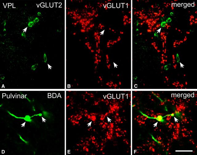
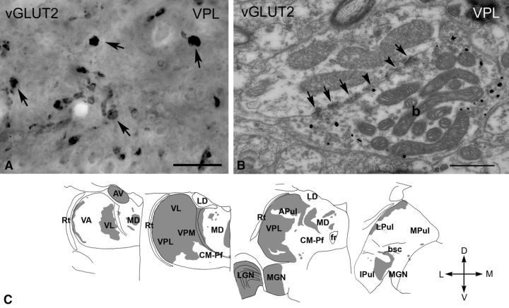
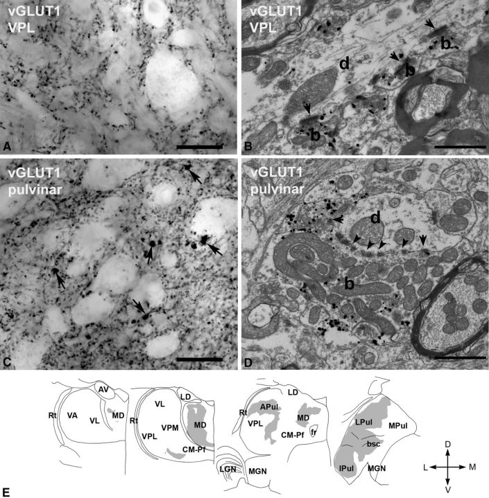
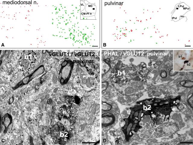
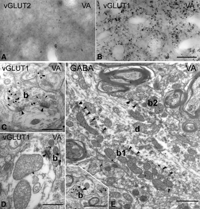
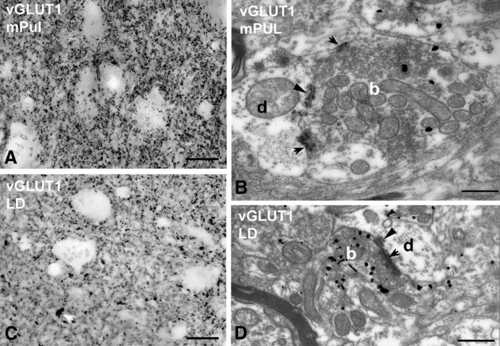
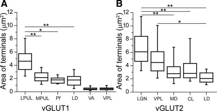
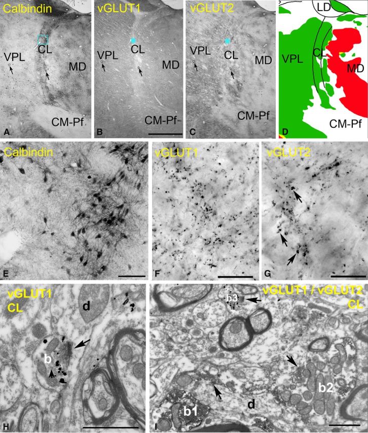
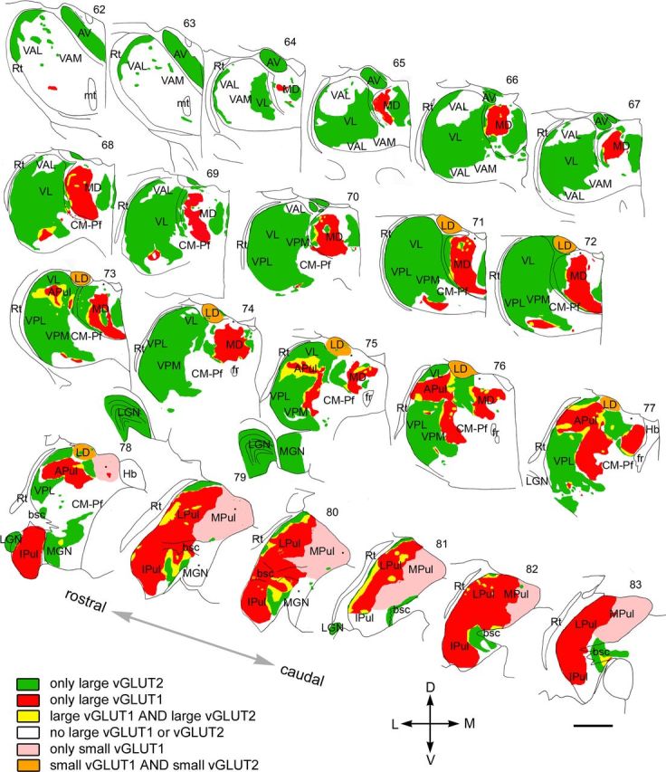
The activity of thalamocortical neurons is primarily determined by giant excitatory terminals, called drivers. These afferents may arise from neocortex or from subcortical centers; however, their exact distribution, segregation, or putative absence in given thalamic nuclei are unknown. To unravel the nucleus-specific composition of drivers, we mapped the entire macaque thalamus using vesicular glutamate transporters 1 and 2 to label cortical and subcortical afferents, respectively. Large thalamic territories were innervated exclusively by either giant vGLUT2- or vGLUT1-positive boutons. Codistribution of drivers with different origin was not abundant. In several thalamic regions, no giant terminals of any type could be detected at light microscopic level. Electron microscopic observation of these territories revealed either the complete absence of large multisynaptic excitatory terminals (basal ganglia-recipient nuclei) or the presence of both vGLUT1- and vGLUT2-positive terminals, which were significantly smaller than their giant counterparts (intralaminar nuclei, medial pulvinar). In the basal ganglia-recipient thalamus, giant inhibitory terminals replaced the excitatory driver inputs. The pulvinar and the mediodorsal nucleus displayed subnuclear heterogeneity in their driver assemblies. These results show that distinct thalamic territories can be under pure subcortical or cortical control; however, there is significant variability in the composition of major excitatory inputs in several thalamic regions. Because thalamic information transfer depends on the origin and complexity of the excitatory inputs, this suggests that the computations performed by individual thalamic regions display considerable variability. Finally, the map of driver distribution may help to resolve the morphological basis of human diseases involving different parts of the thalamus.


Figures

Figure 1.
Figure 1.
High-power fluorescent images of vGLUT1- and vGLUT2-immunoreactive terminals. A–C, High-power fluorescent photomicrographs demonstrating that vGLUT2 (A) and vGLUT1 (B) label distinct sets of afferents in the ventral posterolateral nucleus of the macaque monkey. D–F, High-power fluorescent photomicrographs demonstrating that anterogradely labeled large cortical terminals (green, arrows in D) display vGLUT1 immunoreactivity (red, E) in the pulvinar. Scale bar, 10 μm.
Figure 2.
Figure 2.
vGLUT2 -immunoreactive terminals in the macaque thalamus. A, High-power light microscopic image of a vGLUT2-immunostained section using DAB-Ni as a chromogen from the VPL. vGLUT2 labels large irregularly shaped terminals (arrows). B, High-power electron micrograph of a vGLUT2-positive terminal (b) labeled by silver intensified gold particles (small black dots). The terminal displays the ultrastructural features of RL-type terminals, including large size, multiple mitochondria, puncta adherentia (arrowheads), synapses (arrows). C, Regional distribution of large vGLUT2-positive terminals at four coronal levels of the macaque thalamus arranged from rostral to caudal levels. Note the absence of subcortical driver inputs from large thalamic territories. Scale bars: A, 20 μm; B, 0.5 μm. APul, Anterior pulvinar; AV, anteroventral; bsc, brachium of superior colliculus; CM–Pf, centromedian–parafascicular; fr, fasciculus retroflexus; Ipul, inferior pulvinar; LD, laterodorsal; LGN, lateral geniculate nucleus; LPul, lateral pulvinar; MD, mediodorsal; MGN, medial geniculate nucleus; MPul, medial pulvinar; VA, ventral anterior; VL, ventrolateral; VPL, ventral posterolateral; VPM, ventral posteromedial; Rt, reticular thalamus.
Figure 3.
Figure 3.
vGLUT1-immunoreactive terminals in the macaque thalamus. A, High-power light microscopic image of a vGLUT1-immunostained section from the VPL displaying small vGLUT1-immunoreactive terminals only. B, At the electron microscopic level, these terminals (b, silver-intensified gold) display the features of RS-type terminals, i.e., small size, maximum of one or two mitochodria, no puctum adherens, and a single synapse (arrow). d, Postsynaptic dendrite. C, In the lateral pulvinar, besides the small vGLUT1-immunoreactive terminals, large immunoreactive structures (arrows) can also be distinguished at the light microscopic level. D, At the electron microscopic level, a large vGLUT1-immunoreactive terminal (b, silver-intensified gold) displays all the ultrastructural features of RL terminals. Arrows, Synapses; arrowheads, puncta adherentia; d, postsynaptic dendrite. E, Regional distribution of large vGLUT1-positive terminals at four coronal levels of the macaque thalamus. Note the lack of large vGLUT1-positve terminals from many thalamic nuclei considered as higher order and the heterogeneity of the distribution within the mediodorsal nucleus (MD) and pulvinar. Scale bars: A, C, 20 μm; B, D, 1 μm. APul, Anterior pulvinar; AV, anteroventral; bsc, brachium of superior colliculus; CM–Pf, centromedian–parafascicular; fr, fasciculus retroflexus; Ipul, inferior pulvinar; LD, laterodorsal; LGN, lateral geniculate nucleus; LPul, lateral pulvinar; MD, mediodorsal; MGN, medial geniculate nucleus; MPul, medial pulvinar; VA, ventral anterior; VL, ventrolateral; VPL, ventral posterolateral; VPM, ventral posteromedial; Rt, reticular thalamus.
Figure 4.
Figure 4.
Convergence of large vGLUT1 and vGLUT2 terminals. A, B, Plot of large vGLUT1-immunoreactive (red dots) and vGLUT2-immunoreactive (green dots) terminals using double-immunostained material in the mediodorsal nucleus (A) and pulvinar (B, position shown in the inset). Note clear separation of the two types of terminals in the mediodorsal nucleus and homogeneous mixing in the lateral pulvinar. C, Electron micrograph from the anterior pulvinar displaying close proximity (<3 μm) of a large vGLUT1-immunoreactive terminal (b1, silver-intensified gold particles) and a large vGLUT2-positive terminal (b2, DAB precipitate). D, Correlated light (inset) and electron micrographs in the anterior pulvinar depicting a vGLUT2-immunoreactive large terminal (b1, brown DAB precipitate) adjacent to a large cortical terminal (b2, black DAB-Ni precipitate) anterogradely labeled from the supplementary motor cortex by Phaseolus vulgaris leucoagglutinin (PHAL). Both terminals display RL features. Scale bars: A, B, 50 μm; C, 2 μm; D, 1 μm; inset, 20 μm.
Figure 5.
Figure 5.
Thalamic territories without large vGLUT1 or vGLUT2 terminals. A, B, High-power light microscopic images of vGLUT2-immunostained (A) and vGLUT1-immunostained (B) sections from the ventral anterior nucleus display the lack of large terminal labeling by both markers. C, D, At the electron microscopic level, vGLUT1-positive terminals show the features of RS-type terminals. Most of them form a single synapse or more rarely two synapses and contained a maximum of two mitochondria. E, The large terminals of the ventral anterior nucleus (b1 and b2) are negative for vGLUT1 but show GABA immunoreactivity after postembedding GABA immunogold staining. Inset shows a vGLUT1-immunoreactive terminal at the same magnification. Scale bars: A, B, 20 μm; C, D, 500 nm; E and inset, 1 μm.
Figure 6.
Figure 6.
Small RL-type vGLUT1-positive terminals. AD, High-power light (A, C) and electron (B, D) micrographs of vGLUT1-immunostained material from the medial pulvinar (mPUL; A, B) and the laterodorsal nucleus (LD; C, D). No vGLUT1-positive large terminals can be distinguished at the light microscopic level (a group of small terminals is indicated by the white arrow in C). At the electron microscopic level, however, vGLUT1-positive terminals (b, silver-intensified gold) displaying RL features can be identified (multiple mitochondria, punctum adherens, arrowheads, multiple synapses, arrows) in both the medial pulvinar (B) and the laterodorsal nucleus (D). These terminals are significantly smaller and less complex than RL terminals of the lateral pulvinar or ventral posterolateral nucleus. Scale bars: light micrographs, 20 μm; electron micrographs, 500 nm.
Figure 7.
Figure 7.
Nucleus-specific variability in the size of vGLUT2- and vGLUT1-positive terminals. A, B, Box plots of the median, interquartile, and 5–95% range of the cross-sectional areas of vGLUT1-positve RL (LPUL, MPUL, Pf, LD) and RS (VA, VPL) type (A) and vGLUT2-positive RL (LGN, VPL, MD, CL, LD) type terminals measured in single electron microscopic sections (B). Note the significantly smaller vGLUT1-positive RL-type terminals in MPUL, Pf, and LD compared with lateral pulvinar. In VA, the terminal size does not differ from VPL terminals, which is known to contain only RS-type vGLUT1-positive terminals. Similarly, the size of vGLUT2-positve terminals also displays significant differences. CL, Centrolateral; LD, laterodorsal; LGN, lateral geniculate nucleus; LPUL, lateral pulvinar; MD, mediodorsal; MPUL, medial pulvinar; Pf, parafascicular, VA, ventral anterior; VPL, ventral posterolateral. *p < 0.05, **p < 0.001.
Figure 8.
Figure 8.
Excitatory inputs of the rostral intralaminar nuclei. A–D, Delineation of the intralaminar nuclei. Low-power light micrographs of adjacent sections immunostained for calbindin (A), vGLUT1 (B), and vGLUT2 (C). Note that the borders of the intralaminar nuclei are discernible only in the calbindin immunostaining. Arrows indicate corresponding capillaries. The color map (D) indicate the distribution of large terminals (green, large vGLUT2 terminals; red, large vGLUT1 terminals). Intralaminar nuclei are continuous laterally with VPL but contain no large vGLUT1 terminals, unlike the mediodorsal nucleus. E–G, High-power light microscopic images of the blue boxed regions in A–C, respectively. Note the lack of large vGLUT1-positive terminals and medium-sized vGLUT2 terminals (arrows in G) in the centrolateral nucleus. H, At the electron microscopic level, a vGLUT1-positive terminal (b) displays RS features. I, After double immunostaining, the size difference between RL-type vGLUT2 terminals (b1, b2, DAB precipitate) and RS-type vGLUT1-immunoreactive terminals (b3, silver-intensified gold) are apparent. Scale bars: A–C, 2 mm; E, 100 μm; F, G, 20 μm; H, I, 1 μm.
Figure 9.
Figure 9.
Mosaic of drivers in the primate thalamus. Distribution of cortical and subcortical excitatory terminals in the entire primate thalamus. The map is displayed at 22 coronal levels from anterior (top right) to posterior (bottom left) with 500 μm intervals. It is based on observations from seven monkeys. Six different types of innervation pattern are distinguished based on the light and electron microscopic data. Large thalamic territories receive exclusively subcortical (vGLUT2 green) or cortical (vGLUT1, red) RL inputs. Convergence of the two terminal types (yellow) is restricted to border zones. Basal ganglia-recipient nuclei (VA) receive no discernible excitatory RL driver input (white). Cortical input from smaller RL drivers (pink) characterizes medial pulvinar and CM–Pf. Finally, in the laterodorsal nucleus, the convergence of small cortical and small subcortical RL terminals were observed (orange). Single nuclei, especially the pulvinar and the mediodorsal thalamic nuclei, displayed considerable subnuclear heterogeneity in their driver distribution. Small dots indicate clusters of small vGLUT2-positive nerve endings encountered at the light microscopic level. APul, Anterior pulvinar; AV, anteroventral; bsc, brachium of superior colliculus; CM–Pf, centromedian–parafascicular; fr, fasciculus retroflexus; Hb, habenula; Ipul, inferior pulvinar; LD, laterodorsal; LGN, lateral geniculate nucleus; LPul, lateral pulvinar; MD, mediodorsal; MGN, medial geniculate nucleus; MPul, medial pulvinar; mt, mammillothalamic tract; VA, ventral anterior; VL, ventrolateral; VPL, ventral posterolateral; VPM, ventral posteromedial; Rt, reticular thalamus. Scale bar, 2 mm.

References

    1. Apkarian AV, Shi T. Squirrel monkey lateral thalamus. I. Somatic nociresponsive neurons and their relation to spinothalamic terminals. J Neurosci. 1994;14:6779–6795. - PMC - PubMed
    1. Balaram P, Hackett TA, Kaas JH. VGLUT1 mRNA and protein expression in the visual system of prosimian galagos (Otolemur garnetti) Eye Brain. 2011;2011:81–98. - PMC - PubMed
    1. Balercia G, Kultas-Ilinsky K, Bentivoglio M, Ilinsky IA. Neuronal and synaptic organization of the centromedian nucleus of the monkey thalamus: a quantitative ultrastructural study, with tract tracing and immunohistochemical observations. J Neurocytol. 1996;25:267–288. - PubMed
    1. Barthó P, Freund TF, Acsády L. Selective GABAergic innervation of thalamic nuclei from zona incerta. Eur J Neurosci. 2002;16:999–1014. - PubMed
    1. Beggs J, Jordan S, Ericson AC, Blomqvist A, Craig AD. Synaptology of trigemino- and spinothalamic lamina I terminations in the posterior ventral medial nucleus of the macaque. J Comp Neurol. 2003;459:334–354. - PubMed

Publication types

MeSH terms

Substances

LinkOut - more resources