. 2012 Dec 5;32(49):17921-31.
doi: 10.1523/JNEUROSCI.2664-12.2012.

Rebalance of striatal NMDA/AMPA receptor ratio underlies the reduced emergence of dyskinesia during D2-like dopamine agonist treatment in experimental Parkinson's disease

Affiliations

Rebalance of striatal NMDA/AMPA receptor ratio underlies the reduced emergence of dyskinesia during D2-like dopamine agonist treatment in experimental Parkinson's disease

Vincenza Bagetta et al. J Neurosci. .

Abstract

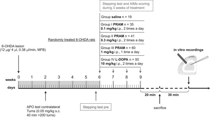
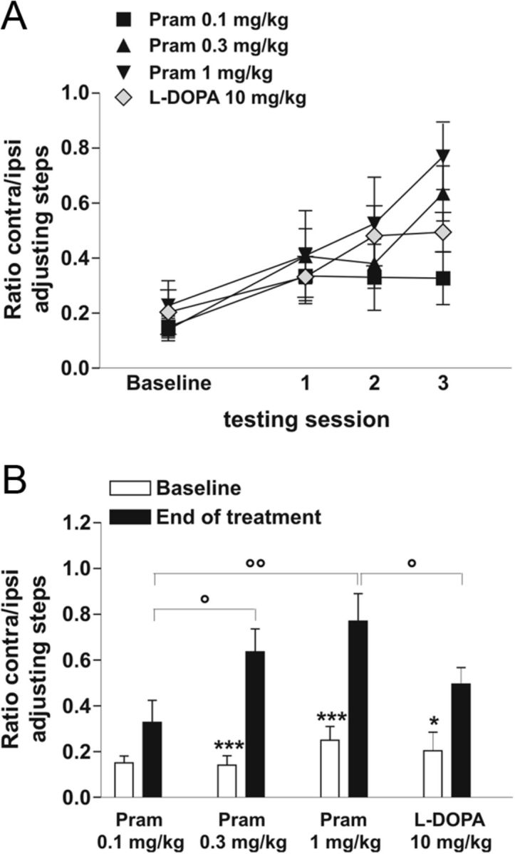
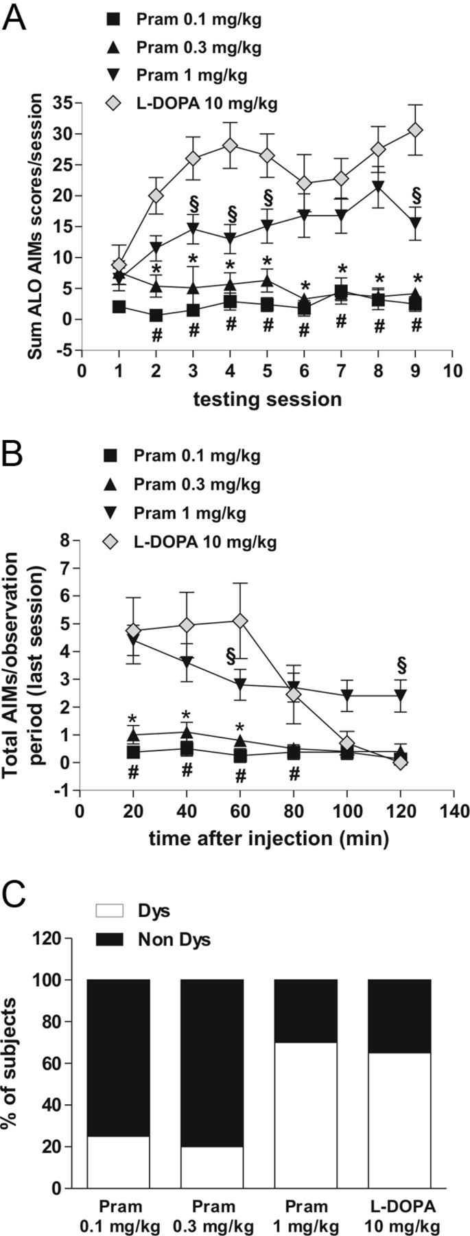
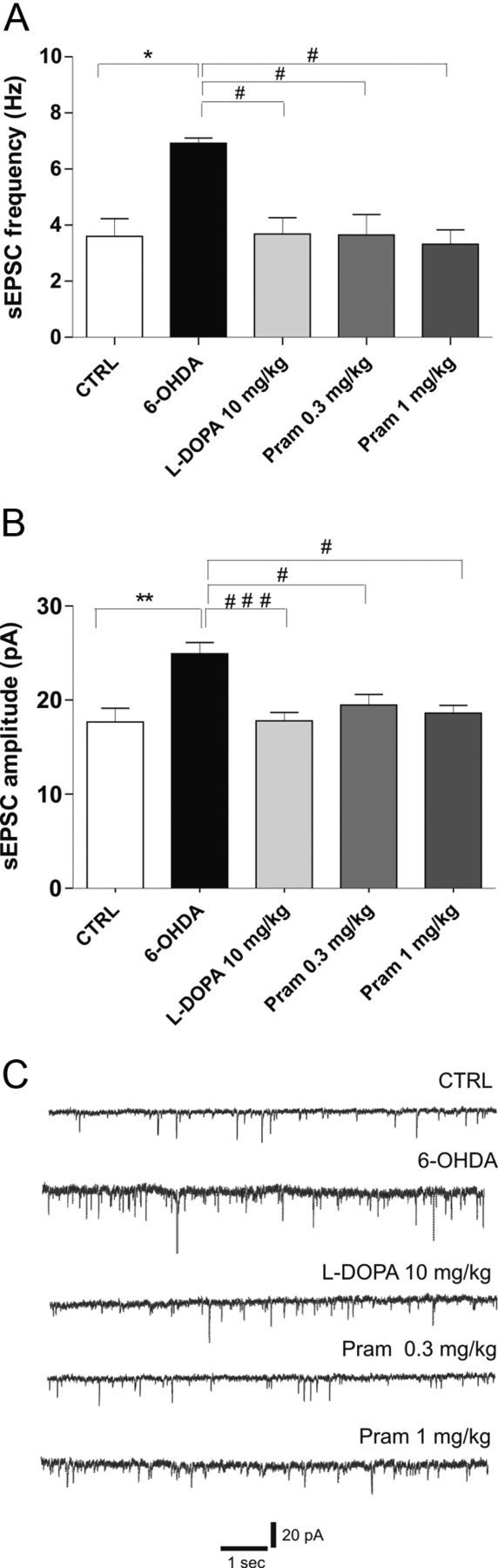
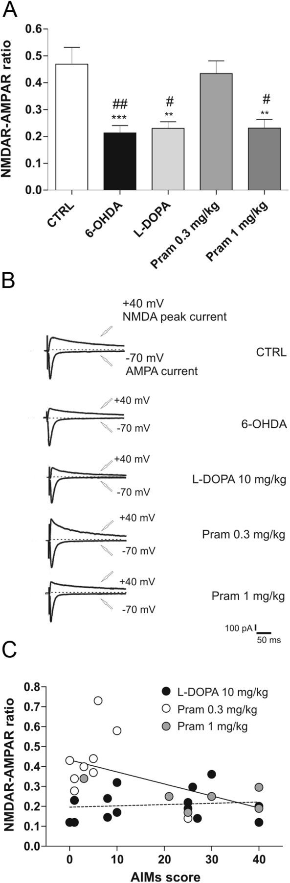
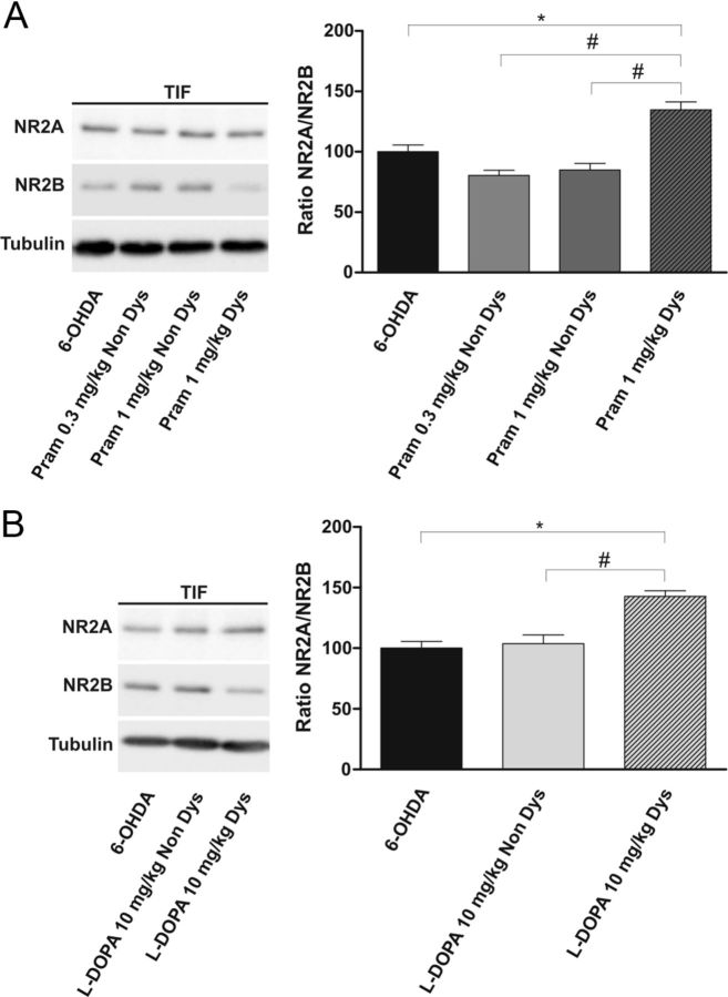
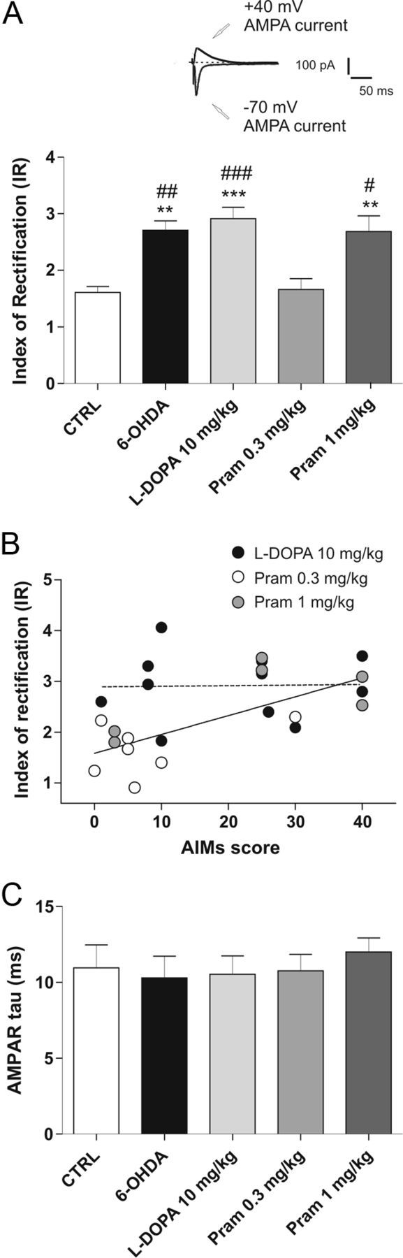
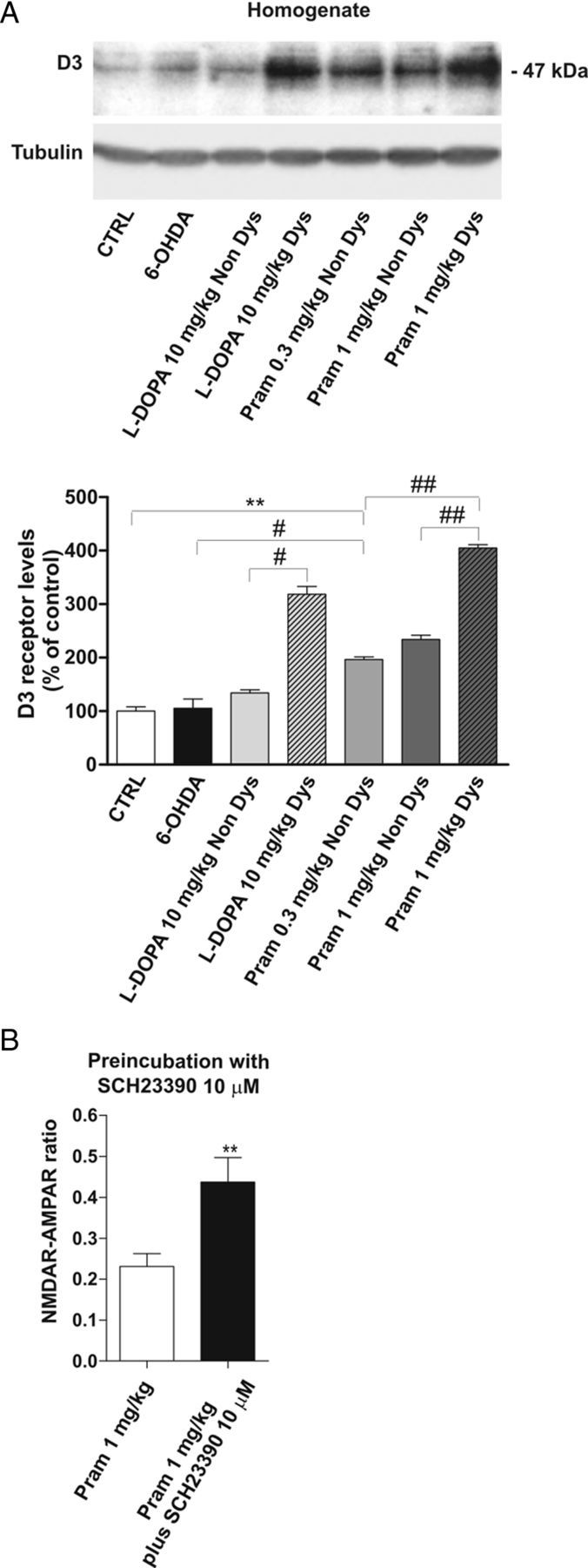
Dopamine replacement with levodopa (L-DOPA) represents the mainstay of Parkinson’s disease (PD) therapy. Nevertheless, this well established therapeutic intervention loses efficacy with the progression of the disease and patients develop invalidating side effects, known in their complex as L-DOPA-induced dyskinesia (LID). Unfortunately, existing therapies fail to prevent LID and very few drugs are available to lessen its severity, thus representing a major clinical problem inPDtreatment. D2-like receptor (D2R) agonists are a powerful clinical option as an alternative to L-DOPA, especially in the early stages of the disease, being associated to a reduced risk of dyskinesia development. D2R agonists also find considerable application in the advanced stages of PD, in conjunction with L-DOPA, which is used in this context at lower dosages, to delay the appearance and the extent of the motor complications. In advanced stages of PD, D2R agonists are often effective in delaying the appearance and the extent of motor complications. Despite the great attention paid to the family of D2R agonists, the main reasons underlying the reduced risk of dyskinesia have not yet been fully characterized. Here we show that the striatal NMDA/AMPAreceptor ratio and theAMPAreceptor subunit composition are altered in experimental parkinsonism in rats. Surprisingly, while L-DOPA fails to restore these critical synaptic alterations, chronic treatment with pramipexole is associated not only with a reduced risk of dyskinesia development but is also able to rebalance, in a dose-dependent fashion, the physiological synaptic parameters, thus providing new insights into the mechanisms of dyskinesia.


Figures

Figure 1.
Figure 1.
Timeline of the experimental plan. Two weeks after the lesion with 6-OHDA, rats were tested with the DA agonist apomorphine (APO) and then selected for successful lesions, based on the number of contraversive turns, counted with automatic rotometers for 40 min as previously reported. Two months after the lesion, the animals were randomly allotted in five different experimentally treated groups: Control group (saline, i.p.); I Pramipexole group (0.1 mg/kg, i.p., twice a day); II Pramipexole group (0.3 mg/kg, i.p., twice a day); III Pramipexole group (1 mg/kg, i.p., once a day); and l-DOPA group (10 mg/kg, twice a day). At the end of chronic pharmacological treatment, animals were killed by cervical dislocation and coronal corticostriatal slices were prepared. After ≥30 min, the slices were used for electrophysiological recordings.
Figure 2.
Figure 2.
Restoration of forelimb akinesia after chronic treatment with dopaminergic drugs: pramipexole versus l-DOPA treatment. A, Graph shows the motor effect of the drugs at the beginning of every week of treatment. All treatments increased the performance in stepping test, without any overall statistically significant differences. B, Graph shows the therapeutic effect of pramipexole versus l-DOPA on the forelimb akinesia, measured prior the beginning of the treatment (Baseline), compared with the last session test (End of treatment). Interestingly, 1 mg/kg doses of pramipexole were more effective than chronic l-DOPA treatment (°p < 0.05), but not statistically different from 0.3 mg/kg pramipexole dose (*p < 0.05, ***p < 0.001; Baseline vs End of treatment) (°p < 0.05, °°p < 0.01 between groups).
Figure 3.
Figure 3.
Therapeutic doses of pramipexole induced fewer dyskinetic movements than l-DOPA. A, l-DOPA but not 0.1 mg/kg and 0.3 mg/kg pramipexole induced the progressive development of AIMs (post hoc test, p < 0.05; #l-DOPA vs pramipexole 0.1 mg/kg; *l-DOPA vs pramipexole 0.3 mg/kg). Pramipexole at the dose of 1 mg/kg induced AIMs. However, AIMs induced by this high dose of pramipexole were significantly fewer than those triggered by l-DOPA (p < 0.05; §l-DOPA vs pramipexole 1 mg/kg). B, Graph shows the development of AIMs measured at the end of the treatment during the last session test. C, Administration of 0.1 mg/kg and 0.3 mg/kg pramipexole induced AIMs in a lower percentage of animals (25% Dys rats for the 0.1 mg/kg group; 20% Dys rats for the 0.3 mg/kg group) than after administration of 1 mg/kg pramipexole (70% Dys rats for the 1 mg/kg group) and l-DOPA (65% Dys).
Figure 4.
Figure 4.
6-OHDA-lesion-related alterations in spontaneous activity of MSNs are rescued by either chronic l-DOPA or pramipexole treatment. A, B, Bar graphs show the averaged values of the frequency (A) and amplitude (B) of spontaneous events recorded in MSNs from the different experimental groups: control (CTRL), 6-OHDA-lesioned rats (6-OHDA), l-DOPA-treated rats, 0.3 mg/kg pramipexole-treated rats, and 1 mg/kg pramipexole-treated rats (n = 8 CTRL vs n = 6 6-OHDA, *p < 0.05 frequency, **p < 0.01 amplitude; n = 14 l-DOPA vs 6-OHDA, #p < 0.05 frequency, ###p < 0.001 amplitude; n = 9 pramipexole 0.3 mg/kg vs 6-OHDA, #p < 0.05 frequency, #p < 0.05 amplitude, n = 6 pramipexole 1 mg/kg vs 6-OHDA, #p < 0.05 frequency, #p < 0.05 amplitude; one-way ANOVA with Bonferroni comparison). C, Representative traces from whole-cell patch-clamp experiments showing glutamatergic sEPSCs recorded from MSNs in control condition (upper trace) and in all the other groups of animals studied.
Figure 5.
Figure 5.
NMDAR/AMPAR ratio reduction after 6-OHDA-lesion and DAergic treatment. A, Summary plot reporting the significant reduction of NMDAR/AMPAR ratio induced by 6-OHDA lesion; neither l-DOPA treatment nor the higher dose of pramipexole rescues this parameter to control levels (n = 10 6-OHDA vs n = 10 CTRL, ***p < 0.001; n = 10 l-DOPA vs CTRL, **p < 0.01; n = 9; pramipexole 0.3 mg/kg vs CTRL, p > 0.05; n = 6, pramipexole 1 mg/kg vs CTRL, **p < 0.01; 6-OHDA vs pramipexole 0.3 mg/kg, ##p < 0.01; l-DOPA vs pramipexole 0.3 mg/kg, #p < 0.05; pramipexole 1 mg/kg vs pramipexole 0.3 mg/kg, #p < 0.05; one-way ANOVA with Bonferroni comparison). B, Representative traces from whole-cell patch-clamp experiments showing AMPAR-evoked and NMDAR-evoked currents, recorded at −70 mV and +40 mV respectively, in the presence of 50 μm picrotoxin. NMDAR component was measured at +40 mV considering the peak amplitude after 50 ms from the beginning of the event. C, Plotting of individual mean AIMs score during the treatment with l-DOPA (filled circles) and pramipexole (open circles) against the individual NMDAR/AMPAR ratio. Significant correlation between the two variables in the group treated with pramipexole (p < 0.05) but not in that treated with l-DOPA is revealed by the linear regression (n = 15 l-DOPA, Spearman's r = 0.1355, p > 0.05; n = 15 pramipexole, Spearman's r = −0.5189, p < 0.05).
Figure 6.
Figure 6.
Alteration of NR2A/NRB subunit ratio in dyskinetic animals treated with l-DOPA or pramipexole. A, B, 6-OHDA-lesioned animals were treated chronically with 0.3 mg/kg pramipexole, with 1 mg/kg pramipexole (A), or with l-DOPA (B) and divided into two groups based on the presence (Dys) or absence (Non Dys) of dyskinesia. Striatal TIFs from the above-indicated experimental groups were analyzed by Western blot (wb) analysis with NR2A, NR2B, and tubulin antibodies (see Materials and Methods). The same amount of proteins was loaded per lane. Histogram in A shows the quantification of Western blotting experiments as NR2A/NR2B ratio after normalization on tubulin levels (Student's t test, 6-OHDA vs pramipexole 1 mg/kg, *p < 0.05; pramipexole 0.3 mg/kg Non Dys vs pramipexole 1 mg/kg Dys, #p < 0.05; pramipexole 1 mg/kg Non Dys vs pramipexole 1 mg/kg Dys, #p < 0.05). B, Histogram shows the NR2A/NR2B distribution between parkinsonian and l-DOPA-treated rats (6-OHDA vs l-DOPA Dys, *p < 0.05; l-DOPA Non Dys vs l-DOPA Dys, #p < 0.05).
Figure 7.
Figure 7.
Modifications of AMPAR subunit complex after 6-OHDA lesion and pharmacological treatments. A, Top of the graph shows example of an individual experiment obtained in control condition to calculate the IR of AMPAR (scale bars, 50 ms and 100 pA). In the bottom, the bar graph shows the IR in the different groups, calculated as the ratio of the AMPAR-mediated EPSCs recorded at −70 mV and +40 mV (n = 7 6-OHDA vs n = 8 CTRL, **p < 0.01; n = 11 l-DOPA vs CTRL, ***p < 0.001; n = 7 pramipexole 1 mg/kg vs CTRL, **p < 0.01; n = 7 6-OHDA vs pramipexole 0.3 mg/kg, ## p < 0.01; l-DOPA vs pramipexole 0.3 mg/kg, ###p < 0.001; pramipexole 1 mg/kg vs pramipexole 0.3 mg/kg, #p < 0.05; one-way ANOVA with Bonferroni comparison). B, IR values obtained from pramipexole-treated and l-DOPA-treated rats were correlated with AIM score. The linear regression revealed a significant correlation between these two variables among the pramipexole-treated group (n = 11 l-DOPA, Pearson's r = 0.0238, p > 0.05; n = 13 pramipexole, Pearson's r = 0.7126, p < 0.01). C, Decay time constants of AMPAR, calculated by fitting with a single exponential, are summarized in the bar graph.
Figure 8.
Figure 8.
Alteration of D3R levels in dyskinetic animals treated with l-DOPA or pramipexole. A, 6-OHDA-lesioned animals were treated chronically with 10 mg/kg l-DOPA, 0.3 mg/kg pramipexole, or 1 mg/kg pramipexole and divided into two groups based on the presence (Dys) or absence (Non Dys) of dyskinesia. Striatal lysates from the above-indicated experimental groups were analyzed by Western blot (wb) analysis with D3 antibodies (see Materials and Methods). The same amount of proteins was loaded per lane. Histograms show the quantification of wb experiments after normalization on tubulin levels (CTRL vs pramipexole 0.3 mg/kg Non Dys, **p < 0.01; 6-OHDA vs pramipexole 0.3 mg/kg Non Dys, #p < 0.05; pramipexole 1 mg/kg Non Dys vs pramipexole 1 mg/kg Dys ##p < 0.01; pramipexole 0.3 mg/kg Non Dys vs pramipexole 1 mg/kg Dys ##p < 0.01; l-DOPA Non Dys vs l-DOPA Dys, #p < 0.05). B, Bar graph compares the NMDAR/AMPAR ratio recorded in vitro from dyskinetic animals after chronic treatment with the high dose of pramipexole, with or without a prolonged incubation of the D1 antagonist SCH23390 (n = 6 pramipexole 1 mg/kg vs n = 5 pramipexole 1 mg/kg plus SCH23390, **p < 0.01).

Similar articles

Cited by

References

    1. Ahlskog JE, Muenter MD. Frequency of levodopa-related dyskinesias and motor fluctuations as estimated from the cumulative literature. Mov Disord. 2001;16:448–458. - PubMed
    1. Ahmed I, Bose SK, Pavese N, Ramlackhansingh A, Turkheimer F, Hotton G, Hammers A, Brooks DJ. Glutamate NMDA receptor dysregulation in Parkinson's disease with dyskinesias. Brain. 2011;134:979–986. - PubMed
    1. Bagetta V, Picconi B, Marinucci S, Sgobio C, Pendolino V, Ghiglieri V, Fusco FR, Giampà C, Calabresi P. Dopamine-dependent long-term depression is expressed in striatal spiny neurons of both direct and indirect pathways: implications for Parkinson's disease. J Neurosci. 2011;31:12513–12522. - PMC - PubMed
    1. Berthet A, Porras G, Doudnikoff E, Stark H, Cador M, Bezard E, Bloch B. Pharmacological analysis demonstrates dramatic alteration of D1 dopamine receptor neuronal distribution in the rat analog of l-DOPA-induced dyskinesia. J Neurosci. 2009;29:4829–4835. - PMC - PubMed
    1. Birkmayer W, Hornykiewicz O. The effect of l-3,4-dihydroxy phenylalanine (=DOPA) on akinesia in parkinsonism. Parkinsonism Relat Disord. 1998;4:59–60. - PubMed

Publication types

MeSH terms