. 2013 Jan 18;288(3):1568-81.
doi: 10.1074/jbc.M112.429548. Epub 2012 Dec 6.

Regulation of cardiac ATP-sensitive potassium channel surface expression by calcium/calmodulin-dependent protein kinase II

Affiliations

Regulation of cardiac ATP-sensitive potassium channel surface expression by calcium/calmodulin-dependent protein kinase II

Ana Sierra et al. J Biol Chem. .

Abstract

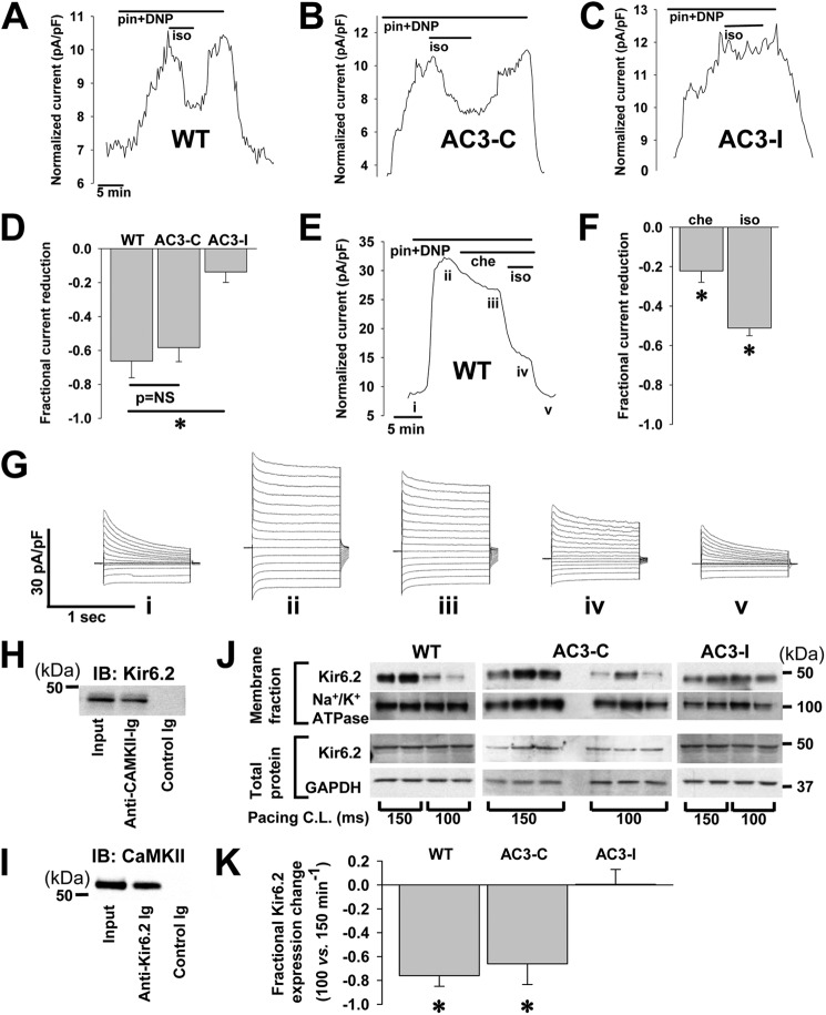
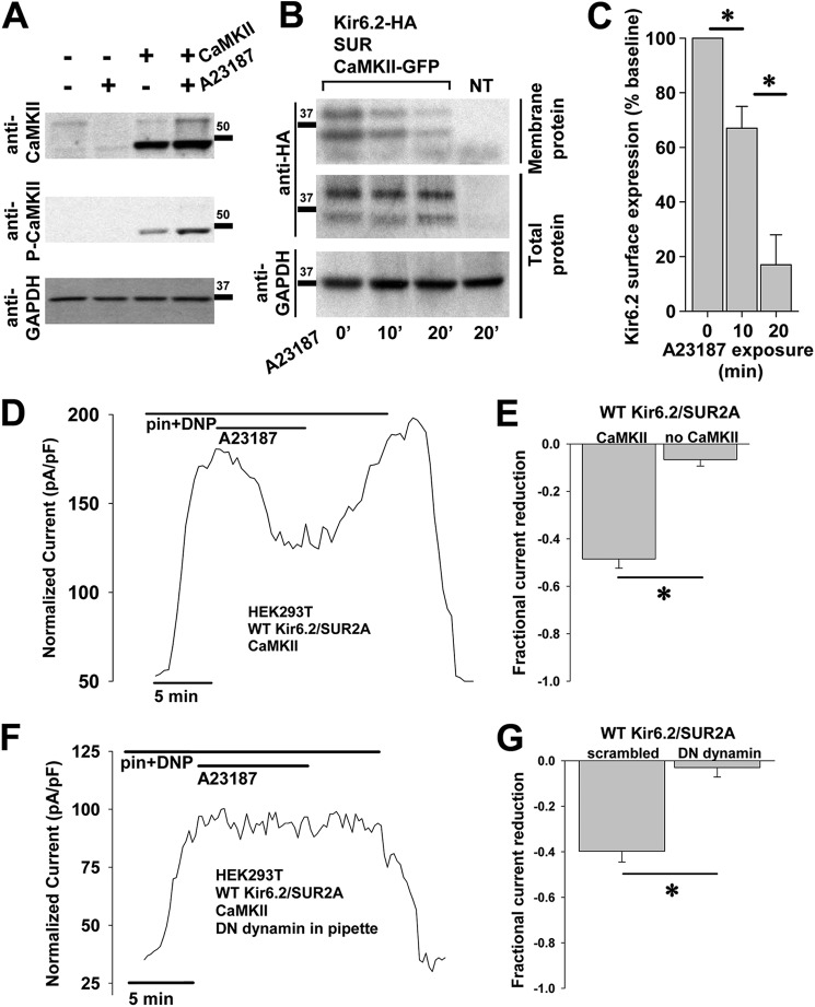
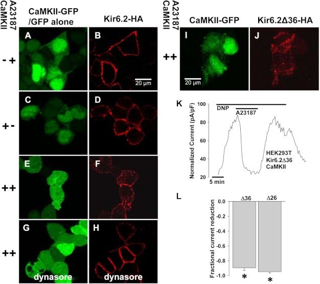
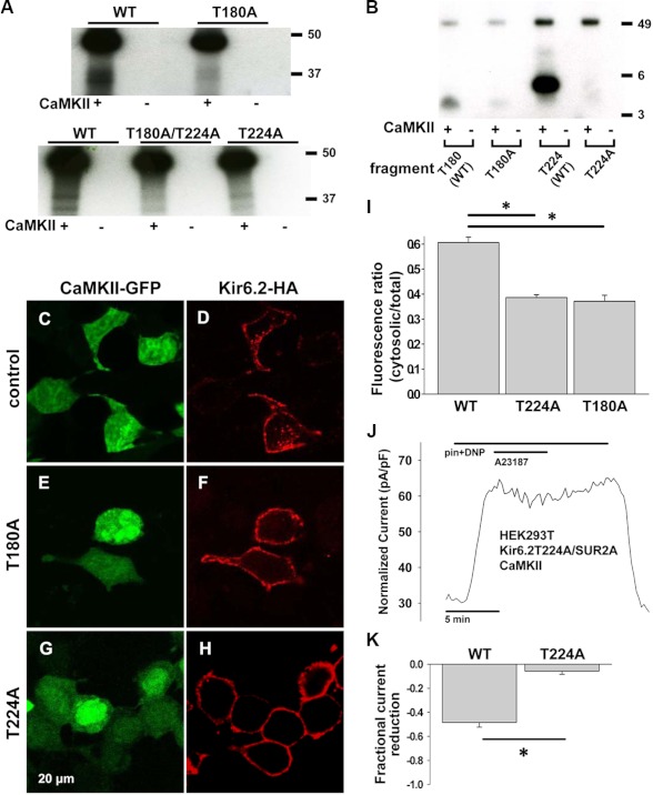
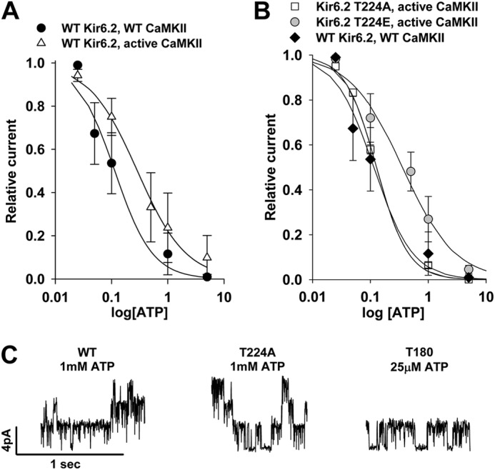
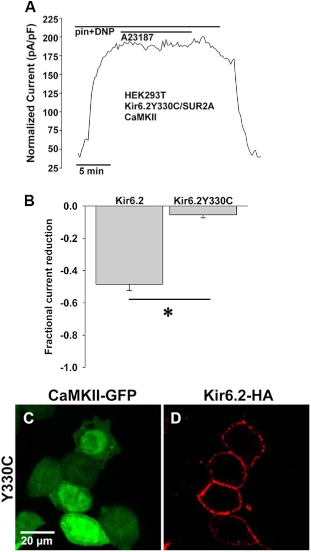
Cardiac ATP-sensitive potassium (K(ATP)) channels are key sensors and effectors of the metabolic status of cardiomyocytes. Alteration in their expression impacts their effectiveness in maintaining cellular energy homeostasis and resistance to injury. We sought to determine how activation of calcium/calmodulin-dependent protein kinase II (CaMKII), a central regulator of calcium signaling, translates into reduced membrane expression and current capacity of cardiac K(ATP) channels. We used real-time monitoring of K(ATP) channel current density, immunohistochemistry, and biotinylation studies in isolated hearts and cardiomyocytes from wild-type and transgenic mice as well as HEK cells expressing wild-type and mutant K(ATP) channel subunits to track the dynamics of K(ATP) channel surface expression. Results showed that activation of CaMKII triggered dynamin-dependent internalization of K(ATP) channels. This process required phosphorylation of threonine at 180 and 224 and an intact (330)YSKF(333) endocytosis motif of the K(ATP) channel Kir6.2 pore-forming subunit. A molecular model of the μ2 subunit of the endocytosis adaptor protein, AP2, complexed with Kir6.2 predicted that μ2 docks by interaction with (330)YSKF(333) and Thr-180 on one and Thr-224 on the adjacent Kir6.2 subunit. Phosphorylation of Thr-180 and Thr-224 would favor interactions with the corresponding arginine- and lysine-rich loops on μ2. We concluded that calcium-dependent activation of CaMKII results in phosphorylation of Kir6.2, which promotes endocytosis of cardiac K(ATP) channel subunits. This mechanism couples the surface expression of cardiac K(ATP) channels with calcium signaling and reveals new targets to improve cardiac energy efficiency and stress resistance.


Figures

FIGURE 1.
FIGURE 1.
CaMKII effect on native KATP current in isolated ventricular cardiomyocytes. Shown is representative whole cell KATP channel current, stimulated by 100 μmol/liter pinacidil (pin) and 50 μmol/liter DNP, measured during application and wash-out of the CaMKII activator 500 nmol/liter isoproterenol (iso) in isolated cardiomyocytes from WT mice (A), trangenic mice expressing a scrambled, control peptide AC3-C (B), and transgenic mice expressing the CaMKII inhibiting peptide, AC3-I (C). pF, picofarads. D, shown are summary data: AC3-I (n = 12) versus WT (n = 4) and AC3-C (n = 3); *, p < 0.05. E, representative whole cell KATP channel current, stimulated by 100 μmol/liter pinacidil and 50 μmol/liter DNP, was measured during application of the PKC inhibitor, 5 μmol/liter chelerythrine (che), and 500 nmol/liter isoproterenol in an isolated WT murine cardiomyocyte. F, shown are summary data for chelerythrine and isoproterenol effects on pinacidil- and DNP-stimulated KATP channel current in isolated WT murine cardiomyocytes (n = 5; *, p < 0.05 versus base line, the isoproterenol bar represents additional reduction beyond that attributed to chelerythrine application). G, shown is an example of whole cell current tracings measured at points marked from the graph in E. H, On left ventricular lysates from WT mice, immunoprecipitation was performed with the anti-Kir6.2 antibody and probed for CaMKII by Western blot (IB) with the anti-CaMKII Ab. I, immunoprecipitation was performed with the anti-CaMKII Ab and probed for Kir6.2 by Western blot with the anti-Kir6.2 Ab. J, shown are Western blots of biotin-labeled membrane and whole cell fractions of ventricular lysates from isolated hearts paced at 150 or 100 ms cycle length (C.L.). Membrane and total protein fractions are probed with anti-Kir6.2 antibody and with antibodies for Na+/K+ ATPase and GAPDH as controls. K, shown are summary data for the fractional change in Kir6.2 expression with pacing cycle length of 100 versus 150 ms (*, p < 0.05, n = 2 hearts under each pacing condition for WT and AC3-I and n = 3 each for AC3-C).
FIGURE 2.
FIGURE 2.
CaMKII effect on recombinant KATP channels in HEK cells. A, HEK cells with/without exogenous overexpression of CaMKII and with/without exposure to A23187 5 μmol/liter were lysed and probed by immunoblot. GAPDH expression served as a control. B, HEK cells expressing Kir6.2-HA, SUR2A, and CaMKII-GFP underwent exposure to 10 μmol/liter A23187 for 0, 10, or 30 min 37 °C. At each time point, groups of cells were cooled to 4 °C and biotinylated. Surface and total protein from lysed cells were assayed for Kir6.2 by anti-HA immunoblot. Cells expressing only CaMKII-GFP without KATP channel subunits served as the negative control. C, shown are summary data of biotinylation experiments in HEK cells, indicating a time-dependent reduction in Kir6.2 surface expression with A23187 exposure (n = 3 each; *, p < 0.05). D, shown is representative whole cell KATP channel current, stimulated by 50 μmol/liter pinacidil (pin) and 50 μmol/liter DNP, measured during application and wash-out of the 5 μmol/liter calcium ionophore A23187 in HEK cells engineered to express Kir6.2, SUR2A, and CaMKII. pF, picofarads. E, shown are summary data from these experiments performed in the presence and absence of CaMKII overexpression (*, p < 0.05). F, the same experiment was performed as in D but with dialysis of the endocytosis inhibitor (DN) dynamin 100μmol/liter through the patch pipette. G, shown are summary data from the experiments performed the presence of dominant negative dynamin or a non-inhibiting, scrambled peptide (100 μmol/liter each, n = 5 each; *, p < 0.05).
FIGURE 3.
FIGURE 3.
CaMKII-dependent internalization of KATP channels and Kir6.2 subunit dependence. Immunofluorescence confocal imaging was performed on HEK cells expressing HA-tagged Kir6.2, WT SUR2A, and GFP-tagged CaMKII or GFP alone without CaMKII. Anti-HA antibody was applied at the beginning of the experiment, and cells were washed. After a 30-min exposure to 10 μmol/liter A23187 to activate CaMKII, cells were cooled, fixed, and permeabilized, and secondary antibody was applied (red) to track surface Kir6.2-HA movement. Shown are cells expressing Kir6.2-HA, SUR2A, and GFP without CaMKII (A and B), Kir6.2-HA, SUR2A, and CaMKII-GFP but not stimulated with A23187 (C and D), Kir6.2-HA, SUR2A, and CaMKII-GFP with application of A23187(E and F), and Kir6.2-HA, SUR2A, and CaMKII-GFP stimulated with A23187 in the presence of the endocytosis inhibitor, dynasore 50μmol/liter (G and H). I and J, immunofluorescence confocal microscopy was performed on HEK cells expressing Kir6.2Δ36-HA (red) and CaMKII-GFP following the same protocol. K, representative whole cell KATP channel current was stimulated by 200 μmol/liter DNP in response to 5 μmol/liter A23187 in a HEK cell expressing Kir6.2Δ36 and CaMKII. L, shown are summary data for DNP-stimulated whole cell KATP channel current from HEK cells expressing CaMKII and Kir6.2Δ26 (n = 4) or Kir6.2Δ36 (n = 5) in response to CaMKII activation by A23187 (*, p < 0.05).
FIGURE 4.
FIGURE 4.
CaMKII effect in Kir6.2T224A and Kir6.2T180A mutants. A, in isolated recombinant cardiac KATP channels with WT or mutated Kir6.2, an in vitro phosphorylation assay was performed in the presence and absence of CaMKII. Kir6.2-HA subunits were mutated from threonine to alanine at Thr-224 or Thr-180 or both. Kir6.2-HA is identified as a double band at ∼37 kDa. The prominent signal near 50 kDA level represents autophosphorylated CaMKII. B, in WT and mutated peptide fragments of Kir6.2, in vitro phosphorylation assay was performed in the presence and absence of CaMKII. Fragments were mutated from threonine to alanine at Thr-224 or Thr-180. Differences in band migration are attributed to different sizes and charge of the fragments. The WT Thr-180 and mutant T180A fragments are identified at ∼3.3 kDa (best seen for Thr-180 + CaMKII and faintly for T180A + CaMKII), whereas the Thr-224 and T224A fragments are identified at ∼4.5 kDa (best seen for Thr-224 + CaMKII). C–H, representative immunofluorescence confocal imaging was performed in HEK cells following the same protocol as in Fig. 3. C and D, cells expressing CaMKII-GFP, Kir6.2-HA, and SUR2A are shown. E and F, cells expressing CaMKII-GFP, Kir6.2T180A-HA, and SUR2A are shown. G and H, cells expressing CaMKII-GFP, Kir6.2T224A-HA and SUR2A. I, summary data for immunofluorescence imaging is shown. For WT, n = 34 cells were from 2 transfections. For T224A, n = 4 transfections, 48 cells; *, p < 0.05 versus WT. For T180A, n = 4 transfections, 42 cells; *, p < 0.05 versus WT. J, representative whole cell KATP channel current was stimulated by 50 μmol/liter pinacidil (pin) and 50 μmol/liter DNP in a HEK cell expressing CaMKII-GFP, Kir6.2T224A, and SUR2A in response to A23187 5 μmol/liter. pF, picofarads. K, summary data of pinacidil- and DNP-stimulated whole cell KATP channel current in response to A23187 in HEK cells expressing CaMKII-GFP, SUR2A, and WT Kir6.2 versus Kir6.2T224A (*, p < 0.05).
FIGURE 5.
FIGURE 5.
CaMKII and mutation effects on biophysical properties of KATP channels. A, shown is KATP channel ATP sensitivity in patches from HEK cells expressing WT Kir6.2, SUR2A, and WT versus constitutively active CaMKII. Symbols indicate raw data. Lines indicate fitted Hill equation. B, KATP channel ATP sensitivity in patches from cells expressing constitutively active CaMKII, SUR2A, and Kir6.2T224A or Kir6.2T224E versus cells expressing WT Kir6.2, SUR2A, and WT CaMKII is shown. Symbols indicate raw data. Lines indicate the fitted Hill equation. C, shown are examples of single-channel inside-out recordings from HEK cells expressing WT CaMKII, SUR2A, and WT Kir6.2 in 1 mmol/liter ATP (left), Kir6.2T224A in 1 mmol/liter ATP (middle), or Kir6.2T180A in 25 μmol/liter ATP (right). The last panel is not entirely representative as the majority of patches from cells expressing Kir6.2T180A had no KATP channels even in the absence of ATP.
FIGURE 6.
FIGURE 6.
Mutation of YXXØ internalization motif interferes with CaMKII-induced KATP channel down-regulation. A, shown is representative whole cell KATP channel current, stimulated by 50 μmol/liter pinacidil (pin) and 50 μmol/liter DNP, measured during application and wash-out of 5 μmol/liter A23187 in HEK cells engineered to express Kir6.2Y330C-HA, SUR2A, and CaMKII-GFP. pF, picofarads. B, shown are summary data from whole cell patch clamp experiments performed with Kir6.2-HA (n = 4) versus Kir6.2Y330C-HA (n = 6; *, p < 0.05). C and D, shown are representative confocal fluorescence images of HEK cells expressing Kir6.2Y330C-HA, SUR2A, and CaMKII-GFP after treatment with A23187.
FIGURE 7.
FIGURE 7.
Homology model of Kir6.2. A, a three-dimensional model of Kir6.2 presented as a solvent-accessible surface (except the phosphatidylinositol diphosphate analog) focused on the ATP binding site indicates the position of Thr-180 (red) within the ATP binding pocket (blue) adjacent to the YSKF internalization motif (green). Thr-224 (also red) is seen in the bottom right corner of the image. B, the same model shows little change in the relationship of Thr-180 or Thr-224 to YSKF with phosphorylation of both Thr-180 and Thr-224. The calculated residue displacements (root mean square deviation) induced by phosphorylation of Kir6.2 are: Kir6.2 + Thr-180(P) = 0.39 Å, Kir6.2 + Thr-224(P) = 0.17 Å, and Kir6.2 + Thr-180(P) + Thr-224(P) = 0.43 Å. The root mean square deviation is slightly larger when Thr-180 is phosphorylated as Thr-180 is embedded in the Kir6.2 homotetramer, poorly accessible to the solvent. As a consequence, the neighbor residues as well as the backbone of Thr-180 adapt their conformation due to introduction of the bulky phosphate group. In contrast, Thr-224 is well exposed on the surface of Kir6.2. The phosphorylation of this amino acid has little consequence on neighbor residues. Both Thr-180 and Thr-224 are remote from the YSKF signal. The closest residue of the YSKF motif to Thr-180 is Tyr-330. The distance between the phosphate/hydroxyl of Thr-180 and the hydroxyl of Tyr-330 is, respectively, 9.3, 9.6, 9.3, and 9.7 Å in Kir6.2 and Kir6.2 phosphorylated at Thr-180, Kir6.2 phosphorylated at Thr-224, and Kir6.2 phosphorylated at both Thr-180 and Thr-224. The closest YSKF residue to Thr-224 is Lys-332. The distance between the phosphate/hydroxyl of Thr-224 and the ammonium of Lys-332 is, respectively, 18.8, 19.0, 19.8, and 19.8 Å in Kir6.2, Kir6.2 phosphorylated at Thr-180, Kir6.2 phosphorylated at Thr-224, and Kir6.2 phosphorylated at both Thr-180 and Thr-224. C, a side view model of Kir6.2, with two subunits removed for clarity, indicates proposed docking with μ2 (large, green). The ATP binding pocket (blue), YSKF (green), Thr-180 (upper red), and Thr-224 (lower red) are also shown. D, an end-on view of the Kir6.2 model shows the relationship of docked μ2 (large, green) with adjacent subunits of Kir6.2. Thr-224 (red) is largely obscured by μ2. Kir6.2 subunits A and C are white, and subunits B and D are gray.

References

    1. Weiss J. N., Venkatesh N. (1993) Metabolic regulation of cardiac ATP-sensitive K+ channels. Cardiovasc. Drugs Ther. 7, 499–505 - PubMed
    1. Noma A. (1983) ATP-regulated K+ channels in cardiac muscle. Nature 305, 147–148 - PubMed
    1. Aguilar-Bryan L., Bryan J. (1999) Molecular biology of adenosine triphosphate-sensitive potassium channels. Endocr. Rev. 20, 101–135 - PubMed
    1. Ashcroft F. M. (1988) Adenosine 5′-triphosphate-sensitive potassium channels. Annu. Rev. Neurosci. 11, 97–118 - PubMed
    1. Lederer W. J., Nichols C. G. (1989) Nucleotide modulation of the activity of rat heart ATP-sensitive K+ channels in isolated membrane patches. J. Physiol. 419, 193–211 - PMC - PubMed

Publication types

MeSH terms