. 2013 Feb 8;288(6):3705-17.
doi: 10.1074/jbc.M112.416651. Epub 2012 Dec 6.

Heparan sulfate 6-O-sulfotransferase isoform-dependent regulatory effects of heparin on the activities of various proteases in mast cells and the biosynthesis of 6-O-sulfated heparin

Affiliations

Heparan sulfate 6-O-sulfotransferase isoform-dependent regulatory effects of heparin on the activities of various proteases in mast cells and the biosynthesis of 6-O-sulfated heparin

Md Ferdous Anower-E-Khuda et al. J Biol Chem. .

Abstract

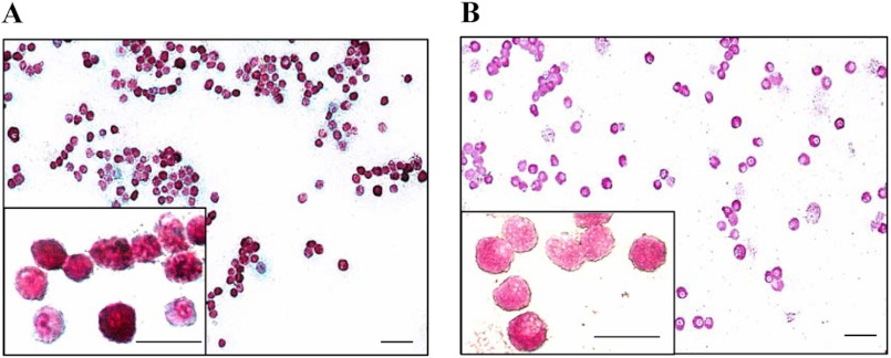
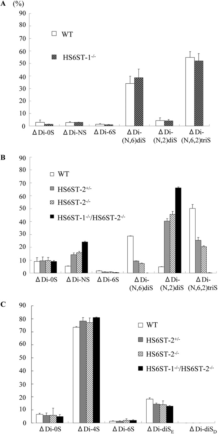
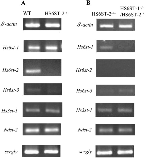
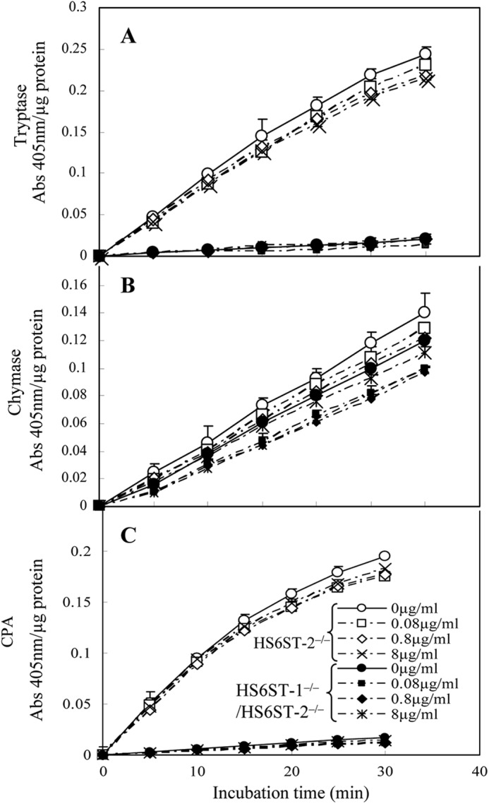
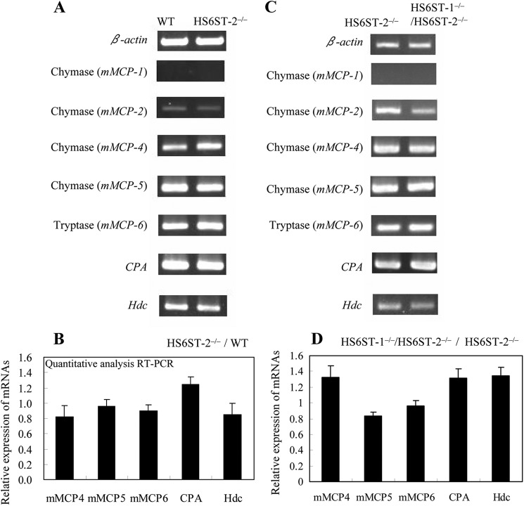
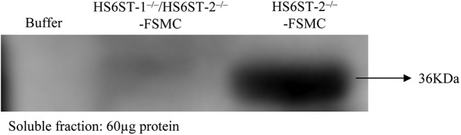
Heparan sulfate 6-O-sulfotransferase (HS6ST) is an enzyme involved in heparan sulfate (HS) biosynthesis that transfers a sulfate residue to position 6 of the GlcNAc/GlcNSO(3) residues of HS, and it consists of three isoforms. Heparin, the highly sulfated form of HS, resides in connective tissue mast cells and is involved in the storage of mast cell proteases (MCPs). However, it is not well understood which isoform(s) of HS6ST participates in 6-O-sulfation of heparin and how the 6-O-sulfate residues in heparin affect MCPs. To investigate these issues, we prepared fetal skin-derived mast cells (FSMCs) from wild type (WT) and HS6ST-deficient mice (HS6ST-1(-/-), HS6ST-2(-/-), and HS6ST-1(-/-)/HS6ST-2(-/-)) and determined the structure of heparin, the protease activity, and the mRNA expression of each MCP in cultured FSMCs. The activities of tryptase and carboxypeptidase-A were decreased in HS6ST-2(-/-)-FSMCs in which 6-O-sulfation of heparin was decreased at 50% of WT-FSMCs and almost lost in HS6ST-1(-/-)/HS6ST-2(-/-)-FSMCs, which lacked the 6-O-sulfation in heparin nearly completely. In contrast, chymase activity was retained even in HS6ST-1(-/-)/HS6ST-2(-/-)-FSMCs. Each MCP mRNA was not decreased in any of the mutant FSMCs. Western blot analysis showed that tryptase (mMCP-6) was almost absent from HS6ST-1(-/-)/HS6ST-2(-/-)-FSMCs indicating degradation/secretion of the enzyme protein. These observations suggest that both HS6ST-1 and HS6ST-2 are involved in 6-O-sulfation of heparin and that the proper packaging and storage of tryptase, carboxypeptidase-A, and chymase may be regulated differently by the 6-O-sulfate residues in heparin. It is thus likely that 6-O-sulfation of heparin plays important roles in regulating MCP functions.


Figures

FIGURE 1.
FIGURE 1.
Characterization of HS/heparin from WT and HS6ST-2−/− ears. GAGs were prepared from the ears of 8-week-old WT and HS6ST-2−/− mice as described under “Experimental Procedures.” A, aliquot of these GAGs was digested with a mixture of heparitinase-I, heparitinase-II, and heparinase and then subjected to reverse phase ion pair chromatography with postcolumn fluorescence labeling as described under “Experimental Procedures.” The histogram shows the percent composition of unsaturated disaccharides in HS/heparin from wild type (open bars) and HS6ST-2−/− ears (filled bars). ΔDi-0S, ΔDi-NS, ΔDi-6S, ΔDi-(N,6)diS, ΔDi-(N,2)diS, and ΔDi-(N,6,2)triS are unsaturated disaccharides derived from the following units in HS/heparin-HexA-GlcNAc-, -HexA-GlcNSO3-, -HexA-GlcNAc(6SO4)-, -HexA-GlcNSO3(6SO4)-, -HexA(2SO4)-GlcNSO3-, and -HexA(2SO4)-GlcNSO3(6SO4)-, respectively. The values shown are the means of triplicate analyses, and the bars represent the standard deviation. B, GAGs from wild type and HS6ST-2−/− ears were digested with chondroitinase ABC; the digested products were removed, and the remaining HS/heparin from wild type and HS6ST-2−/−ears were fractionated into the following six fractions by Mono Q column chromatography as described under “Experimental Procedures”: fraction I (30.5–33.5 min), fraction II (34–36.5 min), fraction III (37–41 min), fraction IV (41.5–44 min), fraction V (44.5–46 min), and fraction VI (46.5–49 min). The % yields of each fraction from WT and HS6ST-2−/− heparin/HS were calculated from the total amounts of unsaturated disaccharides. C, disaccharide composition of each fraction was analyzed as described under “Experimental Procedures.”
FIGURE 2.
FIGURE 2.
Histocytochemical detection of heparin and chloroacetate esterase in WT-FSMCs. A, WT-FSMCs from C57BL/6 mice were cultured for 5 weeks, and then double-stained with Alcian blue and safranin O. Red staining in the FSMCs is heparin. B, WT-FSMCs were also reacted with naphthol AS-D chloroacetate to detect chloroacetate esterase activity. FSMCs containing chymotrypsin-like activity were positively stained (pink). Bar, 50 μm. The granular dot staining outside the cells shows the presence of heparin/CS due to the degranulation of some mast cells. Insets are the 4-fold magnification of some parts of respective stained FSMCs. Bar, 25 μm.
FIGURE 3.
FIGURE 3.
Disaccharide compositions of GAGs from WT, HS6ST-1−/−, HS6ST-2+/−, HS6ST-2−/−, and HS6ST-1−/−/HS6ST-2−/−-FSMCs. FSMCs were prepared from WT, HS6ST-1−/−, HS6ST-2+/−, HS6ST-2−/−, and HS6ST-1−/−/HS6ST-2−/− embryos as described under “Experimental Procedures.” HS/heparin and chondroitin sulfate were isolated from FSMCs, digested with a mixture of heparitinase-I, heparitinase-II, and heparinase, and chondroitinase ABC, and subjected to reverse phase ion pair chromatography as described under “Experimental Procedures.” The histograms show the percentage compositions of the unsaturated disaccharides in the HS/heparin (A and B) and in chondroitin sulfate (C) isolated from the FSMCs of WT (open bar), HS6ST-1−/− (Crossed hatched bar), HS6ST-2+/− (gray bar), HS6ST-2−/− (hatched bar), and HS6ST-1−/−/HS6ST-2−/− (filled bar). A and B, the values (heights of the histograms) shown are the means of triplicate experiments, and the bars represent the standard deviation. ΔDi-0S, ΔDi-NS, ΔDi-6S, ΔDi-(N,6)diS, ΔDi-(N,2)diS, and ΔDi-(N,6,2)triS are the same as described in the legend for Fig. 1A. C, ΔDi-0S, ΔDi-4S, ΔDi-6S, ΔDi-diSE, and ΔDi-diSD are unsaturated disaccharides derived from -HexA-GalNAc-, -HexA-GalNAc(4SO4)-, -HexAGalNAc(6SO4)-, -HexA-GalNAc(4,6)SO4-, and -HexA(2SO4)-GalNAc(6SO4)- units of the GAGs, respectively.
FIGURE 4.
FIGURE 4.
Expression of the mRNAs of various sulfotransferases and serglycin in WT, HS6ST-2−/−, and HS6ST-1−/−/HS6ST-2−/−-FSMCs. The expression of Hs6st-1, Hs6st-2, Hs6st-3, Hs3st-1, Ndst-2, and serglycin mRNAs in WT, HS6ST-2−/−, and HS6ST-1−/−/HS6ST-2−/−-FSMCs was measured by semi-quantitative RT-PCR using the primer pairs shown in Table 1. β-Actin was used as a control. A, expression of mRNA was compared between WT and HS6ST-2−/−-FSMCs. B, expression of mRNA was compared between HS6ST-2−/− and HS6ST-1−/−/HS6ST-2−/−-FSMCs. The expression levels for the left and right panels were normalized to the respective β-actin mRNA expression levels that were different between the left and right panels.
FIGURE 5.
FIGURE 5.
Activities of tryptase (A), chymase (B), and carboxypeptidase A (C) in extracts from HS6ST-2−/− and HS6ST-1−/−/HS6ST-2−/−-FSMCs and the effects of heparin addition. Extracts from HS6ST-2−/− (open symbols) and HS6ST-1−/−/HS6ST-2−/−-FSMCs (closed symbols) (1 μg of protein) were incubated with substrates (S-2288 for tryptase, S-2586 for chymase, and M-2245 for CPA) as described under “Experimental Procedures” in the presence and absence of three different concentrations of heparin (0.08, 0.8, and 8 μg/ml) at 37 °C, and their activities were monitored as absorbance at 405 nm as described under “Experimental Procedures.” The addition of heparin to these extracts did not affect their activities. The values shown represent the mean ± S.D. of triplicate determinations.
FIGURE 6.
FIGURE 6.
Expression levels of mMCPs, CPA, and Hdc mRNAs in WT, HS6ST-2−/−, and HS6ST-1−/−/HS6ST-2−/−-FSMCs. Total RNA was isolated from the FSMCs of WT and HS6ST-2−/− littermates (A and B) or HS6ST-2−/− and HS6ST-1−/−/HS6ST-2−/− littermates (C and D). A and C, expression of each mRNA was measured by semi-quantitative RT-PCR using primer pairs as described under “Experimental Procedures.” The expression level of β-actin was used as a control. The mRNA expression was compared between WT and HS6ST-2−/− FSMCs (A) or between HS6ST-2−/− and HS6ST-1−/−/HS6ST-2−/−-FSMCs (C). B and D, mRNA expression was quantitatively measured using SYBR Green according to the manufacturer's protocol, and expression levels were normalized to β-actin expression as described under “Experimental Procedures.” The mRNA expression was compared between WT and HS6ST-2−/−-FSMCs (B) or between HS6ST-2−/− and HS6ST-1−/−/HS6ST-2−/−-FSMCs (D). Relative mRNA expression values were obtained from two independent experiments that were performed in triplicate, and error bars represent the standard deviation.
FIGURE 7.
FIGURE 7.
Immunoblot analysis of tryptase (mMCP-6) from HS6ST-1−/−/HS6ST-2−/−-FSMC. Proteins were extracted with a high salt buffer containing Triton X-100, EDTA, and a mixture of protease inhibitors. Sixty micrograms of protein was subjected to 10% SDS-PAGE, and the immunoblots were probed using an antibody against mMCP-6 as described under “Experimental Procedures.” The extraction buffer was loaded in the left lane as a control.

References

    1. Esko J. D., Lindahl U. (2001) Molecular diversity of heparan sulfate. J. Clin. Invest. 108, 169–173 - PMC - PubMed
    1. Bernfield M., Götte M., Park P. W., Reizes O., Fitzgerald M. L., Lincecum J., Zako M. (1999) Functions of cell surface heparan sulfate proteoglycans. Annu. Rev. Biochem. 68, 729–777 - PubMed
    1. Perrimon N., Bernfield M. (2000) Specificities of heparan sulphate proteoglycans in developmental processes. Nature 404, 725–728 - PubMed
    1. Grobe K., Ledin J., Ringvall M., Holmborn K., Forsberg E., Esko J. D., Kjellén L. (2002) Heparan sulfate and development: differential roles of the N-acetylglucosamine N-deacetylase/N-sulfotransferase isozymes. Biochim. Biophys. Acta 1573, 209–215 - PubMed
    1. Esko J. D., Selleck S. B. (2002) Order out of chaos. Assembly of ligand binding sites in heparan sulfate. Annu. Rev. Biochem. 71, 435–471 - PubMed

Publication types

LinkOut - more resources