

doi: 10.1039/c2cc37529j.
Epub 2012 Dec 5.
Cadmium-free quantum dots as time-gated bioimaging probes in highly-autofluorescent human breast cancer cells
Affiliations
- PMID: 23223513
- PMCID: PMC3570570
- DOI: 10.1039/c2cc37529j
Item in Clipboard
Cadmium-free quantum dots as time-gated bioimaging probes in highly-autofluorescent human breast cancer cells
Chem Commun (Camb).
.
Abstract
We report cadmium-free, biocompatible (Zn)CuInS(2) quantum dots with long fluorescence lifetimes as superior bioimaging probes using time-gated detection to suppress cell autofluorescence and improve the signal : background ratio by an order of magnitude. These results will be important for developing non-toxic fluorescence imaging probes for ultrasensitive biomedical diagnostics.
Figures
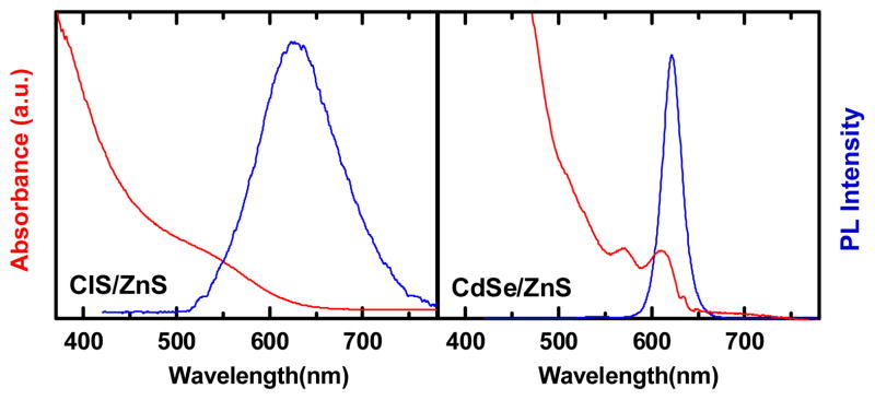
Absorption and photoluminescence spectra of CIS/ZnS and CdSe/ZnS QDs
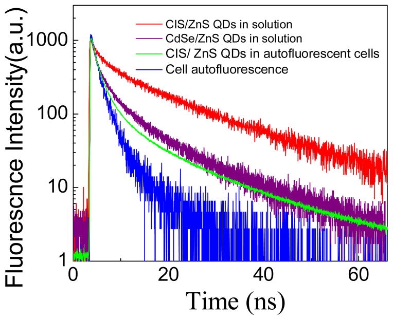
Lifetime decay curves of CdSe/ZnS QDs, CIS/ZnS QDs and SK-BR-3 cells with and without CIS QDs. Average Lifetime is calculated from multi-exponential fitting: τavg = Σi Aiτi, with i = 3, Ai is the fractional amplitude (Σi Ai = 1) and τi are the lifetimes of each decay component
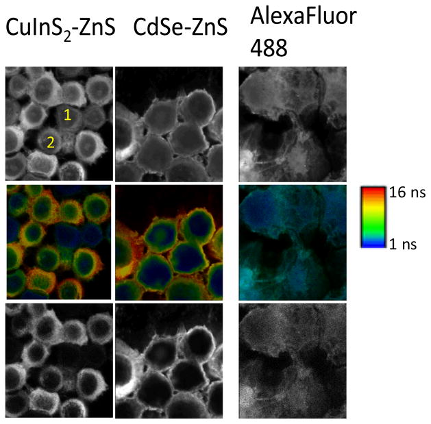
Fluorescence intensity images with two cells labeled 1 and 2 that are difficult to quantify in terms of extent of labeling; Middle row: FLIM images; ranging from 1ns (blue) to 16 ns (red); Bottom row: Image after time-gating the first 10 ns of photons significantly improving contrast, and removes unlabeled cells from the images.
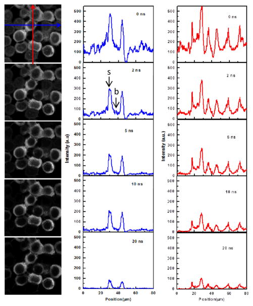
Fluorescence intensity images and cross-sections in x and y to compare the signal (s): background (b) levels of CIS/ZnS-labeled cells as a function of the time-gate applied.
Similar articles
-
Highly luminescent water-soluble quaternary Zn-Ag-In-S quantum dots for tumor cell-targeted imaging.Phys Chem Chem Phys. 2013 Apr 14;15(14):5078-83. doi: 10.1039/c3cp00046j. Phys Chem Chem Phys. 2013. PMID: 23450151
-
Cadmium-Free Quantum Dots as Fluorescent Labels for Exosomes.Sensors (Basel). 2018 Oct 2;18(10):3308. doi: 10.3390/s18103308. Sensors (Basel). 2018. PMID: 30279349 Free PMC article.
-
Amino Acid-Capped Water-Soluble Near-Infrared Region CuInS2/ZnS Quantum Dots for Selective Cadmium Ion Determination and Multicolor Cell Imaging.Anal Chem. 2019 Jul 16;91(14):8987-8993. doi: 10.1021/acs.analchem.9b01183. Epub 2019 Jul 2. Anal Chem. 2019. PMID: 31265249
-
Semiconductor quantum dots for in vivo imaging.J Nanosci Nanotechnol. 2007 Aug;7(8):2567-81. doi: 10.1166/jnn.2007.628. J Nanosci Nanotechnol. 2007. PMID: 17685272 Review.
-
Luminescent copper indium sulfide (CIS) quantum dots for bioimaging applications.Nanoscale Horiz. 2021 Sep 1;6(9):676-695. doi: 10.1039/d1nh00260k. Epub 2021 Jul 15. Nanoscale Horiz. 2021. PMID: 34264247 Review.
Cited by
-
Fluorescence lifetime imaging microscopy in the medical sciences.Protoplasma. 2014 Mar;251(2):293-305. doi: 10.1007/s00709-013-0598-4. Epub 2014 Jan 4. Protoplasma. 2014. PMID: 24390249 Review.
-
Preparation of Photoluminescence Tunable Cu-doped AgInS2 and AgInS2/ZnS Nanocrystals and Their Application as Cellular Imaging Probes.RSC Adv. 2016;6(56):51161-51170. doi: 10.1039/C6RA09494E. Epub 2016 May 19. RSC Adv. 2016. PMID: 27293549 Free PMC article.
-
Applications of functionalized nanomaterials in photodynamic therapy.Biophys Rev. 2018 Feb;10(1):49-67. doi: 10.1007/s12551-017-0383-2. Epub 2018 Jan 2. Biophys Rev. 2018. PMID: 29294258 Free PMC article. Review.
-
Heat-up Synthesis of Ag-In-S and Ag-In-S/ZnS Nanocrystals: Effect of Indium Precursors on Their Optical Properties.J Alloys Compd. 2016 Apr 25;665:137-143. doi: 10.1016/j.jallcom.2016.01.035. J Alloys Compd. 2016. PMID: 26834389 Free PMC article.
-
III-V colloidal nanocrystals: control of covalent surfaces.Chem Sci. 2019 Nov 26;11(4):913-922. doi: 10.1039/c9sc04290c. Chem Sci. 2019. PMID: 34084346 Free PMC article. Review.
References
-
- Weiss S. Science. 1999;283:1676. - PubMed
- Alivisatos P. Nat Biotechnol. 2004;22(1):47. - PubMed
- Zrazhevskiy P, Senawb M, Gao Xiaohu. Chem Soc Rev. 2010;39:4326. - PMC - PubMed
- Barroso MM. J Histochem Cytochem. 2011;59:237. - PMC - PubMed
- Jaiswal JK, Mattoussi H, Mauro JM, Simon SM. Nat Biotechnol. 2003;21:47. - PubMed
-
- Bruchez M, Jr, Moronne M, Gin P, Weiss S, Alivisatos AP. Science. 1998;281:2013. - PubMed
- Chan WCW, Nie S. Science. 1998;281:2016. - PubMed
- Klarreich E. Nature. 2001;413:450. - PubMed
- Mitchell P. Nat Biotechnol. 2001;19:1013. - PubMed
- Yong KT, Roy I, Swihart MT, Prasad PN. J Mater Chem. 2009;19:4655. - PMC - PubMed
- Biju V, Itoh T, Ishikawa M. Chem Soc Rev. 2010;39:3031. - PubMed
- Mieszawska AJ, Gianella A, Cormode DP, Zhao Y, Meijerink A, Langer R, Farokhzad OC, Fayada ZA, Mulder WJM. Chem Commun. 2012;48:5835. - PMC - PubMed
Publication types
MeSH terms
Substances
Grants and funding
LinkOut - more resources
Full Text Sources
Other Literature Sources