. 2012 Dec 26;109(52):21450-5.
doi: 10.1073/pnas.1207754110. Epub 2012 Dec 5.

Microfluidic Western blotting

Affiliations

Microfluidic Western blotting

Alex J Hughes et al. Proc Natl Acad Sci U S A. .

Abstract

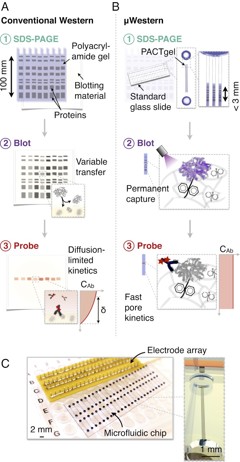
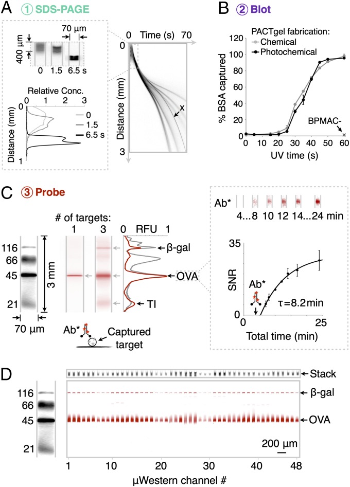
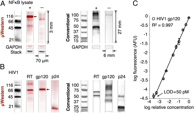
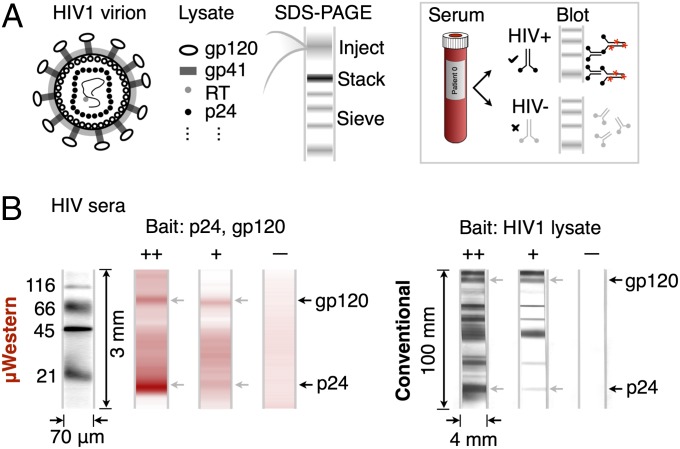
Rapid, quantitative Western blotting is a long-sought bioanalytical goal in the life sciences. To this end, we describe a Western blotting assay conducted in a single glass microchannel under purely electronic control. The μWestern blot is comprised of multiple steps: sample enrichment, protein sizing, protein immobilization (blotting), and in situ antibody probing. To validate the microfluidic assay, we apply the μWestern blot to analyses of human sera (HIV immunoreactivity) and cell lysate (NFκB). Analytical performance advances are achieved, including: short durations of 10-60 min, multiplexed analyte detection, mass sensitivity at the femtogram level, high-sensitivity 50-pM detection limits, and quantitation capability over a 3.6-log dynamic range. Performance gains are attributed to favorable transport and reaction conditions on the microscale. The multistep assay design relies on a photopatternable (blue light) and photoreactive (UV light) polyacrylamide gel. This hydrophilic polymer constitutes both a separation matrix for protein sizing and, after brief UV exposure, a protein immobilization scaffold for subsequent antibody probing of immobilized protein bands. We observe protein capture efficiencies exceeding 75% under sizing conditions. This compact microfluidic design supports demonstration of a 48-plex μWestern blot in a standard microscope slide form factor. Taken together, the μWestern blot establishes a foundation for rapid, targeted proteomics by merging exceptional specificity with the throughput advantages of multiplexing, as is relevant to a broad range of biological inquiry.


Conflict of interest statement

The authors declare no conflict of interest.

Figures

Fig. 1.
Fig. 1.
Single-microchannel μWestern assay design enables high device density formats. Aspects of scale, reagent use, blotting efficiency, and probe-binding kinetics are illustrated by comparative schematics for the conventional (A) and μWestern (B) assays (δ indicates a diffusion boundary layer thickness). The microfluidic workflow is comprised of: (i) analyte stacking and SDS-PAGE within the PACTgel matrix; (ii) band capture (“blotting”) onto the benzophenone-decorated PACTgel in response to UV light (as opposed to transfer to a separate sheet of hydrophobic material in conventional Western blotting); (iii) removal of SDS by brief electrophoretic washing and electrophoretic introduction of fluorescently labeled primary and (optionally) secondary detection antibodies specific to the target. Finally, excess probe is electrophoretically driven out of each device and peak intensities determined by fluorescence micrograph analysis. (C) Modular interfacing of standard microscope slide-sized chips with a scalable electrode array accommodating 48 blots per chip in triplicate (144 microchannels).
Fig. 2.
Fig. 2.
Compact μWestern with integrated high-resolution SDS-PAGE, blot, and detection. (A) SDS-PAGE of fluorescently labeled six protein ladder (black), complete in 60 s (4× magnification; band weights are 155, 98, 63, 40, 32, and 21 kDa). Channel aspect ratios are adjusted to produce gel-like images (see dimensions). (B) Capture efficiency of BSA (± SD, n = 3) for PACTgels fabricated chemically or photochemically. (C, Left) Multiplexed μWestern readout (red) in 40-min total assay times using primary antibodies for (i) OVA, and (ii) β-gal, OVA, and TI; all at 1 μM. (Right) Fluorescence micrographs and plot of SNR (± SD, n = 3) for electrophoretic introduction of red fluorescent primary antibody (Ab*) to OVA band at 4 min total assay time (arrow). (D) Forty-eight concurrent μWesterns of the four-protein fluorescent ladder probed for OVA and β-gal targets (1 μM each) with unlabeled primary and red fluorescent secondary antibodies in 60-min total assay time. At top, total injected (“stack”) fluorescence on weight marker spectral channel at the end of the ITP phase of SDS-PAGE acts as loading control.
Fig. 3.
Fig. 3.
Validation of μWestern for cell lysate and purified proteins. (A) Sixty-minute μWestern of 0.5 mg/mL transfected 293T lysate probed for NFκB with unlabeled primary and fluorescently labeled secondary antibodies (red). Untransfected negative control lysate and loading controls (GAPDH and total injected fluorescence, “stack”) are included. (Right) The corresponding conventional 6- to 8-h Western blot readouts for visual comparison. Note relative dimensions of the conventional blot. (B) Forty-minute μWestern of purified HIV proteins (reverse-transcriptase, 200 nM; gp120, 200 nM; p24, 1 μM) after probing targets with fluorescently labeled primary antibodies (red). (C) Standard curve for gp120 over the 50 pM to 200 nM range (± SD, n = 3) constructed from peak areas of the band indicated by an arrow in B. See Fig. S6 for standard curve of the NFκB p105 peak indicated by an arrow in A.
Fig. 4.
Fig. 4.
Sixty-minute μWestern for HIV antibody detection in human sera. (A) Conventional confirmatory HIV diagnostic assay schematic. (B) Reactivity of 1:100-diluted strongly reactive (++), weakly reactive (+), and nonreactive control (–) human sera to gp120 (200 nM) and p24 (1 μM) “bait” proteins revealed by fluorescently labeled secondary antibody to human IgG (red). At right, the conventional 6- to 18-h HIV Western blot, with gp120- and p24-reactive bands indicated by arrows. The conventional blot employs whole HIV lysate, whereas the μWestern uses specific HIV antigens, accounting for the additional reactive bands visible in the conventional blot.

Similar articles

Cited by

References

    1. Laemmli UK. Cleavage of structural proteins during the assembly of the head of bacteriophage T4. Nature. 1970;227(5259):680–685. - PubMed
    1. Towbin H, Staehelin T, Gordon J. Electrophoretic transfer of proteins from polyacrylamide gels to nitrocellulose sheets: Procedure and some applications. Proc Natl Acad Sci USA. 1979;76(9):4350–4354. - PMC - PubMed
    1. Kurien BT, Scofield RH. Protein Blotting and Detection: Methods and Protocols. New York: Springer; 2009.
    1. Southern E. Southern blotting. Nat Protoc. 2006;1(2):518–525. - PubMed
    1. Alwine JC, Kemp DJ, Stark GR. Method for detection of specific RNAs in agarose gels by transfer to diazobenzyloxymethyl-paper and hybridization with DNA probes. Proc Natl Acad Sci USA. 1977;74(12):5350–5354. - PMC - PubMed

Publication types

LinkOut - more resources