Review
. 2013 Mar;66(3):178-86.
doi: 10.1136/jclinpath-2012-201200. Epub 2012 Dec 8.

Birt-Hogg-Dube syndrome: clinicopathological features of the lung

Affiliations
Free PMC article
Review

Birt-Hogg-Dube syndrome: clinicopathological features of the lung

Mitsuko Furuya et al. J Clin Pathol. 2013 Mar.
Free PMC article

Abstract

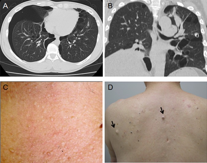
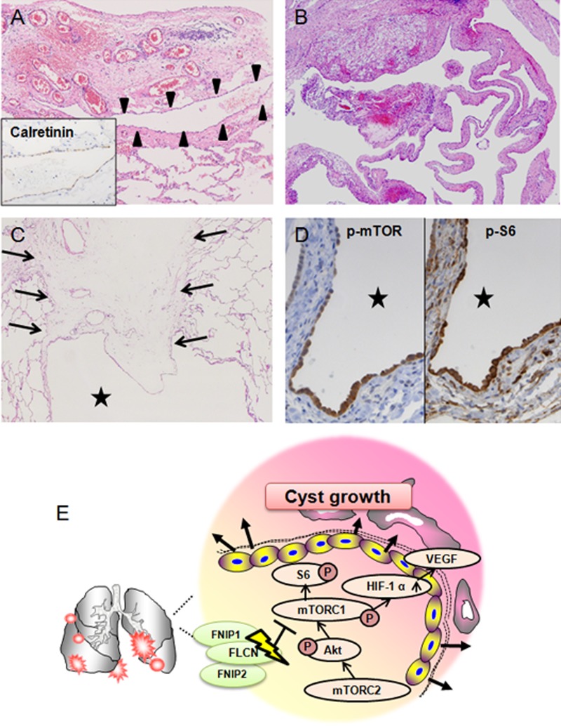
Birt-Hogg-Dubé syndrome (BHD) is an autosomal dominant inherited disorder characterised by fibrofolliculomas, renal tumours, pulmonary cysts and pneumothorax. The pulmonary cysts and repeated episodes of pneumothorax are the clinical hallmarks for discovering families affected by the syndrome. This disorder is caused by mutations in the gene coding for folliculin (FLCN). FLCN forms a complex with FLCN-interacting protein 1 (FNIP1) and FNIP2 (also known as FNIPL), and the complex cross-talks with signalling molecules such as 5'-AMP-activated protein kinase (AMPK) and the mammalian target of rapamycin (mTOR). Heterozygous Flcn knockout mice and rats with Flcn gene mutations develop renal cysts, adenomas and/or carcinomas. These findings suggest that FLCN functions as a tumour suppressor that inhibits renal carcinogenesis. However, the mechanisms of the formation of pulmonary cysts and pneumothorax associated with heterozygous mutations in FLCN are poorly understood. Resected lung specimens from patients with BHD are often misdiagnosed by pathologists as non-specific blebs or bullae or emphysema, and patients with BHD who have pulmonary cysts and repeated pneumothorax frequently do not receive appropriate medical investigations. This review discusses the clinical and pathological features of lungs of patients with BHD, focusing on the diagnostic pathology and possible mechanisms of cyst formation.


Figures

Figure 1
Figure 1
Thoracic CT scans and skin tumours of patients with Birt-Hogg-Dubé syndrome. (A) A 33-year-old woman who had a pneumothorax six times showing cysts localised in the perimediastinal subpleura. (B) A 53-year-old woman who had a pneumothorax twice showing most of the cysts in contact with the interlobular septum. (C) A 69-year-old woman with renal cell carcinoma who had multiple skin papules on the neck. (D) A 76-year-old man with a few flat-topped fibrous papules on the back (arrows).
Figure 2
Figure 2
Histopathological features of lungs affected by Birt–Hogg–Dubé syndrome (BHD): H&E staining of resected lungs from patients diagnosed with BHD by genetic testing. Cysts are indicated by stars. (A) A resected cyst from a 41-year-old woman. The cyst wall partially incorporates the pleura (arrowheads) and interlobular septum (arrows). (B) A double-spaced microscopic cyst from the same patient. The inner surface is lined by pneumocytes (inset) and is partially embedded in an interlobular septum (reproduced from Koga et al with permission of the publisher). (C) Innermost layer of a cyst: the lining cells show neither aberrant proliferation nor pleomorphic features. They are immunostained for proSP-C, suggesting pneumocytes. (D) A few alveoli abut on the innermost layer and are anastomosed to the cyst lining cells. (E) Finger-like protrusion of a vein into the cystic space. (F) Epithelial spores lining the inner layer of the cyst wall (left), which is highlighted by immunostaining for cytokeratin CAM5.2 (right).
Figure 3
Figure 3
Histopathological features of ruptured cysts and hypothetical mechanism of cyst growth. (A) Ruptured visceral pleura thickened with fibrosis and inflammation. Mesothelial invagination (arrowheads) is observed. Inset shows the invaginated cells immunostained for calretinin. (B) Elongated cysts form a complicated structure. (C) Interlobular septum (arrows) associated with a cyst shows oedematous features. (D) The cyst lining epithelial cells are strongly immunostained for p-mammalian target of rapamycin (mTOR) (left) and p-S6 (right). (E) A hypothetical signalling pathway involved in pulmonary cyst growth. Folliculin insufficiency in cyst composing cells leads to mTOR activation and accelerates downstream molecules such as p-S6 and vascular endothelial growth factor (VEGF). FNIP, FLCN-interacting protein; HIF-1α, hypoxia-inducible factor 1α; mTORC1 and 2, mTOR complex 1 and 2.
Figure 4
Figure 4
Schemes of mutation patterns of 19 Asian families with Birt–Hogg–Dubé syndrome (BHD). The responsible mutation sites of the 19 Asian families are indicated. F6 is excluded because genetic testing was done in another institute. F5 is Taiwanese and the others are Japanese families with BHD. The most frequent mutations are 4 bp deletion in exon 13 (c.1533_1536delGATG) and duplication of cytosine in exon 11 (c.1285dupC).

References

    1. Birt AR, Hogg GR, Dube WJ. Hereditary multiple fibrofolliculomas with trichodiscomas and acrochordons. Arch Dermatol 1977;113:1674–7 - PubMed
    1. Hornstein OP, Knickenberg M. Perifollicular fibromatosis cutis with polyps of the colon: a cutaneo-intestinal syndrome sui generis. Arch Dermatol Res 1975;253:161–75 - PubMed
    1. Happle R. Hornstein-Birt-Hogg-Dube syndrome: a renaming and reconsideration. Am J Med Genet A 2012;158A:1247–51 - PubMed
    1. Khoo SK, Bradley M, Wong FK, et al. Birt-Hogg-Dube syndrome: mapping of a novel hereditary neoplasia gene to chromosome 17p12-q11.2. Oncogene 2001;20:5239–42 - PubMed
    1. Schmidt LS, Warren MB, Nickerson ML, et al. Birt-Hogg-Dube syndrome, a genodermatosis associated with spontaneous pneumothorax and kidney neoplasia, maps to chromosome 17p11.2. Am J Hum Genet 2001;69:876–82 - PMC - PubMed

Publication types

MeSH terms