. 2013 Feb;24(3):194-209.
doi: 10.1091/mbc.E12-07-0565. Epub 2012 Dec 5.

A novel Rac1 GAP splice variant relays poly-Ub accumulation signals to mediate Rac1 inactivation

Affiliations

A novel Rac1 GAP splice variant relays poly-Ub accumulation signals to mediate Rac1 inactivation

Timothy Y Huang et al. Mol Biol Cell. 2013 Feb.

Abstract

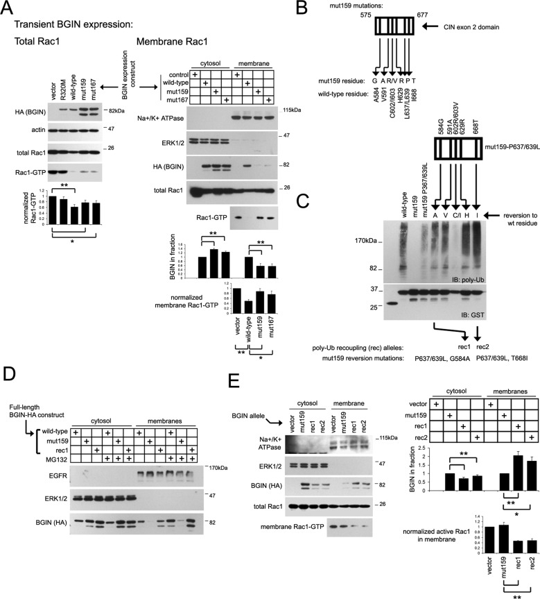
Spatial control of RhoGTPase-inactivating GAP components remains largely enigmatic. We describe a brain-specific RhoGAP splice variant, BARGIN (BGIN), which comprises a combination of BAR, GAP, and partial CIN phosphatase domains spliced from adjacent SH3BP1 and CIN gene loci. Excision of BGIN exon 2 results in recoding of a 42-amino acid N-terminal stretch. The partial CIN domain is a poly-ubiquitin (poly-Ub)-binding module that facilitates BGIN distribution to membranous and detergent-insoluble fractions. Poly-Ub/BGIN interactions support BGIN-mediated inactivation of a membranous Rac1 population, which consequently inactivates membrane-localized Rac1 effector systems such as reactive oxygen species (ROS) generation by the Nox1 complex. Given that Ub aggregate pathology and proteotoxicity are central themes in various neurodegenerative disorders, we investigated whether BGIN/Rac1 signaling could be involved in neurodegenerative proteotoxicity. BGIN/Ub interactions are observed through colocalization in tangle aggregates in the Alzheimer's disease (AD) brain. Moreover, enhanced BGIN membrane distribution correlates with reduced Rac1 activity in AD brain tissue. Finally, BGIN contributes to Rac1 inhibition and ROS generation in an amyloid precursor protein (APP) proteotoxicity model. These results suggest that BGIN/poly-Ub interactions enhance BGIN membrane distribution and relay poly-Ub signals to enact Rac1 inactivation, and attenuation of Rac1 signaling is partially dependent on BGIN in a proteotoxic APP context.


Figures

FIGURE 1:
FIGURE 1:
BGIN is an alternative splice product from SH3BP1/CIN loci and comprises a recoded N-terminal BAR domain. (A) Schematic of the human chromosome 22 SH3BP1/CIN locus. Three transcripts have been identified from this locus: 3BP1, CIN, and BGIN (AK126873). (B) Purification and sequence determination of the BGIN N-terminus. The BGIN BAR-domain doublet was expressed, purified, and subjected to mass spectrometry analysis. A peptide demarcating the minimal BGIN N-terminal BAR region was identified (blue sequence), and potential non-AUG initiation codons upstream are indicated. (C) The recoded N-terminal BGIN BAR region is conserved among various mammalian species. CLUSTALW alignment of the recoded reading frame from the CUG (L) start signal between the species indicated; an in-frame stop was detected within this sequence in rat. (D) Multiple tissue Western blots (IMGENEX) were probed with a C-terminal (CIN exon 2) BGIN/CIN antibody. The CIN exon 2 antibody detects both BGIN (73.59 kDa) and CIN (31 kDa). Arrowheads indicate nonspecific bands. (E) Identification of Ub in BGIN complexes by mass spectrometry. BGIN-HAhis6 was purified as described in the Supplemental Methods, and elutions were resolved by SDS–PAGE and stained with Coomassie blue or immunoblotted for HA. Peptides identified for BGIN or Ub in BGIN eluates by mass spectrometry are indicated in gray.
FIGURE 2:
FIGURE 2:
BGIN interacts with poly-ubiquitin through the CIN exon 2 module. (A) HeLa cells were transfected with GST or BGIN-GST constructs and treated with dimethyl sulfoxide (DMSO) or 10 μM MG132 or 17AAG for 5 h, and GST precipitates generated from lysates were immunoblotted for GST or Ub. (B) GST-tagged BGIN constructs were expressed in HeLa cells, and GST precipitates were generated from cells treated with DMSO or MG132 (5 μM, 5 h). Precipitates were immunoblotted for GST or Ub. (C) Recombinant purified GST-Ub chains of the indicated lengths were incubated with recombinant purified CIN exon 2-GFP and precipitated with glutathione–Sepharose. Right, Coomassie stain of the binding reaction input. (D–F) Comparison of Ub-binding modules in vitro and in vivo. (D) A 20-μg amount of recombinant GST-tagged Ub-binding domains immobilized on glutathione–Sepharose was incubated with 20 μg of his6-Ub2 or Ub5, precipitated, and analyzed for GST/Ub by immunoblot. Right, Coomassie stain of binding inputs. (E) GST-tagged Ub-binding domains in pRK5 were transfected into HeLa cells, precipitated with glutathione–Sepharose, and analyzed for GST/Ub. (F) Graphs represent relative Ub binding in vitro (top) and in vivo (bottom; highest value set to 1.0) from two experiments (mean ± SD). Saturated band intensity with Ub5 in vitro binding as indicated will diminish differences detected in CIN exon 2 interactions compared with other Ub-binding domains.
FIGURE 3:
FIGURE 3:
MG132 enhances BGIN-dependent Rac1 inactivation. (A) BGIN expression enhances Rac1/Cdc42 inactivation. Myc-tagged Rac1 or Cdc42 was coexpressed with GFP-tagged BGIN wild-type or R320M constructs in HEK293T cells as indicated, and lysates were subjected to PBD-GST pull-down. Lysates were immunoblotted for GFP and myc-GTPase expression, and PBD-GST precipitates were immunoblotted for GTP-loaded myc-Rac1/Cdc42. (B) Stable BGIN expression inactivates Rac1. Lysates from HeLa cells stably expressing long or short GFP-tagged BGIN isoforms were subjected to PBD pull-down assays. (C) Schematic of BGIN siRNA-targeting oligos. Control HeLa cells or BGIN-expressing clones were transfected with control, CIN1, or CIN4 siRNA oligos as indicated, and GTP-Rac1 was assayed by PBD pull-down assay. (D) BGIN expression results in MG132-responsive Rac1 inactivation. Control or stably expressing BGIN-GFP HeLa cells were treated with MG132 for the time indicated, and Rac1-GTP was quantified by PBD pull-down assay. All graphs displayed (A–D) depict Rac1-GTP levels normalized for total Rac1 and compiled from a minimum of three experiments (mean ± SEM, **p < 0.006, *p < 0.05).
FIGURE 4:
FIGURE 4:
Interactions between BGIN and poly-ubiquitin promote BGIN partitioning to membrane fractions. (A) Poly-ubiquitin accumulation triggers BGIN distribution to detergent-soluble membrane fractions. Stably expressing BGIN-GFP HeLa cells were treated with MG132, and lysates were separated into cytosolic and detergent-soluble/insoluble membrane fractions. Quantification of Rac1 in cytosolic and detergent-soluble membrane fractions are presented as mean ± SEM from three independent experiments, where the peak intensity is set to 1.0 (**p < 0.001). (B) CIN exon 2 mut159, 167, and 168 alleles were subcloned into full-length GST-BGIN constructs, expressed in HeLa cells, and assayed for poly-Ub interactions under steady-state and presence of MG132 (5 μM, 5 h) by GST pull-down assay. (C) GST-tagged wild-type BGIN and two poly-Ub refractory mutants (mut159 and mut167) from B were expressed in untreated or MG132-treated (5 μM, 5 h) HeLa cells and separated into the subcellular fractions indicated. Equal protein quantities were immunoblotted from each fraction and quantified as a ratio in comparison to the wild-type BGIN (set to 1.0). Values of mean ± SEM from three independent experiments are presented (**p < 0.003). (D) Morphology of cells stably expressing wild-type or mut159 BGIN-GFP. A total of 105 cells seeded on 15-mm coverslips were stained for F-actin and scored for a multipolar morphology (more than two apexes) or an elongated/bipolar phenotype (primarily two apexes). Infrequent appearance of rounded cells were excluded from scoring. Images shown are inverted phalloidin-stained images. The graph displayed are cumulative percentages of multipolar/bipolar cells from four experiments (mean ± SD, **p < 0.0005). (E) Cell spreading on fibronectin. HeLa cells transiently expressing GFP alone or constitutively active GFP-Rac1 Q61L or stably expressing wild-type or mut159 BGIN-GFP were seeded onto 0.0001% fibronectin coverslips for 45 min and costained for actin and paxillin. Total cell area was measured under each condition from three independent experiments (mean ± SD shown in the adjacent graph, **p < 3 × 10⁻⁹).
FIGURE 5:
FIGURE 5:
Interactions with poly-Ub promote BGIN-mediated Rac1 inactivation at the membrane. Uncoupling poly-Ub interactions attenuates BGIN-dependent Rac1 inactivation. (A) HEK293T cells coexpressing the myc-Rac1 and BGIN-HA constructs indicated were subjected to PBD pull-down assay as described in the Supplemental Methods to determine total (left) or membrane-specific Rac1-GTP levels (right). Graphs depict BGIN levels normalized to wild-type BGIN (set to 1.0) or normalized Rac1-GTP levels from four experiments (mean ± SEM, *p < 0.05, **p < 0.005). (B) Schematic of mutated residues in the mut159 allele within the BGIN aa 575–677 region. (C) Reversion scheme of the mut159 allele. The mut159 allele with an L637/639P reversion pair was coupled to an additional reversion to wild-type residue, and GST-tagged BGIN575-677 constructs were assayed for poly-Ub recoupling. Two mut159 mutational reversion combinations demonstrating poly-Ub recoupling, rec1 (P637/639L, G584A) and rec2 (P637/639L, T668I), were selected for further analysis. (D) Recoupling BGIN/poly-Ub interactions restores BGIN membrane distribution. HeLa cells were transfected with HA-tagged full-length BGIN wild-type, mut159, or rec1 alleles, treated with 5 μM MG132 for 5 h as indicated, and subjected to cytosol/membrane fractionation and analysis by immunoblotting. (E) Recoupling BGIN/poly-Ub interactions restores BGIN-dependent Rac1 inhibition in detergent-soluble membranes. HEK293T cells coexpressing myc-Rac1 and the full-length HA-tagged BGIN constructs were subjected to membrane fractionation and PBD pull-down assay as described in Materials and Methods. Quantification of BGIN in cytosol and membrane fractions and normalized membrane Rac1-GTP levels are shown as averages ± SEM of four experiments (**p < 0.006, *p < 0.02).
FIGURE 6:
FIGURE 6:
BGIN inhibits ROS generation through the membrane-associated Nox1 complex. (A) The Nox1 complex and its activator/organizer components NoxA1 and NoxO1 (and the ubiquitous p22 subunit) mediate ROS generation through Rac1. A membrane-associated BGIN population may modulate membranous Rac1 activity to attenuate Nox1-dependent ROS generation. (B) Uncoupling BGIN/poly-Ub interactions attenuates BGIN inhibition of Nox1-dependent ROS generation. NoxO1/NoxA1/Nox1 and BGIN constructs were transfected in HEK293T cells as described earlier and processed for chemiluminescence/immunoblotting. (C) Recoupling BGIN/poly-Ub interactions restores BGIN inhibition of Nox1-dependent ROS generation. HEK293T cells were transfected and processed for chemiluminescence/immunoblotting as described. For B and C, a portion of the transfected cells was assayed for ROS activity, and lysates from the remainder were probed for myc-NoxA1/O1 and BGIN expression. Quantification of ROS generation by chemiluminescence assay is shown below with transfection of all three Nox (O1/A1/Nox1) components (last lane) set to 1.0, with all other samples expressed as a comparative ratio (mean ± SE of triplicate samples). A kinetic curve measurement from a representative sample is shown in the line graph (ROS is measured in relative light units [RLU]). (D) siRNA-directed depletion of BGIN in SH-SY5Y cells. Cells transfected with 20 nM siRNAs in stably expressing BGIN SH-SY5Y (left) or normal SH-SY5Y cells (right) were immunoblotted for the components indicated. (E) BGIN depletion increases ROS generation. SH-SY5Y cells transfected with the BGIN-targeting CIN4 oligo result in a slight elevation of ROS generation. Bar graphs from B, C, and E are derived from average ± SE of mean values from at least four independent experiments (**p < 0.002, *p < 0.02) and normalized as described, with a representative kinetic ROS measurement depicted in adjacent line graphs. (F) BGIN overexpression results in colocalization with Ub at perinuclear granules. BGIN-GFP–expressing cells were costained for Ub, and BGIN-GFP perinuclear tubules were examined for Ub staining. Magnified view of perinuclear BGIN-tubule clusters are numbered in merged color images (bottom) and displayed adjacently in numbered magnified panels.
FIGURE 7:
FIGURE 7:
BGIN localizes to fibrillary tangles and partitions to membranes in AD. (A) BGIN is present in dense-staining pathological aggregates in AD brain. Cortical control/AD sections from human patients (top) or control/APP transgenic mice (bottom) were stained with an affinity-purified BGIN antibody recognizing the unique BGIN N-terminus (bar,10 μm). (B) BGIN and Rac1 localize to neurofibrillary tangles in AD. Human AD brain sections were stained using antibodies labeling BGIN (top) or Rac1 (bottom; green) and costained with a fluorescent antibody for pTau (PHF; red). Merged panels are displayed adjacently (bar, 10 μm). (C) Cytosolic, detergent-soluble membranes and insoluble fractions were generated from human brain tissue by ultracentrifugation as described in the Supplemental Methods. A 30-μg amount of protein from cytosol, membranes, and insoluble fractions from a control sample was immunoblotted for ERK1/2, Na+/K+ ATPase, and actin to determine the fractionation quality. (D) Left, signaling schematic of the BGIN/Rac1 pathway probed. Cytosolic (D) and detergent-soluble membrane fractions (E) from brain tissue of seven AD (numbered 1–7) and four control (1–4) patients were immunoblotted for the components indicated. BGIN, Rac1-GTP (normalized for total Rac1) and p-cofilin (normalized with total cofilin) bands are expressed as averages ± SE between groups, with the control mean set to 1.0 (*p < 0.05, ***p < 0.002).
FIGURE 8:
FIGURE 8:
Determining a role for BGIN in Rac1 signaling with APP proteotoxicity. (A) Probing poly-Ub/CIN exon 2 interactions in APP-expressing SH-SY5Y cells. GST or GST-CIN exon 2 was expressed and precipitated from untransfected or APP-expressing SH-SY5Y cells as indicated and immunoblotted for GST or Ub. Graph indicates poly-Ub smear band density normalized to GST-CIN exon 2 precipitates from three experiments. (B) Stable APP expression attenuates Rac1 activity. Top, normal or APP-expressing SYSH5Y cells were processed to generate total, cytosolic, membrane, or insoluble fractions and immunoblotted. Bottom, total lysates were assayed for Rac1 activity by PBD pull-down assay or cofilin phosphorylation with or without APP expression. (C) Long-term BGIN siRNA treatment elevates Rac1-GTP levels in APP-expressing cells. Normal or APP-expressing SH-SY5Y cells were transfected for three rounds of siRNA as described in the Supplemental Methods and subjected to Rac1-GTP analysis by PBD pull-down assay. Fold change in Rac1-GTP levels with long-term BGIN siRNA treatment in comparison to controls (set to 1.0) were calculated in the lower bar graphs. (D) Cells were transfected as described in C and subjected to chemiluminescence assay for ROS output. Kinetic ROS output values are shown in the upper line graph, and relative ROS values from three experiments measured in triplicate are depicted in the lower bar graph. All bar graph values (B–D) represent mean ± SE from a minimum of three experiments (**p < 0.01, ***p < 0.002). Paired t test values were tabulated for C (*p < 0.02). (E) Left, schematic summary of poly-Ub/BGIN interactions and consequent effects on Rac1/ROS generation. Right, experimental summary of results supporting the key features of the model.

Similar articles

Cited by

References

    1. Alves-Rodrigues A, Gregori L, Figueiredo-Pereira ME. Ubiquitin, cellular inclusions and their role in neurodegeneration. Trends Neurosci. 1998;21:516–520. - PubMed
    1. Arnaud E, Touriol C, Boutonnet C, Gensac MC, Vagner S, Prats H, Prats AC. A new 34-kilodalton isoform of human fibroblast growth factor 2 is cap dependently synthesized by using a non-AUG start codon and behaves as a survival factor. Mol Cell Biol. 1999;19:505–514. - PMC - PubMed
    1. Bedard K, Krause KH. The NOX family of ROS-generating NADPH oxidases: physiology and pathophysiology. Physiol Rev. 2007;87:245–313. - PubMed
    1. Benard V, Bohl BP, Bokoch GM. Characterization of rac and cdc42 activation in chemoattractant-stimulated human neutrophils using a novel assay for active GTPases. J Biol Chem. 1999;274:13198–13204. - PubMed
    1. Billuart P, et al. Oligophrenin-1 encodes a rhoGAP protein involved in X-linked mental retardation. Nature. 1998;392:923–926. - PubMed

Publication types

MeSH terms