

Mitogen-activated protein kinase phosphatase 2 regulates histone H3 phosphorylation via interaction with vaccinia-related kinase 1
- PMID: 23223570
- PMCID: PMC3564537
- DOI: 10.1091/mbc.E12-06-0456
Mitogen-activated protein kinase phosphatase 2 regulates histone H3 phosphorylation via interaction with vaccinia-related kinase 1
Abstract
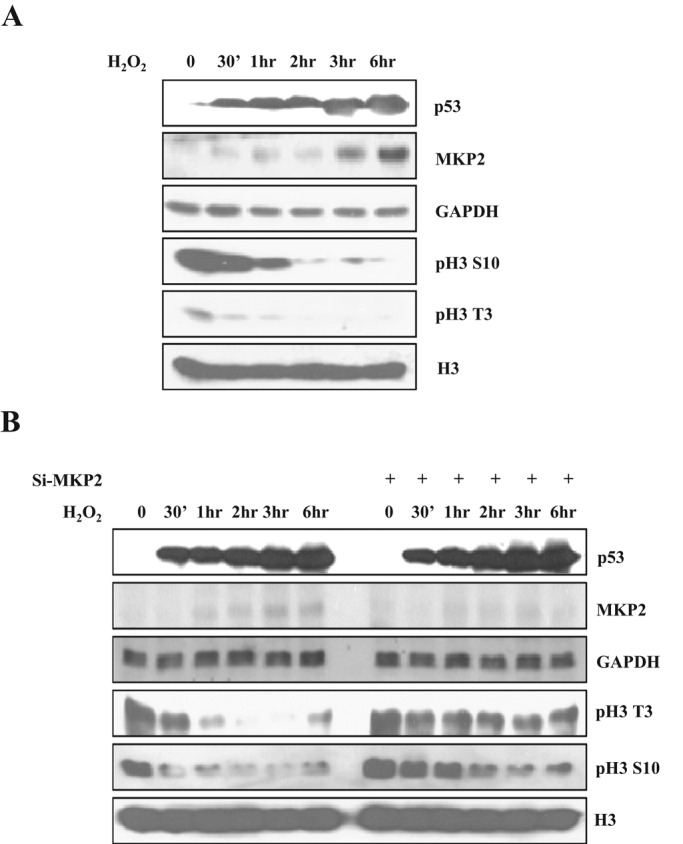
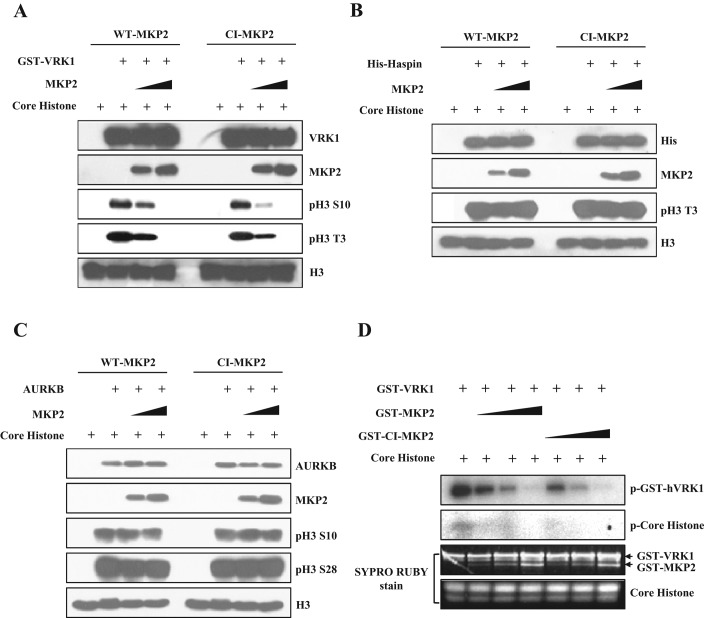
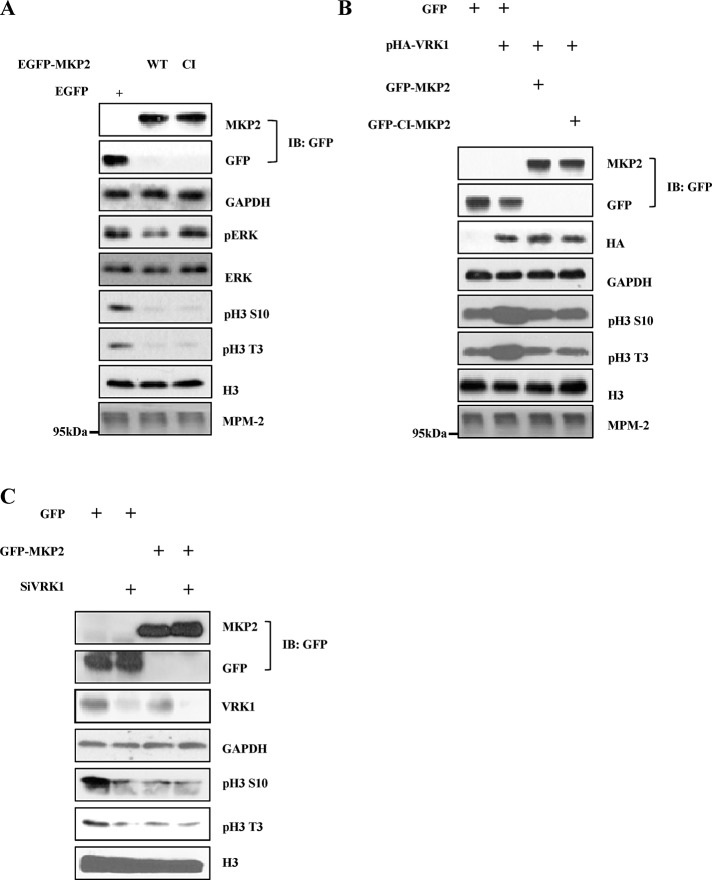
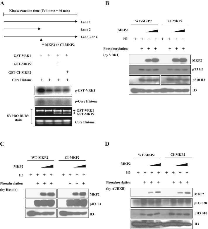
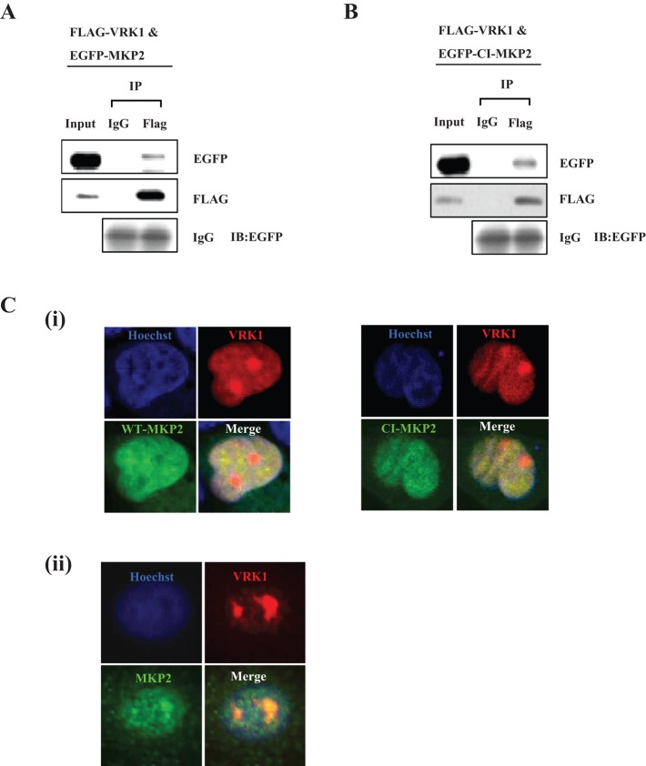
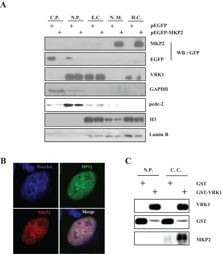
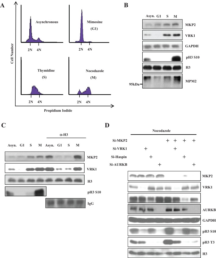
Mitogen-activated protein kinase phosphatase 2 (MKP2) is a member of the dual-specificity MKPs that regulate MAP kinase signaling. However, MKP2 functions are still largely unknown. In this study, we showed that MKP2 could regulate histone H3 phosphorylation under oxidative stress conditions. We found that MKP2 inhibited histone H3 phosphorylation by suppressing vaccinia-related kinase 1 (VRK1) activity. Moreover, this regulation was dependent on the selective interaction with VRK1, regardless of its phosphatase activity. The interaction between MKP2 and VRK1 mainly occurred in the chromatin, where histones are abundant. We also observed that the protein level of MKP2 and its interaction with histone H3 increased from G1 to M phase during the cell cycle, which is similar to the VRK1 profile. Furthermore, MKP2 specifically regulated the VRK1-mediated histone H3 phosphorylation at M phase. Taken together, these data suggest a novel function of MKP2 as a negative regulator of VRK1-mediated histone H3 phosphorylation.
Figures
Similar articles
-
VRK1 and AURKB form a complex that cross inhibit their kinase activity and the phosphorylation of histone H3 in the progression of mitosis.Cell Mol Life Sci. 2018 Jul;75(14):2591-2611. doi: 10.1007/s00018-018-2746-7. Epub 2018 Jan 16. Cell Mol Life Sci. 2018. PMID: 29340707 Free PMC article.
-
Mitotic histone H3 phosphorylation by vaccinia-related kinase 1 in mammalian cells.Mol Cell Biol. 2007 Dec;27(24):8533-46. doi: 10.1128/MCB.00018-07. Epub 2007 Oct 15. Mol Cell Biol. 2007. PMID: 17938195 Free PMC article.
-
Macro histone H2A1.2 (macroH2A1) protein suppresses mitotic kinase VRK1 during interphase.J Biol Chem. 2012 Feb 17;287(8):5278-89. doi: 10.1074/jbc.M111.281709. Epub 2011 Dec 22. J Biol Chem. 2012. PMID: 22194607 Free PMC article.
-
Nuclear functions regulated by the VRK1 kinase.Nucleus. 2024 Dec;15(1):2353249. doi: 10.1080/19491034.2024.2353249. Epub 2024 May 16. Nucleus. 2024. PMID: 38753965 Free PMC article. Review.
-
The human VRK1 chromatin kinase in cancer biology.Cancer Lett. 2021 Apr 10;503:117-128. doi: 10.1016/j.canlet.2020.12.032. Epub 2021 Jan 29. Cancer Lett. 2021. PMID: 33516791 Review.
Cited by
-
Vaccinia-related kinase 1 (VRK1) confers resistance to DNA-damaging agents in human breast cancer by affecting DNA damage response.Oncotarget. 2014 Apr 15;5(7):1770-8. doi: 10.18632/oncotarget.1678. Oncotarget. 2014. PMID: 24731990 Free PMC article.
-
Elevated VRK1 levels after androgen deprivation therapy promote prostate cancer progression by upregulating YAP1 expression.J Cancer Res Clin Oncol. 2025 Mar 20;151(3):116. doi: 10.1007/s00432-025-06168-z. J Cancer Res Clin Oncol. 2025. PMID: 40111564 Free PMC article.
-
VRK1 and AURKB form a complex that cross inhibit their kinase activity and the phosphorylation of histone H3 in the progression of mitosis.Cell Mol Life Sci. 2018 Jul;75(14):2591-2611. doi: 10.1007/s00018-018-2746-7. Epub 2018 Jan 16. Cell Mol Life Sci. 2018. PMID: 29340707 Free PMC article.
-
Advances in synthetic lethality modalities for glioblastoma multiforme.Open Med (Wars). 2024 Jun 10;19(1):20240981. doi: 10.1515/med-2024-0981. eCollection 2024. Open Med (Wars). 2024. PMID: 38868315 Free PMC article. Review.
-
Whole Genome Microarray Analysis of DUSP4-Deletion Reveals A Novel Role for MAP Kinase Phosphatase-2 (MKP-2) in Macrophage Gene Expression and Function.Int J Mol Sci. 2019 Jul 12;20(14):3434. doi: 10.3390/ijms20143434. Int J Mol Sci. 2019. PMID: 31336892 Free PMC article.
References
-
- Ajiro K, Yasuda H, Tsuji H. Vanadate triggers the transition from chromosome condensation to decondensation in a mitotic mutant (tsTM13) inactivation of p34cdc2/H1 kinase and dephosphorylation of mitosis-specific histone H3. Eur J Biochem. 1996a;241:923–930. - PubMed
-
- Ajiro K, Yoda K, Utsumi K, Nishikawa Y. Alteration of cell cycle-dependent histone phosphorylations by okadaic acid. Induction of mitosis-specific H3 phosphorylation and chromatin condensation in mammalian interphase cells. J Biol Chem. 1996b;271:13197–13201. - PubMed
-
- Baek SH. When signaling kinases meet histones and histone modifiers in the nucleus. Mol Cell. 2011;42:274–284. - PubMed
-
- Boutros T, Chevet E, Metrakos P. Mitogen-activated protein (MAP) kinase/MAP kinase phosphatase regulation: roles in cell growth, death, and cancer. Pharmacol Rev. 2008;60:261–310. - PubMed
Publication types
MeSH terms
Substances
LinkOut - more resources
Full Text Sources
Other Literature Sources
Miscellaneous
