. 2013 Feb 1;288(5):3346-58.
doi: 10.1074/jbc.M112.418046. Epub 2012 Dec 7.

Thioredoxin and thioredoxin reductase control tissue factor activity by thiol redox-dependent mechanism

Affiliations

Thioredoxin and thioredoxin reductase control tissue factor activity by thiol redox-dependent mechanism

Pei Wang et al. J Biol Chem. .

Abstract

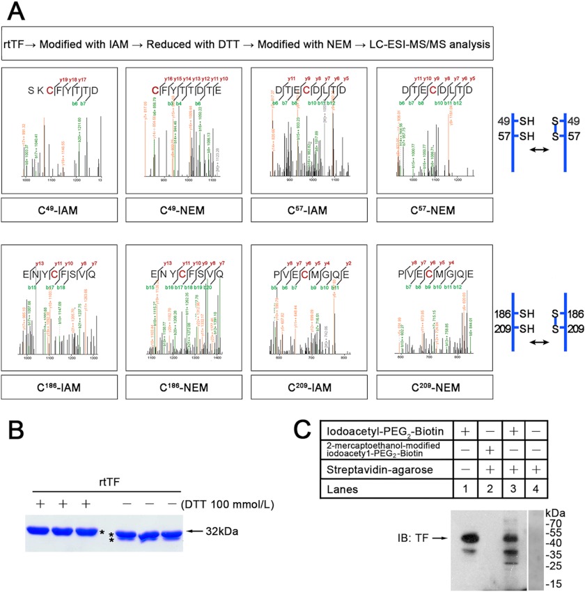
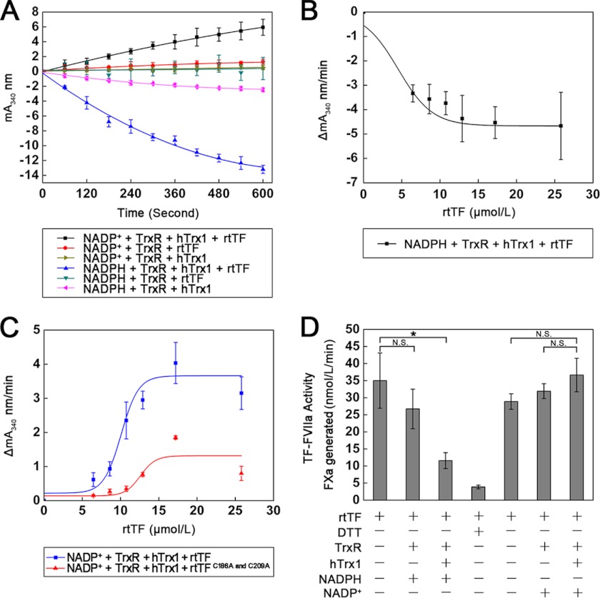
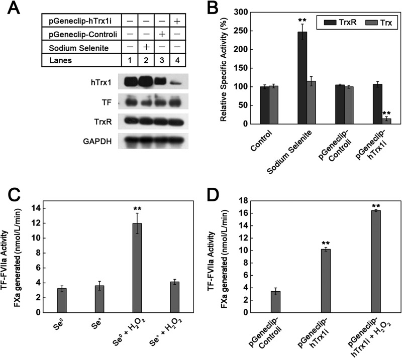
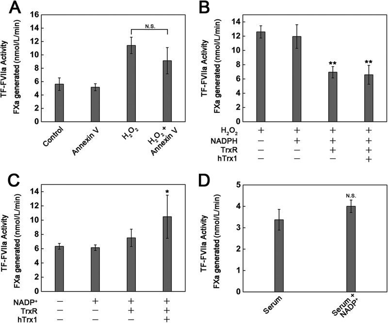
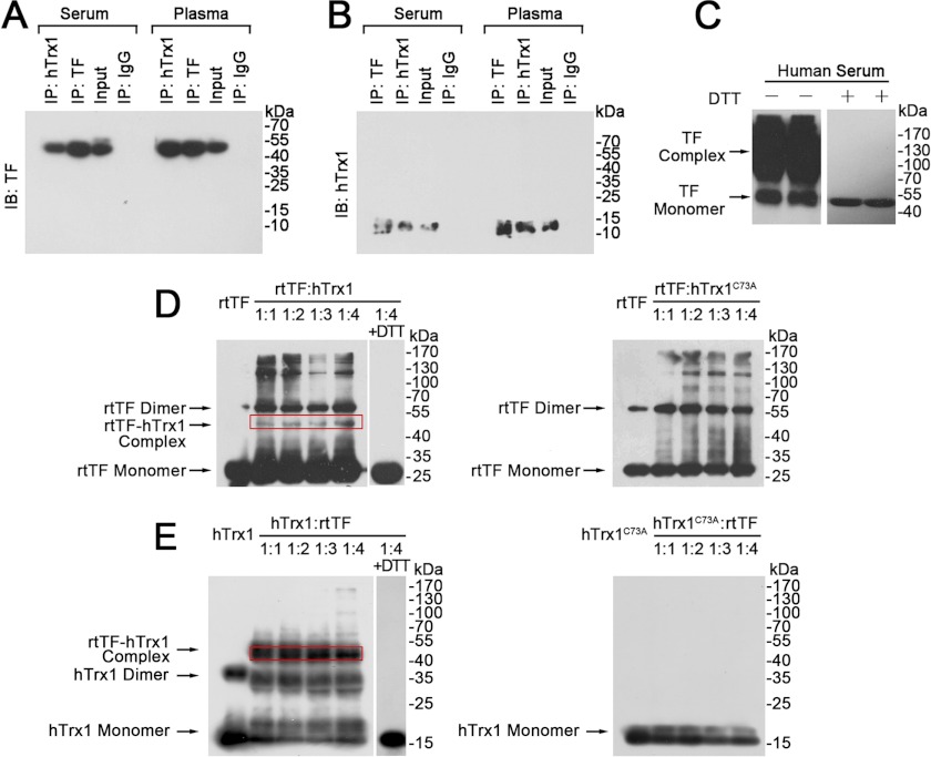
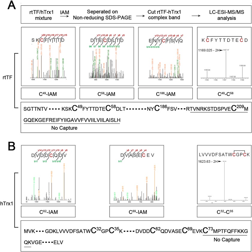
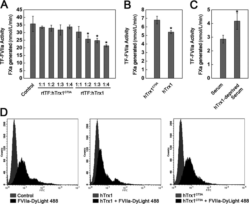
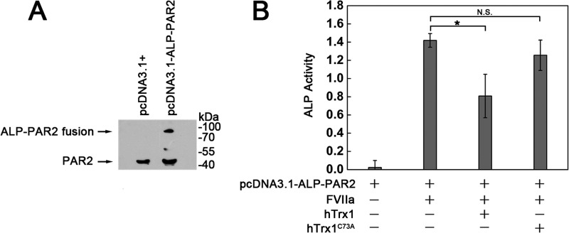
Abnormally enhanced tissue factor (TF) activity is related to increased thrombosis risk in which oxidative stress plays a critical role. Human cytosolic thioredoxin (hTrx1) and thioredoxin reductase (TrxR), also secreted into circulation, have the power to protect against oxidative stress. However, the relationship between hTrx1/TrxR and TF remains unknown. Here we show reversible association of hTrx1 with TF in human serum and plasma samples. The association is dependent on hTrx1-Cys-73 that bridges TF-Cys-209 via a disulfide bond. hTrx1-Cys-73 is absolutely required for hTrx1 to interfere with FVIIa binding to purified and cell-surface TF, consequently suppressing TF-dependent procoagulant activity and proteinase-activated receptor-2 activation. Moreover, hTrx1/TrxR plays an important role in sensing the alterations of NADPH/NADP(+) states and transducing this redox-sensitive signal into changes in TF activity. With NADPH, hTrx1/TrxR readily facilitates the reduction of TF, causing a decrease in TF activity, whereas with NADP(+), hTrx1/TrxR promotes the oxidation of TF, leading to an increase in TF activity. By comparison, TF is more likely to favor the reduction by hTrx1-TrxR-NADPH. This reversible reduction-oxidation reaction occurs in the TF extracellular domain that contains partially opened Cys-49/-57 and Cys-186/-209 disulfide bonds. The cell-surface TF procoagulant activity is significantly increased after hTrx1-knockdown. The response of cell-surface TF procoagulant activity to H(2)O(2) is efficiently suppressed through elevating cellular TrxR activity via selenium supplementation. Our data provide a novel mechanism for redox regulation of TF activity. By modifying Cys residues or regulating Cys redox states in TF extracellular domain, hTrx1/TrxR function as a safeguard against inappropriate TF activity.


Figures

FIGURE 1.
FIGURE 1.
Cys redox states of TF. A, shown is identification of Cys redox states in rtTF by LC-ESI-MS/MS. Free thiols are labeled with IAM, and DTT-induced thiols are labeled with NEM. B, shown are SDS-PAGE and the effect of DTT on electrophoretic mobility of rtTF. C, shown is a Western blot (IB) with anti-TF monoclonal antibody. To analyze free thiols in extracellular domain of cell-surface TF, intact cells were modified with membrane-impermeable thiol-reactive biotinylating reagent. Then the biotinylated proteins were separated using streptavidin-agarose beads. Lane 1, cell lysate control; lane 2, reagent control; lane 3, eluted proteins from the agarose; lane 4, agarose control. These results are representative of at least two independent experiments.
FIGURE 2.
FIGURE 2.
Effect of Trx system on rtTF redox states and activity. A, hTrx1/TrxR-mediated oxidation and reduction of rtTF by NADP(H) is shown. The ratio of hTrx1/rtTF was ∼0.6:1. The formation or consumption of NADPH was measured by following an increase or a decrease in absorbance at 340 nm. B, shown is reduction of rtTF by hTrx1-TrxR-NADPH in a TF concentration-dependent manner. C, shown is oxidation of prereduced rtTF by hTrx1-TrxR-NADP+. The rtTF was treated with 10 mmol/liter DTT followed by dialysis to remove DTT before assay of NADPH formation. D, shown is the effect of hTrx1-TrxR-NADP(H) on rtTF-precoagulant activity. rtTF was incubated with hTrx1-TrxR-NADP(H) at room temperature for 2 h before assay for TF activity. The ratio of hTrx1/rtTF was ∼7:1. All data points in A–D are expressed as the mean ± S.D. (n = 3). *, p < 0.05; N.S., not significant.
FIGURE 3.
FIGURE 3.
Effects of endogenous hTrx1/TrxR on cell-surface TF-procoagulant activity. A, a Western blot tests the effects of hTrx1 knockdown and selenium supplementation on the expression of hTrx1, TrxR, and TF. GAPDH was used as an internal control. B, shown are specific activities of cellular TrxR and Trx. Cell extracts had a protein concentration of 3 mg/ml, 20 μl of which was used in measuring TrxR/Trx activity via super-insulin assay. C, shown is the effect of elevating TrxR activity by selenium supplementation on the basal activity of cell-surface TF as well as on the response of cell-surface TF activity to H2O2. The concentration of H2O2 was 1 mmol/liter, and the treatment time of H2O2 was 15 min. D, shown is the effect of hTrx1 knockdown on cell-surface TF-procoagulant activity. All data points in B–D are expressed as the means ± S.D., n = 5. **, p < 0.01.
FIGURE 4.
FIGURE 4.
Redox-related changes in cell-surface TF-procoagulant activity. A, shown is the effect of Annexin-V on H2O2-stimulated cell-surface TF activity. The cells were exposed to 200 nmol/liter Annexin-V for 15 min after they were treated with 1 mmol/liter H2O2 for 15 min. B, shown is the effect of hTrx1-TrxR-NADPH on the response of cell-surface TF activity to H2O2. The cells were pretreated with 1 mmol/liter H2O2 for 15 min followed by washing twice with PBS, then incubated with 5 μmol/liter hTrx1, 20 nmol/liter TrxR, and 0.2 mmol/liter NADPH at 37 °C for 1 h, which was washed away before the assay of TF activity. C, shown is the effect of hTrx1-TrxR-NADP+ on cell-surface TF activity. The cells were treated with 5 μmol/liter hTrx1, 20 nmol/liter TrxR, and 0.2 mmol/liter NADP+ at 37 °C for 1 h, which was washed away before assay TF activity. D, shown is the effect of human serum with elevated NADP+ levels on cell-surface TF activity. The cells were incubated with 200 μl of human serum or serum plus 0.2 mmol/liter NADP+ for 1 h. All data points in A–D are expressed as the means ± S.D., n = 5. *, p < 0.05; **, p < 0.01; N.S., not significant.
FIGURE 5.
FIGURE 5.
Association of hTrx1 with TF. TF (A) and hTrx1 (B) were detected by Western blotting (IB) after co-immunoprecipitation (IP) from human serum and plasma. C, shown is a Western blot of human serum TF. The same amount of serum protein (140 μg) was loaded per well and separated on SDS-PAGE under non-reducing (left, without DTT) or reducing (right, with DTT) conditions. D, shown is Western blotting analysis of rtTF status in rtTF-hTrx1/hTrx1C73A mixture under non-reducing condition. The immunoblots were detected with anti-TF monoclonal antibody. E, shown is Western blotting analysis of hTrx1 or hTrx1C73A status in rtTF-hTrx1/hTrx1C73A mixture under non-reducing condition. The immunoblots were detected with anti-hTrx1 monoclonal antibody. These results are representative of at least three independent experiments.
FIGURE 6.
FIGURE 6.
hTrx1-Cys-73 bridges rtTF-Cys-209. The mixture of hTrx1 and rtTF was incubated with IAM to block free thiols and then separated by non-reducing SDS-PAGE. The protein band corresponding to TF-hTrx1 complex was cut out for in-gel tryptic digestion and LC-ESI-MS/MS analysis. A, MS/MS signals showed those corresponding to IAM-labeled Cys-47, Cys-59, and Cys-189 from rtTF. MS signals revealed a fragment containing Cys-47/-59 disulfide of rtTF. A peptide containing rtTF-Cys-209 was missing. B, MS/MS signals showed those corresponding to IAM-labeled Cys-62 and Cys-69 from hTrx1. MS signals contained a fragment containing Cys-32/-35 disulfide of hTrx1. A peptide containing Cys-73 was missing.
FIGURE 7.
FIGURE 7.
hTrx1 is specifically involved in interfering with FVIIa binding to TF. A, the inhibitory effect of hTrx1 on rtTF activity was absent using hTrx1C73A mutant instead of wild-type hTrx1. hTrx1 (or hTrx1C73A) and rtTF were incubated at room temperature for 1 h. B, hTrx1-Cys-73 was required for hTrx1 to inhibit cell-surface TF activity. C, a positive effect of hTrx1-deprived serum on cell-surface TF activity shown is shown. The cells were treated with human serum or hTrx1-deprived serum at 37 °C for 30 min. D, shown is flow cytometry analysis. hTrx1-Cys-73 was implicated in reducing FVIIa binding to cell-surface TF. These results are representative of three independent experiments. The results shown in A–C are expressed as the means ± S.D., n = 5. *, p < 0.05.
FIGURE 8.
FIGURE 8.
hTrx1-Cys-73 is required for hTrx1 to inhibit TF·FVIIa-dependent activation of PAR2. A, Western blotting analysis of PAR2 expression is shown. Transient expression of ALP-PAR2 fusion protein in MDA-MB-231 cells was examined using anti-PAR2 monoclonal antibody. B, shown is determination of PAR2 cleavage (activation) by measuring increased ALP activity in culture medium. The cells bearing ALP-PAR2 were treated with serum-free medium containing hTrx1 or hTrx1C73A. Then the cells were washed once with PBS followed by the addition of FVIIa. After 1 h of incubation, the cell culture medium was collected and used for analyzing ALP activity. The results are expressed as the means ± S.D., n = 5. *, p < 0.05; N.S., not significant.

Similar articles

Cited by

References

    1. Cimmino G., Golino P., Badimon J. J. (2011) Pathophysiological role of blood-borne tissue factor. Should the old paradigm be revisited? Intern. Emerg. Med. 6, 29–34 - PubMed
    1. Rao L. V., Rapaport S. I. (1988) Activation of factor VII bound to tissue factor. A key early step in the tissue factor pathway of blood coagulation. Proc. Natl. Acad. Sci. U.S.A. 85, 6687–6691 - PMC - PubMed
    1. Riewald M., Ruf W. (2003) Science review. Role of coagulation protease cascades in sepsis. Crit. Care 7, 123–129 - PMC - PubMed
    1. Riewald M., Ruf W. (2001) Mechanistic coupling of protease signaling and initiation of coagulation by tissue factor. Proc. Natl. Acad. Sci. U.S.A. 98, 7742–7747 - PMC - PubMed
    1. Belting M., Ahamed J., Ruf W. (2005) Signaling of the tissue factor coagulation pathway in angiogenesis and cancer. Arterioscler. Thromb. Vasc. Biol. 25, 1545–1550 - PubMed

Publication types

MeSH terms

LinkOut - more resources